Introduction
I would first like to thank the Kavli Foundation and the Materials Research Society for giving me the opportunity to present this talk. I will show the ways in which we are fighting cancer with nanoparticle medicines (nanomedicines). This is an area where the nanoscale is very important, and I will provide some illustrations to demonstrate it. First, I will provide some background about cancer, and then second, I will discuss nanoparticles and their potential to create new ways to treat cancer. Nano-sized particles can have a major effect in trying to attack solid tumors, and I will show data from the clinic where we have been treating patients since 2006.
People have been trying to find a cure for cancer for a long time; there is evidence of writings from ancient Egypt that describe cancer in papyrus manuscripts from 1600 BC. Toward the end of the last century, cancer became the number one killer of Americans (in the United States) under the age of 85, replacing heart disease.Reference Jemal, Siegel, Xu and Ward1 The good news is that deaths due to heart disease have declined significantly over the past few decades, whereas deaths from cancer have remained relatively constant. As one might expect, based on data from the World Bank,2 this phenomenon is not confined to the United States, but is a worldwide problem: the total number of deaths from cancer is larger than the total number of deaths from malaria, HIV, and tuberculosis, and this number is predicted to increase significantly as the population increases. The costs of therapies to treat cancer have increased dramatically over the last decade or so, and this increase is unsustainable in the long term. The cost of cancer to society is not only the cost of the drugs used, but also the loss of life of young people, and the consequent loss of productivity. Healthcare costs for cancer now surpass that for all other diseases or injuries, including those from road accidents and heart disease;Reference O’Callaghan3thus, this is a very large problem, and one that will increase.
Most of us know somebody in our neighborhood, or in our family, who is currently undergoing cancer therapy. Patients suffer a significant loss in quality of life, whether it is acute or long-term side effects. We therefore have a number of reasons for developing new therapeutics that can lower the death rate and maintain a high quality of life for patients. In order to do this, we need to attack at least two problems: metastatic disease and drug-resistant disease. Cancer is a metastatic disease; it can spread from its original site to multiple sites simultaneously. If we wish to attack metastatic disease, we require therapies that act throughout the body. Additionally, cancer can “fight back” when being treated by chemotherapeutics. When cancer is treated with conventional small-molecule chemotherapeutics, one of the ways it can “fight back” is to put proteins on the surface of the cell that act as chemical pumps. When the molecules used to treat the patient are exposed to the cancer cells, the cells pump the drug out, and the treatment is ineffective. This phenomenon is called “multidrug resistance”; the cell can pump drugs out even if they have never been used on the patient before, thus, whole classes of drugs become ineffective. When this happens to a patient, after numerous treatments for example, very little can be done. To find a solution to this problem, (1) we need to develop new therapies that can be administered systemically and can treat metastatic disease; and (2) it will not be sufficient to apply the therapy to the tumor itself, but we will need to move it through the tumor and into the cancer cells before it is released to carry out its cell-killing properties. This problem is much more difficult than just getting drugs to the tumor.
Current cancer treatments
Since 1955, the major cancer treatment has been chemotherapy. The first drug used for metastatic cancer was methotrexate; which was followed by a series of small molecules (e.g., Adriamycin, Carboplatin, and Taxol) through the 1970s to 1990s. These small-molecule treatments are used frequently; for example, billions of dollars’ worth of Taxol is sold each year. Some of these drugs are administered orally, but most of them are administered intravenously (IV); they move through the whole body by diffusion and convection, both into and throughout cells in the various areas they can reach. The primary function of these molecules is to inhibit cell division, but most of the dose quickly exits via the kidney and into the urine. The small amount of the dose that remains in the body enters all types of cells, and this leads to a number of different side effects. If the drug enters the hair follicles, it kills those cells, and the patient’s hair or eyebrows fall out; if it enters the cells in the gastrointestinal tract, then it causes vomiting; and if it enters the bone marrow, it causes loss of cells that make up the immune system and other blood cells. Hopefully, some of the drug also kills the cancer cells, but if the cancer has become multidrug resistant, this can result in a number of harmful side effects, with no effect on the cancer.
More recent treatments use “targeted, molecular medicines.” These can still be small molecules, such as Gleevec, but now antibody molecules that are ∼1–5 nm in size are being used. They still can have significant side effects, but they are more selective in hitting targets.
Nanoscale treatment of cancer
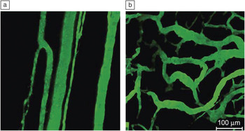
I would like to first briefly discuss some aspects of tumor biology. When either primary tumors or metastatic tumors become larger than ∼1 mm in size—roughly the thickness of a credit card—they need to create new blood vessels to be able to bring oxygen and nutrients to continue to grow, so they send out signals to the current blood vessels to grow new ones into the tumor mass. These grow very quickly and are different from the normal, mature blood vessels in the body. Figure 1 shows a comparison of normal blood vessels and those in a tumor.Reference Dreher, Liu, Michelich, Dewhirst, Yuan and Chilkoti4The vasculature for normal cells is leaky to molecules that are ∼1 nm or smaller in size; these are the molecules needed for nourishment. In a tumor, however, these vessels are formed quickly, and they are not completely closed, as in the case of a mature vessel, so entities that are tens or even hundreds of nanometers in size can leak out. The question is whether we can exploit this difference to introduce nanoscale entities into the tumor. Of course, this requires an injection into the circulatory system of a patient: we want to move nanoparticles into a tumor, but they also pass through the circulatory system and enter other organs in the body. When we started this work in the 1990s, there was very little information known about the way nanoscale objects interact with these organ systems. Our aim is to minimize the interaction with these organ systems and ensure the nanoparticles enter the tumor.
Comparison of (a) normal vasculature (impermeable to entities larger than ca. 2–4 nm) and (b) tumor vasculature (impermeable to entities larger than ca. 400–700 nm). Reprinted with permission from Reference 4. ©2006, National Cancer Institute.

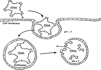
This is illustrated in a schematic from my 1996 patent application (Figure 2).Reference Davis5Our idea was to create a stable colloid (these were not called nanoparticles at that time) that could be built with the right properties in order to introduce them into tumor cells. These particles would contain chemical sensors that would recognize that they were inside of the cell and allow them to perform certain functions to release the drug. These were our objectives, and 15 years later, we have met all of them in treating patients.
Initial schematic for cancer treatment with nanoparticles. Issues from the initial design that have carried through to the clinic are cyclodextrin polymer-based colloid (nanoparticle), sizes below 100 nm in diameter, targeted colloid (R in diagram), and intracellular delivery and active mechanism of drug release. Reprinted with permission from Reference 5. ©2009, American Society for Pharmacology and Experimental Therapeutics.

We wanted to try and create new therapies to treat the metastatic drug-resistant problem in a physician’s office, instead of a research hospital, and provide a high quality of life for patients. Of course, to be viable, this treatment needed to be highly efficient. If we can create high quality of life therapeutics, we might be able to treat a patient for a much longer period of time. Also, we wanted to have a robust system, while hopefully keeping the costs under control. At the California Institute of Technology (Caltech) in the mid-1990s, we started with two approaches: one way was to try and build systems that could ultimately obtain FDA approval to treat patients; the other was to build model systems, from which we could understand some of the fundamental principles about the way these nanoparticles interact with various tissues. Very little was known about the way nanoscale entities interacted with the various organs in the body. One of the model systems we used was gold nanoparticles functionalized with polyethylene glycol (PEGylated gold). The research started in 1996, and in 2006, we were able to treat the first patients, which illustrates the time and effort required.
These colloidal particles are now referred to as “nanoparticles,” and they started to become significant in the early 2000s. I believe that one of the main reasons for this was the National Nanotechnology Initiative (NNI), which was started at that time. I was fortunate to be in the audience at Caltech when President Clinton announced the initiation of this program. In the early 2000s, the National Cancer Institute started their program in nanotechnology, and they defined nanoparticles to be between 1 nm and 100 nm in size. In our efforts to attack solid cancers from systemic administrations, we found that the range is much more restrictive, but it is in the middle of this region.
Many of us were very positive about this approach, but others were concerned about the negative aspects of injecting nanoparticles into patients. I was asked to testify in front of a US Senate subcommittee on the safety of nanoparticles, and I explained nanoparticles to the senators by saying that the ratio of a hundred-nanometer particle to that of a soccer ball is of the same order of magnitude change as that of the soccer ball to planet Earth. The senators understood this; they knew a nanoparticle is supposed to be small. It is also interesting to note that a nanoparticle is very large relative to a molecule, so a molecule that is smaller than 1 nm, compared to a 100-nm nanoparticle, has the same ratio as that of the soccer ball to the Goodyear blimp. I do not think that the senators had an appreciation for this point. We learned over that last decade or so that the “Goodyear blimp” is too large for treating solid cancers, and we require something like the size of a hot air balloon relative to a soccer ball.
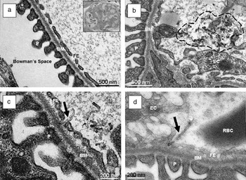
We are therefore trying to learn the rules for making nanoparticles on the order of 50 nm, and I will show why I believe this is approximately the right size. We need to ensure that these highly multifunctional systems perform correctly in the right place and at the right time. These are not passive entities; they must be very dynamic in order to perform the tasks required of them. We will consider size first. As an engineer, I always think of bounds, so what is the lower bound? For example, animal and human kidneys all contain holes, ∼10 nm in diameter, to allow molecules to pass into urine. A nanoparticle larger than 10 nm in diameter, with the right properties, can circulate in the blood for a number of hours, whereas a small molecule would escape directly into the urine. As I mentioned earlier, most small-molecule drugs are delivered directly into the urine, because they are smaller than 10 nm. This is a very fixed bound if we are dealing with a non-deformable spherical particle, but for different types of morphologies and aspect ratios, different results may occur. Figure 3 shows a carbon nanotube passing through one of the pores in a kidney.Reference Lacenda, Herrero, Venner, Bianco, Prato and Kostarelos6, Reference Ruggiero, Villa, Bander, Rey, Bergkvist, Batt, Manova-Todorova, Deen, Scheinberg and McDevitt7Joe DeSimone, of the University of North Carolina at Chapel Hill, has shown that it is possible to make nanoparticles of any shape, and therefore the rules will change depending on both the shape and the aspect ratio. However, the rules we will discuss here are for spherical nanoparticles that are not very deformable.
Excretion of carbon nanotubes by a mouse kidney. (a) Glomerular filtration barrier. Inset: section of renal glomerulus (scale bar is 10 mm); (b) bundled MWNT agglomerate (circled by dashed black line) in the glomerular capillary. Individual MWNTs (black arrow) (c) 5 min and (d) 30 min after IV injection crossing the filtration membrane. P, podocyte; BM, basal membrane; FE, fenestrated endothelium; EC, endothelial cell; RBC, red blood cell. Reprinted with permission from Reference 6. ©2008, Wiley.

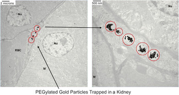
What happens if we make particles greater than 10 nm in size? They will distribute themselves throughout the body and we need to make sure they disassemble; otherwise they will be present in the body for a long time. Figure 4 is an image of 70 nm gold nanoparticles inside a kidney, where they will remain forever. Fitzpatrick et al. showed that even two years after an injection, nanoparticles that have neither disassembled nor dissolved are still present in the kidney.Reference Fitzpatrick, Andreko, Ernst, Waggoner, Ballou and Bruchez8This is unacceptable for treating patients, so we need to make nanoparticles greater than 10 nm in size that circulate but also disassemble when required.
Gold nanoparticles functionalized with polyethylene glycol (PEGylated gold) trapped in a kidney. ZnS capped CdSe quantum dots coated with PEG injected into Balb/c mice were detected two years after injection. The nanoparticles are larger than ca. 10 nm. Figure courtesy of Chung Hang J. Choi (Caltech).

Now consider the upper bound. As one would expect, the smaller the size, the farther these particles can move from the vessel into the tumor tissue. It is now known that 100 nm is definitely far too large, and our preference is 50 nm +/– 20 nm.
All tumors are different, of course, and they are all heterogeneous in nature, but it can be shown that for very permeable tumors, there is not much difference in the 100- to 30-nm range for penetration into the tumors. However, for tumors that are not very permeable, there is great discrimination in size range.Reference Cabral, Matsumoto, Mizuno, Chen, Murakami, Kimura, Terada, Kano, Miyazono, Uesaka, Nishiyama and Kataoka9
Very small particles have a large relative surface area compared to volume, and there are cells in the body whose function is to scavenge nanoparticles from nature, such as viruses and fungi, whose surfaces are all highly electrically negative. It was known in the 1980s, from microspheres, that if these surfaces are made to be almost neutral, the scavenging is minimized, and as they become negative and are more similar to nature’s particles, the scavenging is increased. We must avoid creating positive surfaces in the body, because all other surfaces are negative.
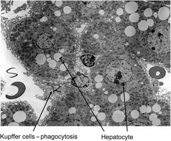
Figure 5 shows a transmission electron microscopy (TEM) image of a section of a liver showing the liver cells; the Kupffer cells marked with a K are the cells that scavenge particles.Reference Popielarski, Hu-Lieskovan, French, Triche and Davis10We made a series of nanoparticles with a constant zeta potential close to neutral and with increasing sizes from 25 nm to 160 nm. These nanoparticles were injected into the tail vein of a mouse, and the TEM images of single cells showed that the difference in scavenging in the liver cells between 100 nm and 70 nm was dramatic. The smaller the particles, the fewer remain in the cells; thus, it is clear that smaller is better, because we do not want to waste valuable therapeutics by allowing them to be taken up in these cells. In essence, the rule that applies at the microscale level is the same rule that applies at the nanoscale level; however, the smaller the nanoparticle, the less scavenging occurs by these cells.
Cells in liver uptake particles; cells marked with K are the cells that scavenge particles. Figure courtesy of S.R. Popielarski (Caltech).

Another point is that these particles need to engage various molecules on the surface of the cells they enter. If the particles are too small, they cannot engage enough of these molecules to cause the membrane to wrap around them; if they are too large, the membrane cannot wrap around them completely. The optimum size for this membrane wrapping is about 40–50 nm.Reference Jiang, Kim, Rutka and Chan11
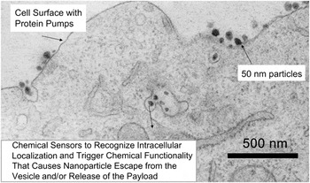
In summary, over the last decade we have learned that these nanoparticles must have sizes between 10 nm and 100 nm. If they are entering cells, they should be as close to electrically neutral as possible, but it is preferable to err on the side of negative charge. This gives us a basis for the design rules we require, independent of the type of drug or payload. Figure 6 shows a TEM image of nanoparticles with desirable properties entering the cells and localizing into vesicles.Reference Mishra, Webster and Davis12It is interesting to note that the pH is near neutral in these vesicles and becomes acidic as they move toward the nucleus of the cell. Therefore, we create chemical entities on the nanoparticles that can recognize this acidity and trigger a number of changes that allow the drug molecules to be released. If we are using big molecules like RNA, we have to actively take them out of vesicles, as they do not diffuse through the vesicle membranes like small-molecule drugs. These systems are highly multifunctional; they stay together during circulation and release the payload in the right place.
Nanoparticles enter cells as individual particles, bypass surface pumps, and deliver their payloads. Reprinted with permission from Reference 12. ©2004, Elsevier.

Patient treatment using nanoparticles
I will now describe some specifics about two nanoparticles that we have been using to treat patients. The first one involves a small-molecule drug, and the second one involves RNA. We started to build new polymers based on a molecule called “cyclodextrin”; this is a ring of sugar. The reason I chose this molecule is primarily based on human data. The human dose of cyclodextrin is 8 grams in the therapeutic called Sporanox; for comparison, a typical dose of Advil or Motrin is 100 mg to 200 mg—up to 1 gram or higher for arthritic patients. These cyclodextrin molecules have low toxicity, and my idea was to use these as molecular building blocks to build polymeric materials with high functionality. We started creating linear polymers, where the cyclodextrin was part of the backbone for a variety of reasons. The first nanoparticle that we created using cyclodextrin-containing polymers carried a small-molecule drug called “camptothecin,” that inhibits a particular protein called “topoisomerase I,” and its mechanism of action would favor having the drug continually bound to topoisomerase I. Camptothecin itself is so toxic that it was never commercialized, although it is a very potent drug for a wide variety of different cancers. However, there are two commercial drugs based on the camptothecin core, but they have other organic functional groups on them to assist in their function in humans. Sales of these commercially available molecules have been over a billion dollars a year, but they have many side effects.
We built a polymer with repeating units of cyclodextrin and a PEG molecule along the backbone. This can be thought of as a long, flexible rope with knots in it; the knots are the cyclodextrins, and the rope in between is the PEG. We attached the camptothecin molecules to this polymer chain. When this polymer is placed in water, the camptothecin molecules hide in the cyclodextrin; they form what is called an “inclusion complex.” Some of the molecules on this polymer chain hide in the cyclodextrins of the same chain, while others also enter cyclodextrins on other chains When this is done correctly, it forms a nanoparticle of ∼30 nm diameter with a slightly negative zeta potential that contains about 5–10 polymer chains. Based on our design rules, this is the right size range and the right charge. This nanoparticle was originally called IT-101, but is now denoted CRLX101. The nanoparticle must also be able to disassemble at the right time, because it is too large to escape through the kidney. Since the drug-cyclodextrin interaction holds the nanoparticle together, it disassembles into single polymer strands when the drug is released; these polymer strands are made of sufficient molecular weight or size so that an individual polymer strand is small enough to escape the body through the kidney. The nanoparticles will circulate, enter the tumor, enter cancer cells, release the drug, and disassemble into single strands that escape from the body through the urine.
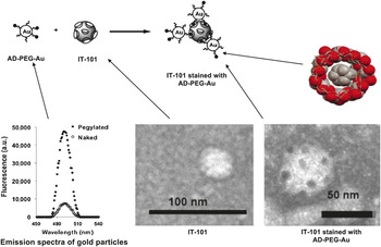
We also used nanotechnology in order to observe these nanoparticles as they accumulate in tumors. We take a 5 nm Au nanoparticle and attach PEG molecules with adamantane at their termini to the gold surface, forming Au-PEG-AD. The adamantane molecule fits snuggly inside the cyclodextrin so that it can bind to the CRLX101 nanoparticle. Since the Au-PEG-AD is a fluorophore, it can be used to locate the positions of the CRLX101 nanoparticles in a tissue sample. Figure 7shows both a cryo TEM image of the therapeutic particle and a cryo TEM image of the gold particles decorating the surface of the therapeutic particle. When we take sections of a tumor, we can use the Au-PEG-AD “stain” to follow the nanoparticles as they move into the tissue and into the cells.
Schematic representation and TEM of the interaction between the 5 nm Au-PEG-AD and 30–40 nm IT-101. The TEM images show the therapeutic particle (left) and gold particles decorating the therapeutic particle surface.

As already mentioned, we can develop the chemistry such that the nanoparticle releases the drug in a pre-programmed way; this particular drug should be released over a long period of time. This means we can retain a slow release agent over a period of days in the tumor only.
After years of work, we started treating patients in the summer of 2006, and the first trial was performed at the City of Hope. We made a freeze-dried product that was then reformulated in water in an IV bag. The fluid in the bag was then infused into the patient. Several patients had survival times of a year or more using this treatment.
It is interesting to note that, while these nanoparticles circulate with a half-life of approximately a day in rats and dogs, they circulated even longer in human patients—with almost a two-day half-life. Patient reproducibility was excellent, which we believe is due to the circulation of nanoparticles not bound to blood components.
From the biopsies of patients 14 days after a dose, we could recognize from that gold stain that there were nanoparticles still in the tumor and releasing the drug. It was also pleasing to note that the side effect profile was extremely low; patients had a high quality of life, and there were no new side effects due to the presence of the nanoparticles.
We started to observe encouraging activity over a variety of different cancers, and the mechanism of long circulation and long drug release we observed in animals has been extended to humans. It is encouraging to find that the rules learned from animals may help us try to learn the rules for application to humans.
CRLX101, a product of Cerulean Pharma, is currently undergoing a randomized Phase II trial for a particular type of lung cancer; the trial involves 150 patients at more than 25 sites, and there are a variety of other Phase II trials that will start in 2012.
RNA as a therapeutic
I now discuss ways of delivering a piece of RNA as a therapeutic. The understanding of the molecular biology of cancer is advancing extremely rapidly. It is now known that cells use many different pathways as they pass through cell growth, cell division, and cell death. In cancer, these pathways become altered, and there is an imbalance such that cells continue to grow all the time. We would like to develop a way to selectively attack multiple pathways in a patient-by-patient way. A new technology is available that may help us do this, called “RNA interference (RNAi),” in which we use a small piece of RNA called a “duplex.” An RNA duplex used for RNAi consists of two small pieces of RNA (around 20–25 base pairs), and when they enter cells, they can interact with some proteins within the cell. The proteins that incorporate one of these strands shuttle it to what is called “messenger RNA” (mRNA), line it up appropriately, and cut that mRNA at a very specific spot. If the mRNA is cut, then the protein that is normally synthesized by that RNA message cannot be created. The understanding of this technology won the Nobel Prize in Medicine and Physiology in 2006 for Craig Mello and Andrew Fire, and they conducted their study using worms. It requires a large transition from worms to human patients, but my colleagues and I reported the first proof of RNAi in patients in 2010. Fire said, “If a person has a tumor, why not take a gene that’s essential for that tumor and administer double stranded RNA corresponding to that gene to shut down growth of that tumor?”Reference Fire13We did just that.
Most drugs work by binding to proteins, and since proteins have many different functions, these drugs must be very specific for each protein and each function. But if you attack the mRNA instead, the molecules you can use are essentially the same as the mRNA. The only thing you are doing is changing the orders of the letters on the duplex RNA. Thus, in principle, we can attack any gene with any function, whereas in the case of drugs, there are a number of proteins that are “undruggable” because we do not know how to drug or attack them. This technology, if it can be developed as a therapeutic, essentially changes the methodology from primarily chemistry to informatics. We can examine which genes have gone wrong and dial in appropriate sequences, but the chemistry is essentially the same.
This process is analogous to a bathtub with faucets. We normally turn the water on, but when we have enough water, we turn it off. This is similar to the way a cell knows how to turn functions on and off before they have mutated. When they mutate, the faucet is on all the time, and the cell is constantly making protein and the cells continue to grow; we are simply creating water, and the water flows out over the tub all of the time. Traditional drugs work at the protein level, and they are spending all their time mopping up the water. The proteins keep being created, and the drugs keep mopping up as much as they can. Using RNAi, we simply turn the faucet off, which can be a much more efficient system to stop the production of protein, and this is independent of the protein being stopped. This method has very high potential to be broadly applicable.
Consequently, we developed a nanoparticle carrying the RNA duplexes, again using a polymer, and some other materials to decorate the surface of the nanoparticle to help it target cancer cells. These pieces of RNA are large relative to chemotherapeutics. We can construct a particle carrying about 2000 RNA molecules as a payload. Once again, we can use the Au-PEG-AD stain because we are using a cyclodextrin particle. Thus, when we examine the tissues, we can see the particles as well as the position of the RNA in order to see that it is being delivered throughout the tumor. We can examine individual cells and again show individual particles circulating from the point where the animal is dosed to the inside of the cancer cell. We started treating patients with these nanoparticles in the spring of 2008, and in 2010, we published the first results showing that this technology can actually be used in a living human being.Reference Davis, Zuckerman, Choi, Seligson, Tolcher, Alabi, Yen, Heidel and Ribas14
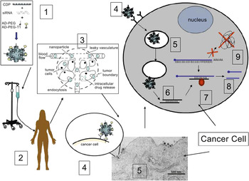
Figure 8 illustrates what we believe happens when we infuse these nanoparticles into the patient: (step 3) they circulate, (step 4) enter the tumor, and (step 5) move into the tumor cells. These nanoparticles contain chemical sensors that can recognize that they have entered the vesicles, and we have built in a mechanism to (step 6) bring them out of the vesicles and release the RNA that then will be (step 7) taken up by the protein machinery, guide it to the mRNA, and (step 8) cut the mRNA to (step 9) stop the production of protein.Reference Davis5 In principle, if this mechanism was acting in the way we believe it is acting, we should see a decrease in the mRNA, a decrease in the protein, and a new fragment of RNA. We were fortunate to obtain biopsies from patients at three different dose levels: 18, 24, and 30 mg-siRNA/m2. When we examined the tissues, we saw that for the lowest dose, using the Au-PEG-AD stain, we saw no nanoparticles. When we examined the intermediate dose, we started to see some nanoparticles, and then at the higher dose, we saw many nanoparticles. After a month, we examined biopsies of one of the patients where we saw the nanoparticles, and we observed that the nanoparticles had all disassembled (the disassembled components are sufficiently small to escape the body via the kidney). After a repeat dose, we saw the nanoparticles returning again, in a very reproducible manner. Thus, for the first time, we have seen a dose-dependent accumulation of these nanoparticles within tumor cells from a systemic administration: this is the first example of this using nanoparticles of any type.
Schematic of the delivery and function of treating a patient with targeted nanoparticles containing RNA.

A very encouraging result was to find that we did not see any nanoparticles in the tissue adjacent to the tumor. We believe that this is good evidence to suggest that these nanoparticles accumulate in tumors through the leaky vessels, but cannot accumulate in the healthy tissue next to the tumor.
When we examined the tissue through staining, we observed reductions in the protein we were trying to inhibit. When we looked for either the protein or the level of the mRNA, we found it is reduced. Additionally, we showed the presence of RNA fragments after dosing, and by sequencing these fragments, we revealed that the mRNA was cut at exactly the right position by the RNAi mechanism. This was the first example showing that we could perform RNAi in a patient. Thus, we have demonstrated that RNAi can be successful in a human patient and gives a high quality of life during treatment.
The future
We are now learning a great deal about design rules and how to control the properties of these nanoparticles to make them more biocompatible and more effective. The newer particles I have just shown have ever-increasing functionality in order to perform the required function at the right place and at the right time. There is no doubt that these nanoparticles will be complex, but hopefully, this will be worth the effort. We can now focus on ways to create very effective therapeutics for solid tumors and give patients a high quality of life.
One thing I am very proud of is that we have been able to stop the production of an individual gene in the tumor of the patient, thus there is no reason we cannot stop the production of multiple genes at the same time. We could take a biopsy from a patient, find which genes are causing their disease, create RNAs to treat the patient, and be able to follow how the treatment of the disease is progressing, maybe by simply taking a prick of blood. In the future, one could envision that there will be an app on a smartphone that will read the information from a prick of blood, call up a physician, and report the results. This will enable the physician to know how the disease is either regressing or progressing, and this information could be used dynamically to decide the next dose, what genes it should attack, and when it should be administered to the patient. This is really a dream, but the basic science and engineering for every step of this process has already been worked out. It has yet to be integrated, but the basic principles of such a system are all in place, and every step of the way required new nanoscience and nanoscale engineering. My hope is that at least some fraction of this will be achieved in the near future.
In conclusion, I hope I have been able to convincingly show very high potential for the use of nanoparticles to create new types of therapies for treatment of solid tumors that provide patients with a high quality of life. It is very encouraging for me to see patients in these early clinical trials having a high quality of life when treated with these therapies.
I have had the great privilege of working with wonderful people everywhere—at Caltech, and with various companies. I would very much like to thank all the patients who have been treated in these trials. It has been a pleasure for me to be present in the treatment rooms with them in a number of different trials. Finally, I would once again like to thank the Kavli Foundation, and MRS, for giving me the opportunity to speak with you tonight.

Mark E. Davis is the Warren and Katharine Schlinger Professor of Chemical Engineering at the California Institute of Technology and a member of the Experimental Therapeutics Program of the Comprehensive Cancer Center at the City of Hope. His research efforts involve materials synthesis in two general areas: namely, zeolites and other solids that can be used for molecular recognition and catalysis and polymers for the delivery of a broad range of therapeutics. He is the founder of Insert Therapeutics Inc. and Calando Pharmaceuticals Inc. He also has been a member of the scientific advisory boards of Symyx and Alnylam. Davis has more than 375 scientific publications, 2 textbooks, and more than 50 patents. He is a founding editor of CaTTech and has been an associate editor of Chemistry of Materials and the AIChE Journal. He also is the recipient of the Colburn and Professional Progress Awards from the American Institute of Chemical Engineers (AIChE), the Ipatieff, Langmuir, Murphree, and Gaden Prizes from the American Chemical Society (ACS), and the National Science Foundation (NSF) Alan T. Waterman Award. He was elected to the National Academy of Engineering in 1997, the National Academy of Sciences in 2006, and the Institute of Medicine of the National Academies in 2011. Davis can be reached by email at mdavis@cheme.caltech.edu.