Acute rheumatic fever occurs as a result of a complex interaction between group A streptococcus, a susceptible host, and the environment. An abnormal immune response to group A streptococcus infection leads to an acute inflammatory illness that most commonly affects the joints, brain, heart, or skin. Although the other manifestations are self-limiting and resolve without sequelae, carditis may result in significant morbidity and mortality.Reference Carapetis, McDonald and Wilson 1 , Reference Narula, Chandrasekhar and Rahimtoola 2 The degree of cardiac involvement is quite variable, ranging from mild, asymptomatic valvulitis to severe carditis, with significant acute mitral and/or aortic regurgitation resulting in heart failure.Reference Ravisha, Tullu and Kamat 3
Left atrial enlargement may occur as a result of valvular insufficiency in acute rheumatic fever.Reference Kocaoglu, Sert and Aypar 4 The resultant electrophysiologic and electromechanical abnormalities of left atrial enlargement are associated with a higher risk of atrial fibrillation.Reference Daubert, Pavin, Jauvert and Mabo 5 These abnormalities include increased atrial electromechanical delay and increased P-wave dispersion. With recent developments in tissue Doppler echocardiography, it is possible to evaluate atrial electromechanical delay with high temporal resolution.Reference Cui, Zhang and Wang 6 , Reference Omi, Nagai and Takamura 7 P-wave dispersion, which is associated with inhomogeneous and discontinuous propagation of sinus impulses, can be evaluated by electrocardiography as the difference between the shortest and longest P-wave durations.Reference Dilaveris, Gialafos and Sideris 8 Increased atrial electromechanical delay and increased P-wave dispersion carry a risk of atrial fibrillation.Reference Cui, Zhang and Wang 6 , Reference Dilaveris and Gialafos 9
In acute rheumatic fever patients, atrial electromechanical delay has not been investigated previously for the detection of atrial conduction abnormalities. The hypothesis of this study is that there might be an increased risk of atrial arrhythmia in patients with acute rheumatic fever, as the conduction system is affected and there is left atrial enlargement in these patients. Therefore, we aimed to evaluate atrial electromechanical delay and P-wave dispersion non-invasively in acute rheumatic fever patients.
Materials and methods
Study population
The study had a prospective design. The study was initiated upon approval from the Hospital Ethics Committee. Patients in the age group of 8–18 years who were in follow-up at Akdeniz University Medical Faculty Pediatric Cardiology Policlinic due to acute rheumatic fever (disease duration: 1–12 years) were included in this study. The diagnosis of all the patients included in the study was made according to the Jones and World Health Organization criteria. 10 In all, 48 patients with acute rheumatic fever, including 26 boys and 22 girls, and a control group of 40 healthy subjects, including 20 boys and 20 girls, were included in the study. Acute rheumatic fever patients without a diagnosis of carditis were excluded from the study.
The acute rheumatic fever patients included in the study were receiving benzathine penicillin G every 21 days. The patients were receiving anticongestive treatment according to the degree of heart failure. Of the 48 acute rheumatic fever patients included in the study, three did not have mitral insufficiency. In all, 16 patients had grade 1 mitral insufficiency, 9 patients had grade 2, 10 patients had grade 3, and 10 patients had grade 4 mitral insufficiency. Furthermore, there was aortic insufficiency varying from grade 1 to 4 in patients with acute rheumatic fever. Aortic valve insufficiency was not identified in 12 of the patients.
Patients with hypertension, left ventricular wall motion abnormality, primary cardiomyopathy, bundle branch block, and mitral stenosis were not included in the patient and control groups. Apart from these, those with technically insufficient echocardiographic images and cases where clear-cut beginning or ending of the P-wave could not be discerned were also excluded from the study.
Echocardiographic examination
In all subjects, two-dimensional, M-mode, pulsed, and colour flow Doppler echocardiographic examinations (Vivid 7 pro, GE, Horten, Norway, 3 MHz transducer) were performed by a cardiologist who was blinded to the clinical details and results of the other investigations of each patient and controls. During echocardiography, a 1-lead electrocardiography was recorded continuously. The systolic function of the left ventricle was evaluated using M-mode echocardiography in the parasternal long-axis view.Reference Kimball and Michelfelder 11 Left atrial dimension determined by M-mode echocardiography was assessed in a plane parallel to the mitral valve annulus in the parasternal long-axis view during atrial end diastole.Reference Patel, Ren and Marchlinski 12
The mitral insufficiency and aortic insufficiency detected with colour Doppler in patients with acute rheumatic fever were performed on the basis of the measurement of jet length. This value was considered as grade 1 when it was equal to or below 1.5 cm, as grade 2 when it was between 1.5 and 2.9 cm, as grade 3 when it was between 3.0 and 4.4 cm, and as grade 4 when it was above 4.5 cm. Grade 1 was accepted as mild insufficiency, grade 2 was accepted as moderate, and grades 3 and 4 were accepted as severe insufficiency.Reference Otto 13
The World Health Organization recommends the following criteria to differentiate pathologic from physiologic mitral and aortic regurgitation: (a) colour jet longer than 1 cm, (b) colour jet evident in at least two imaging planes, (c) colour jet mosaic with a peak velocity >2.5 m/s, and (d) Doppler signal holosystolic for mitral regurgitation and holodiastolic for aortic regurgitation. 14 Pathological valvular insufficiencies were evaluated in accordance with The World Health Organization recommendation.
Atrial electromechanical delay
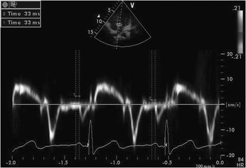
Tissue Doppler echocardiography was performed by transducer frequencies of 3 MHz, adjusting the spectral pulsed Doppler signal filters until a Nyquist limit of 15–20 cm/s was reached, and using the minimal optimal gain. The monitor sweep speed was set at 50–100 mm/s to optimise the spectral display of myocardial velocities. In the apical four-chamber view, the pulsed Doppler sample volume was subsequently placed at the level of left ventricular lateral mitral annulus, septal mitral annulus, and right ventricular tricuspid annulus. The sampling window was positioned as parallel as possible with the myocardial segment of interest to ensure the optimal angle of imaging. Atrial electromechanical delay was defined as the time interval from the onset of the atrial electrical activity (P-wave on surface electrocardiography) to the beginning of the mechanical atrial contraction (late diastolic A-wave; Fig 1). All values were averaged over three consecutive beats. It was measured from the lateral mitral annulus, septal mitral annulus, and right ventricle tricuspid annulus. The difference between lateral mitral annulus atrial electromechanical delay and right ventricle tricuspid annulus atrial electromechanical delay was defined as interatrial electromechanical delay, and the difference between septal mitral annulus atrial electromechanical delay and right ventricle tricuspid annulus atrial electromechanical delay was defined as intra-atrial electromechanical delay.Reference Ozer, Yavuz and Can 15 , Reference Dogdu, Yarlioglues and Kaya 16
Measurement of the time interval from the onset of P-wave on surface electrocardiogram to the beginning of A-wave interval with tissue Doppler imaging.

P-wave dispersion measurements on 12-lead electrocardiography
All standard 12-lead electrocardiography was obtained simultaneously using a recorder set at a 50-mm/s paper speed and 2-mV/cm standardisation (Nihon Kohden electrocardiogram, Cardiofax GEM, Model 9022 K, Tokyo, Japan). Electrocardiogram tracings of all the children were blindly analysed by two investigators. The arithmetic mean of these two measurements was taken. To improve accuracy, measurements were performed with calipers and magnifying lens. The onset of the P-wave was defined as the junction between the isoelectric line and the beginning of the P-wave deflection, and the offset of the P-wave was defined as the junction between the end of the P-wave deflection and the isoelectric line. Maximum P-wave duration and minimum P-wave duration were measured from the 12-lead surface electrocardiogram. Dispersion of the P-wave was calculated as the difference between the maximum and minimum P-wave durations.Reference Köken, Demir, Sen, Kundak, Oztekin and Alpay 17 , Reference Ozyilmaz, Eroğlu and Güzeltaş 18 An acceptable electrocardiogram was determined by its ability to measure P-wave duration in at least 8 of the 12 electrocardiographic leads recorded simultaneously.
Statistical analysis
SPSS 15.0 statistical program (SPSS Inc., Chicago, Illinois, United States of America) was used for statistical study. All values are given as median, means ± standard deviations. Pearson Chi-Square was used for gender, and the normal distribution test, Shapiro–Wilk test, was used for all other variables. The non-parametric test, Mann–Whitney U-test, was used when variables did not comply with normal distribution. Spearman correlation analysis was used for the correlations. A p < 0.05 was considered to be statistically significant.
Result
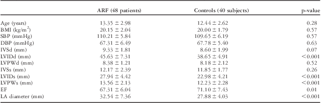
Clinical characteristics and echocardiographic findings of the two groups are shown in Table 1. The two groups were found to be similar in terms of age, body mass index, systolic blood pressure, diastolic blood pressure, interventricular septal wall thickness (diastolic), left ventricular posterior wall thickness (diastolic), and interventricular septal wall thickness (systolic; p > 0.05 for all parameters). Left ventricle end-diastolic diameter, left ventricle end-systolic diameter, left ventricular posterior wall thickness (systolic), and left atrium diameter were significantly increased in patients with acute rheumatic fever (p < 0.001 for all parameters).
Clinical characteristics of the study population.

ARF = acute rheumatic fever; BMI = body mass index; DBP = diastolic blood pressure; EF = Ejection fraction; IVSd = interventricular septal wall thickness (diastolic); IVSs = interventricular septal wall thickness (systolic); LA = Left atrium; LVIDd = left ventricular internal dimension (diastolic); LVIDs = left ventricular internal dimension (systolic); LVPWd = left ventricular posterior wall thickness (diastolic); LVPWs = left ventricular posterior wall thickness (systolic); SBP = systolic blood pressure
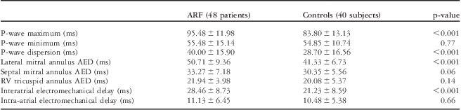
P-wave measurements were shown in Table 2. Maximum P-wave duration and P-wave dispersion were significantly higher in patients with acute rheumatic fever compared with the control group (p < 0.001 for both parameters). No difference was found between the groups regarding minimum P-wave duration (p > 0.05).
Comparison of the electrocardiographic and atrial electromechanical parameters.

AED = atrial electromechanical delay; ARF = acute rheumatic fever; RV = right ventricle
The atrial electromechanical parameters are reported in Table 2. Lateral mitral annulus atrial electromechanical delay was significantly higher in patients with acute rheumatic fever compared with the control group (p < 0.001). Septal mitral annulus atrial electromechanical delay and right ventricle tricuspid annulus atrial electromechanical delay were found to be similar. When compared with the control group, interatrial electromechanical delay was significantly higher in patients with acute rheumatic fever (p < 0.001). However, there was no difference in terms of intra-atrial electromechanical delay (p > 0.05).
A significant correlation was found between the left atrium diameter and the maximum P-wave duration, P-wave dispersion and interatrial electromechanical delay (r = 0.660 and p < 0.001, r = 0.524 and p < 0.001, r = 0.351 and p = 0.014, respectively). On the other hand, no correlation was found between the left atrium diameter and the intra-atrial electromechanical delay (r = 0.049 and p > 0.05). Furthermore, a significant correlation between the P-wave dispersion and the interatrial electromechanical delay was observed in our study (r = 0.494 and p < 0.001).
Discussion
In developing countries, acute rheumatic fever is still an important health issue and rheumatic valvular heart diseases are the most important complication of it. Myocardial inflammation, atrial fibrosis, mitral valve insufficiency, and atrial dilation may be observed in rheumatic carditis. These changes may cause increased P-wave dispersion and atrial arrythmia by affecting the atrial conduction system. The possible reasons for P-wave prolongation are intra-atrial or interatrial conduction defects.Reference Kocaoglu, Sert and Aypar 4 , 14 , Reference Ozer, Yavuz and Can 15 There is no previous study about interatrial or intra-atrial electromechanical delay at patients with rheumatic carditis. The prolongation of intra-atrial and interatrial conduction times and the inhomogeneous propagation of sinus impulses are well-known electrophysiological characteristics of the atrium prone to fibrillate.Reference den Uijl, Gawrysiak and Tops 19 , Reference Daubert, Pavin, Jauvert and Mabo 20 Recently, it has been possible to perform precise analysis of atrial motion among different regions at high temporal resolution with tissue Doppler imaging.Reference Ozer, Yavuz and Can 15
In our study, interatrial electromechanical delay was found to be significantly higher in patients with acute rheumatic fever compared with the control group. However, there was no difference regarding the intra-atrial electromechanical delay. Internodal conduction pathways were observed on the right atrium. These pathways are the anterior, median, and posterior bundles. There are numerous conduction pathways that enable interatrial conduction. The most important among these is the Bachmann's bundle. Any effect on the intra-atrial and interatrial conduction system will lead to an increase in the atrial electromechanical delay.Reference Daubert, Pavin, Jauvert and Mabo 20 The increase in interatrial electromechanical delay identified during our study might be associated with the increased left atrial effects in patients with acute rheumatic fever.
Increased P-wave duration and P-wave dispersion in 12-lead standard surface electrocardiography is an indicator of atrial conduction disorder.Reference Baspinar, Sucu and Koruk 21 Kocaoglu et alReference Kocaoglu, Sert and Aypar 4 found higher P-wave dispersion in patients with acute rheumatic fever and ongoing, newly diagnosed inflammation compared with the control group. In our study, we found significantly higher maximum P-wave duration and P-wave dispersion in patients with acute rheumatic fever. In our study, we identified an increase in P-wave dispersion in the late stage of rheumatic valvular disease. Increased atrial fibrillation risk has been reported in patients with rheumatic mitral stenosis, and this increased risk has been associated with left atrial enlargement.Reference Ozaydin, Turker and Varol 22 , Reference Kabukçu, Arslantas, Ates, Demircioglu and Ersel 23 In our study, it can be concluded that the risk of atrial fibrillation is increased even in the absence of mitral stenosis in patients with acute rheumatic fever.
We identified a significant correlation in our study between left atrial enlargement and P-wave dispersion, P-wave duration and interatrial electromechanical delay. The correlation between P-wave dispersion and left atrial diameter has been demonstrated in previous studies.Reference Kocaoglu, Sert and Aypar 4 , Reference Yagmur, Cansel and Acikgoz 24 Larger left atrial dimension predisposes to a greater risk of atrial fibrillation development and accounts for an independent predictive value for determining the risk of atrial fibrillation.Reference Parkash, Green and Kerr 25 Increased non-uniform anisotropy and conduction delay are the main characteristics of the structural and electropysiological changes in dilated atria, and these changes are associated with a higher risk of paroxysmal atrial tachyarrhythmias.Reference Cui, Zhang and Wang 6
The major limitation is that this is a cross-sectional design of the study. Patients could not be followed prospectively for arrhythmic episodes. Therefore, we do not know whether prolongation P-wave dispersion and interatrial electromechanical delay predict atrial arrhythmias in acute rheumatic fever patients. In addition, interatrial electromechanical delay was not investigated by invasive electrophysiological techniques, and the correlation between interatrial electromechanical delay obtained by tissue Doppler echocardiography and invasive interatrial electromechanical delay was not investigated in this study.
In conclusion, we observed in our study a prolonged interatrial electromechanical delay, increased P-wave dispersion, and increased maximum P-wave duration in the patients with acute rheumatic fever. We identified in our study a significant correlation between the left atrial enlargement and the P-wave dispersion, maximum P-wave duration and interatrial electromechanical delay. Therefore, there might be an increased risk of atrial dysrhythmias in patients with acute rheumatic fever. To determine the risk of atrial dysrhythmias in patients with acute rheumatic fever, it will be necessary to perform prospective studies with larger populations, as well as the long-term follow-up of patients for dysrhythmias.