INTRODUCTION
The liver fluke, Fasciola hepatica, is the etiological agent of fasciolosis. These important neglected zoonoses affect livestock and humans and have a worldwide distribution. Cattle and sheep infection is responsible for economic losses, which are estimated globally in US$2.6 billion/year, with more pronounced impact in rural areas of developing countries (Spithill et al. Reference Spithill, Smooker, Copeman and Dalton1999; Furst et al. Reference Furst, Keiser and Utzinger2012). Moreover, in the last decade, the human infection has been increasingly detected mainly in parts of South America and thus fasciolosis was recognized as re-emerging zoonoses by the World Health Organization (Mas-Coma et al. Reference Mas-Coma, Bargues and Valero2005; Garcia et al. Reference Garcia, Moro and Schantz2007). The human infection is acquired by the ingestion of vegetable and water contaminated with metacercaria. This quiescent stage is activated during its passage through the digestive tract and excysts at the duodenum. Then, the emerging newly excysted juvenile (NEJ) crosses the gut wall and reaches the liver, where it develops into the adult fluke, which finally locates at the bile duct (Andrews, Reference Andrews and Dalton1999).
The understanding of molecular aspects of host parasite cross-talk is critical in order to develop rational strategies for disease control. Particularly interesting are the parasitic stages that affect mammalian hosts, because they cause significant damage to the host. Mucins are highly O-glycosylated proteins widely distributed among the animal kingdom. In mammalians and other metazoans, mucins are important in normal and pathological conditions. For example, they are secreted by epithelial cells to protect tissue against chemical products, bacterial, viral and helminth infection and also play an important role in cell–cell interaction (Fukuda, Reference Fukuda2002; Hasnain et al. Reference Hasnain, Gallagher, Grencis and Thornton2013). Aberrant expression of mucin is a hallmark of diverse type of cancerous cell and is associated with cancer progression and metastasis (Ponnusamy et al. Reference Ponnusamy, Seshacharyulu, Lakshmanan, Vaz, Chugh and Batra2013). A typical mucin is composed of a polypeptide backbone (apomucin) with a central domain formed of variable number of tandem repeats, rich in serine and threonine and highly glycosylated. Up to 85% of the dry weight corresponds to O-linked glycans, commonly with N-acetylgalactosamine (GalNAc) attached to the hydroxyl group of serine or threonine residues (Berninsone, Reference Berninsone2006).
In various protozoan and helminth parasites mucin-like proteins were previously reported (Hicks et al. Reference Hicks, Theodoropoulos, Carrington and Corfield2000; Theodoropoulos et al. Reference Theodoropoulos, Hicks, Corfield, Miller and Carrington2001). For instance, the surface of Trypanosoma cruzi is covered by mucins, which contribute to parasite protection and establishment of persistent infection. In T. cruzi, mucins are encoded by a large family of genes and the coordinated expression of this large gene repertoire during host infection suggests that it is a possible strategy to subvert host immune responses (Buscaglia et al. Reference Buscaglia, Campo, Frasch and Di Noia2006; dos Santos et al. Reference dos Santos, Freitas, Lobo, Rodrigues-Luiz, Mendes, Oliveira, Andrade, Chiari, Gazzinelli, Teixeira, Fujiwara and Bartholomeu2012). Polymorphism in mucin genes were also described in the opportunist protozoan Cryptosporidium parvum where mucins are important molecules for attachment and parasite host cell invasion (O'Connor et al. Reference O'Connor, Burns, Ha-Ngoc, Scarpato, Khan, Kang and Ward2009). In the cestode Echinococcus granulosus, the etiological agent of cystic echinococcosis, the acellular laminated layer that protect hydatid cyst from host immune response is formed by a complex mesh of mucins (Diaz et al. Reference Diaz, Casaravilla, Irigoin, Lin, Previato and Ferreira2011). In the parasitic trematode, Schistosoma mansoni, a highly polymorphic mucin gene family was identified (Roger et al. Reference Roger, Gourbal, Grunau, Pierce, Galinier and Mitta2008a ). These mucins were predominantly expressed in the larval stage (miracidium), and they were identified as key factors for the compatibility of schistosomes interacting with the intermediate host Biomphalaria glabrata (Roger et al. Reference Roger, Mitta, Mone, Bouchut, Rognon, Grunau, Boissier, Theron and Gourbal2008c ). Previous transcriptomic analysis of the F. hepatica invasive NEJ stage, showed a set of highly redundant transcripts, one of which encoded a putative mucin-like protein. Real time polymerase chain reaction (qPCR) confirmed that mucin transcript was predominantly expressed by NEJ comparing with adult stage suggesting that this glycoprotein is involved in host–parasite relationship (Cancela et al. Reference Cancela, Ruetalo, Dell'Oca, da Silva, Smircich, Rinaldi, Roche, Carmona, Alvarez-Valin, Zaha and Tort2010).
In the present work, we describe the identification of a diverse repertory of mucin transcripts expressed by F. hepatica NEJ. At least six different clusters were identified, with open reading frames (ORFs) encoding secreted serine-threonine rich proteins. Moreover, we cloned and sequenced a complete apomucin-encoding gene and identified two allelic forms in the F. hepatica adult fluke population by PCR and high-resolution melting (HRM) analysis.
MATERIALS AND METHODS
Parasite
Metacercaria were obtained from experimentally infected Lymnaea viatrix snails, and excystment were performed as described previously (Carmona et al. Reference Carmona, Dowd, Smith and Dalton1993). After excystment, NEJ were washed with Roswell Park Memorial Institute (RPMI)-1640 medium and used for RNA extraction. Adults of F. hepatica were collected from bovine livers obtained at local abattoirs from metropolitan region of Porto Alegre, Brazil. Liver flukes were washed for 1 h at 37 °C in 10 mm phosphate-buffered saline pH 7·4 and then frozen in liquid nitrogen and stored at −80 °C until use.
DNA and RNA extraction and cDNA synthesis
Genomic DNA was extracted from individual F. hepatica adult fluke using the Pure Link Genomic DNA extraction kit (Life Technologies, USA). Briefly, 50 mg of tissue from one fluke was chopped and incubated for at least 3 h and then DNA was extracted following manufacturer instructions.
Total RNA from NEJ was extracted using the micro-midi RNA extraction kit (Life Technologies) following manufacturer's protocol. DNA and RNA quality was assessed by 0·8% agarose gel stained with GelRed™ (Biotium, Inc, Hayward, CA) after quantification using Qubit Fluorometric quantification kit (Life Technologies, USA).
First strand cDNA was synthesized using the RevertAid™ First Strand cDNA Synthesis Kits (Fermentas). Total RNA was treated with DNase I-RNase free enzyme (Fermentas) and approximately 50 ng of RNA from NEJ were incubated with 100 pmol of oligo(dT)18 primer, 1 U µL−1 of RiboLock RNase inhibitor, 1 mM dNTPs, and 10 U µL−1 of RevertAid M-MuLV Reverse Transcriptase. cDNA synthesis was performed for 1 h at 42 °C and stored at −20 °C until use.
Amplification, cloning and sequencing of mucin cDNAs from NEJ of F. hepatica
In order to detect mRNA diversity in NEJ mucin cDNAs, we performed a PCR amplification using 10 ng of NEJ cDNA as template, 0·2 mm each dNTPs, 1·5 mm MgCl2, 0·05 U µL−1 HiFi Taq Platinum DNA polymerase (Life Technologies, USA), and mucin specific primers MucF1 (5′-ATGGTGCGAACGCTAAG-3′) and MucR1 (5′-TCAGAAGAACGCAACGCC-3′) at a final concentration of 0·3 µ m each one. Temperature conditions were as follows: denaturation at 94 °C for 180 s followed by 35 cycles at 94 °C for 60 s; 58 °C for 45 s and 72 °C for 60 s. The resulting PCR was gel-purified using the glass fiber matrix (GFX) column kit (Amersham Pharmacia Biotech, UK) and ligated into pCR2·1 TA cloning vector (Life Technologies, CA, USA). Recombinant clones were identified by PCR using universal primers M13 forward and reverse to evaluate the quality of the library and insert size. Selected clones were grown in Circle Grow medium in 96-well plates and were stored at −20 °C in medium with 15% Glycerol. Ninety-six random clones were selected for plasmid DNA extraction and both strands were sequenced using Terminator cycle sequencing kit for MEGABACE DNA Analysis Systems (Amersham Pharmacia Biotech, UK) according to the manufacturer's instructions.
PCR amplification and sequencing of F. hepatica mucin gene from genomic DNA
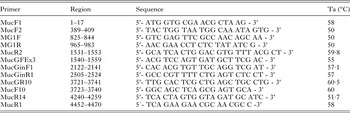
To determine mucin gene organization, PCR reactions were prepared using 25 ng of genomic DNA (gDNA) extracted from one adult F. hepatica, 1·5 mm MgCl2, 0·4 µ m of each MucF1 (5′-ATGGTGCGAACGCTAAG-3′) and MucR1 primer (5′-TCAG AAGAACGCAACGCC-3′), 0·2 mm of each dNTPs and 0·05 U µL−1 High Fidelity PCR enzyme mix (Fermentas). Temperature conditions were as follows: denaturation at 94 °C for 300 s followed by 35 cycles at 94 °C for 30 s; 58 °C for 45 s and 72 °C for 300 s. PCR products were separated by electrophoresis in 1% agarose gels and after confirmation of expected size, PCR fragment of 4470 bp was gel-purified using the GFX kit (Amersham Pharmacia Biotech, UK) and ligated into pCR2·1 TA vector (Life Technologies, CA, USA). Plasmid DNA from 96 recombinant clones were extracted and sequenced using sequencing kit for MegaBACE DNA and universal M13 Forward and Reverse primers to confirm the identity of clones. Complete sequencing of 4470 bp insert was performed using primers shown in Table 1.
Mucin-specific oligonucleotides, annealing region, primer sequence and annealing temperature (Ta)

Analysis of polymorphism in mucin gene of F. hepatica
To verify mucin gene variation between adult worms, we performed a PCR amplification using gDNA from 20 individual adult flukes (FH1–FH20) collected from five different livers (four flukes from each bovine liver). PCR reactions were performed in 25 µL using 20 ng of gDNA, 1·5 mm MgCl2, 0·4 µ m of each MG1F (5′- GTCGAGTTCGCCAACAGCAA -3′) and MG1R primer (5′-AACGAACCTCTCTAT ATCG-3′), 0·2 mm of each dNTPs and 0·05 U µL−1 High Fidelity PCR enzyme mix. MG1F and MG1R primers amplify an internal region of fhemuc-1 intron-2. Temperature conditions were as follows: denaturation at 94 °C for 300 s followed by 35 cycles at 94 °C for 45 s; 50 °C for 45 s and 72 °C for 60 s. Amplicons obtained were analysed in 1% agarose gel electrophoresis and the amplicons derived from two individual adult fluke were ligated in pCR2·1 TA vector. From each ligation, 96 random clones were selected and sequenced. To species confirmation of F. hepatica adult worm isolates, the cytochrome c oxidase subunit 1 gene (cox-1) gene was amplified, sequenced and analysed as previously described (Nguyen et al. Reference Nguyen, Van De, Vercruysse, Dorny and Le2009).
HRM analysis
In order to investigate the degree of variability in F. hepatica mucin gene, we also performed a HRM analysis using gDNA from 20 adult flukes, as described previously (Santos et al. Reference Santos, Espinola, Ferreira, Margis and Zaha2014). The same genomic region was amplified by PCR using the primers MG1F and MG1R (Table 1), following the reaction and thermal conditions as described in the section ‘Analysis of polymorphism in mucin gene of F. hepatica’. We performed the PCR reactions, in the presence of 5 µ m SYTO 13 (Life technologies, USA), a DNA dye that does not preferentially bind to GC- or AT-rich sequences, does not increase the melting temperature, and has a minimal inhibitory effect on PCR. The PCR reactions were carried out in technical duplicate with non-template control. Melting was conducted by increasing the temperature from 60 to 99 °C at ramping increments from 0·1 °C s−1. The HRM analysis was carried out using the HRM software from Applied Biosystem (version 3.0·1) with normalization regions between 77·3–77·8 and 83·0–83·5 °C. As reference curves for HRM, we used two samples that correspond to the known allelic variants described in this work, FH5 and FH6.
Estimation of mucin gene copy number by qPCR
In order to estimate mucin copy number in F. hepatica genome,we performed a qPCR assay using gDNA extracted from individual F. hepaticaas described in the section ‘DNAand RNA extraction and cDNA synthesis’. Different amounts of gDNA were tested in order to determine PCR efficiency (15, 30 and 60 ng of gDNA). PCR reaction were performed in 20 µL volume containing 3·0 mm MgCl2, 50 µ m dNTPs, 0·2 µ m each specific primers, 0·25 UPlatinum Taq DNA polymerase (Life Technologies, USA), 0·2× SyBr Green. Primers sequences for fhemuc-1 amplification were MucF1 and MucR1 andfor stefin-I (Forward 5′-ATGATGTGCGGCGGCTG-3′ and Reverse 5′-AATATTGCA ATGCGTCCGT-3′). Reactions were performed in triplicate using the following thermal cycling parameters: 1 cycle (5 min at 94 °C) and 40 cycles (15 s at 94 °C, 10 s at 50 °C, 15 s at 72 °C). For determination of mucin copy number, Ct amplification of mucin were determined for different dilution and compared with a reference gene (F. hepatica stefin-I), described as a single copy gene by southern blot in F. gigantica (Tarasuk et al. Reference Tarasuk, Vichasri Grams, Viyanant and Grams2009) and by bioinformatics analysis in F. hepatica.
Bioinformatic analysis
Reads were processed with the Phred, Pregap (version 1.5) and GAP4 program using the Staden Package (Staden, Reference Staden1996). Assembled contigs were compared and aligned with mucin cDNAs previously reported by using ClustalX program (Thompson et al. Reference Thompson, Gibson, Plewniak, Jeanmougin and Higgins1997). Genomic sequences were also assembled with the Staden Package and compared with mucin cDNA sequences. Signal sequence prediction was performed using SignalP 4·0 program (Petersen et al. Reference Petersen, Brunak, von Heijne and Nielsen2011). Prediction of trans-membrane domains were conducted using TMHMM 2·0 software (Krogh et al. Reference Krogh, Larsson, von Heijne and Sonnhammer2001). MEME software was used to identify motifs (highly conserved region) in the mucin cDNAs identified (http://meme.nbcr.net/meme/).
Fasciola hepatica draft genome was mined using mucin gene sequence identified in this work as a query for blast searches and genomic sequences deposited as PRJEB6687 and PRJNA179522 at Wormbase Parasite v 2·0 (http://parasite.wormbase.org). Contigs containing hits with an e value <10−5 were selected for further analysis in order to detect sequence similarity between fhemuc-1 gene and F. hepatica genome.
RESULTS
mRNA diversity encoding putative mucin-like proteins were detected in the invasive stage of F. hepatica
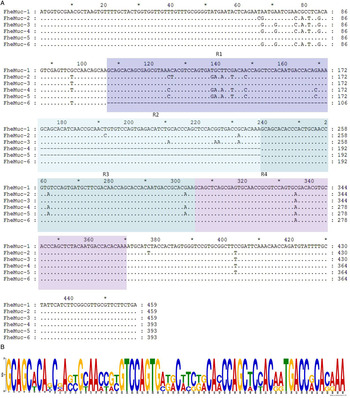
To analyze diversity in mucin transcript expressed by NEJ after metacercariae excystment, we amplified full length cDNA encoding mucin-like proteins by PCR, ligated the amplicons to pCR21 vector and sequenced 96 clones randomly picked. We found the presence of point mutations, deletions and insertions between the sequences identified. Clustering using GAP4 software showed that at least six different cDNAs encoding mucin-like proteins were expressed by F. hepatica NEJ (Fig. 1A). These six cDNAs differed in size and nucleotide composition. According to the ORF size, the clones were organized in two groups, those with ORFs of 459 bp and those with 393 bp. Nucleotide sequences showed more variability at the 5′ region of mucin coding sequences (CDSs) than at the 3′ region. Notably, we observed that differences in cDNA lengths were due to an insertion/deletion of 66 nucleotides and up to four repeats (R1, R2, R3 and R4) were recognized. Shorter mucin cDNAs had three repeats whereas larger mucin cDNAs had four repeats (Fig. 1A) and highly conserved regions between R1, R2, R3 and R4 were found (Fig. 1B). The translation of the cDNAs obtained resulted in 152 and 130 amino acid residues proteins (online Supplementary Fig. 1A). Each cDNA variant identified in NEJ had a putative ORF encoding a serine/threonine rich protein with an N-terminal signal peptide (online Supplementary Fig. 1B). No transmembrane domain was detected in the apomucin ORFs analysed. Theoretical molecular mass ranged from 13·1 to 15·4 kDa between the different mucin ORFs and pI between 4·77 and 9·0. Ser + Thr content was >50% in some ORFs encoding mucin-like proteins. Some aromatic amino acid residues like His, Trp and Tyr and the basic amino acid Lys were absent in all variants identified.
Mucin transcripts identified in Fasciola hepatica NEJ. (A) Six different cDNAs were identified by DNA sequencing. Alignment showed tandem repeat (R) R1, R2, R3 and R4 (boxed in blue) and two groups of mucin were defined (one group with 3 and the other with 4 repeats). Dashed lines indicate sequence gaps. (B) Sequences of each repeat (66 bp) found in the six distinct mucin cDNAs were aligned and motifs detected using the Meme logo. Some degree of conservation was observed. Abbreviation: NEJ, newly excysted juvenile.

Mucin gene structure
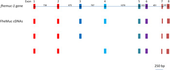
In order to determine F. hepatica mucin gene sequences, genomic DNA was extracted from a single adult fluke and used as template for PCR amplification using primers to amplify full-length coding sequence. To analyse intron/exon organization, we aligned the genomic and mucin cDNAs sequences. We found that the 4470 bp of genomic sequence had significant similarity (E value < 3 e-31) with mucin cDNA sequences found in this work. The genomic sequence obtained was named fhemuc-1 and started at the initiation codon (ATG) and finished at position 4470 (TGA) (online Supplementary Fig. 2). Eight exons (E) were detected (E1 = 55, E2 = 51, E3 = 66, E4 = 66, E5 = 66, E6 = 66, E7 = 18, E8 = 71 bp) interrupted by seven introns (I) (I1 = 738, I2 = 675, I3 = 767, I4 = 1076, I5 = 163, I6 = 451, I7 = 141 bp). The exon–intron boundary sites, determined by comparison with the cDNA sequence, are all consistent with the GT-AG rule, except for the seventh exon and intron. Some transcripts obtained from F. hepatica NEJ can be explained by alternative splicing of this gene (Fig. 2).
Schematic representation of mucin gene and transcripts identified in Fasciola hepatica. Mucin gene sequence (fhemuc-1 gene) has 4470 bp and consists of eight exons and seven introns. cDNAs transcribed by NEJ (FheMuc cDNAs) consisted of exons 1 and 2 encoding signal peptide sequence (boxed in red), a central tandem repeat region composed of 3 or 4 exons (blue-scale boxes – exons 3–6) and two additional exons (brown boxes – exons 7 and 8). Only cDNAs transcripts with differential exon usage are schematic shown. Abbreviation: NEJ, newly excysted juvenile.

Mucin gene variability between different isolates of F. hepatica
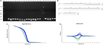
Variability at the fhemuc-1 gene was assessed by analysing DNA samples extracted from 20 liver fluke isolates, collected from five different bovine livers by PCR. A region of intron-2 was PCR amplified resulting in two different fragments of 159 and 192 bp, respectively. Eight individual flukes presented both amplicons, whereas only the 192 bp fragment was observed in the remaining 12 samples (Fig. 3A), Two samples, FH5 (single band) and FH6 (double band), were selected to confirm the identity of amplicons obtained by cloning and sequencing. While the shorter 159 bp fragment (allele B) was identical to the fhemuc1 gene region targeted, the 192 bp variant (allele A) resulted from a 32 bp insertion (Fig. 3B). High resolution melting analysis of the samples confirmed homozygocity (A/A) in the 12 samples while two patterns consistent with heterozygous (A/B) genotype (Fig. 3C and D). The Tm obtained from the HRM analysis, showed a difference of 0·75 °C between the two genotypes and a standard deviation of 0·1 °C. This data confirm the presence of two alleles of fhemuc-1 between F. hepatica isolates. We also observed genetic diversity in fhemuc1 gene between F. hepatica collected from the same bovine liver. Interestingly, neither HRM nor agarose gel electrophoresis demonstrated the presence of the third genotype expected (homozygous B/B).
Analysis of Fasciola hepatica mucin gene variability in different adult liver fluke isolates. (A) PCR was performed using 20 different genomic DNA samples (FH1 to FH20) extracted from individual F. hepatica. After amplification (using an internal region of intron-2) samples were run in 1·5% agarose gel and two different bands of 159 and 192 bp were detected. M, molecular weight marker, 1–20 amplicons obtained from FH gDNA, N: negative control. (B) Amplicons obtained from sample five (FH5) and six (FH6) were cloned and DNA sequences were clustered to distinguish sequence difference (see section Materials and Methods). Only one sequence was identified in sample FH5 whereas two distinct sequences were found in FH6. These sequences are represented by A and B and were aligned with fhemuc-1 to show differences between them. (C–D) HRM analysis of fhemuc-1 using genomic DNA from 20 individual adult flukes. (C) Difference curves derived from the normalization data using the homozygous (AA) as baseline. (D) Normalization curves derived from the raw data plots. In blue the curves that match with the homozygous (AA) and in green the curves that match with heterozygous (AB). Abbreviation: HRM, high-resolution melting; PCR, polymerase chain reaction.

Analysis of mucin copy number in F. hepatica genome
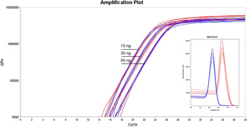
By qPCR we observed that amplification curves for stefin-I and fhemuc-1 were overlapped using different dilutions of genomic DNA (Fig. 4). The identical Cts values obtained for these genes at different gDNA concentrations is suggestive of similar copy number of mucin and stefin-I in F. hepatica genome.
qPCR analysis for detection of mucin gene copy number in Fasciola hepatica. Amplification curves for fhemuc-1 (blue) and stefin-I (red) genes using different gDNA concentrations (15, 30 and 60 ng) are shown. Inlet represents the dissociation curves for mucin and stefin-I amplicons obtained after qPCR. Abbreviation: qPCR, real time polymerase chain reaction.

By mining the F. hepatica draft genome using the fhemuc-1 gene sequence as bait, we observed sequence match using PRJEB6687 and PRJNA179522 databases. We found some contigs with significant similarity with fhemuc-1 but there were truncated (with <3000 bp) or with low quality. For this reason, a more complete analysis of mucin gene organization in F. hepatica will be possible with deeper and better genome coverage.
DISCUSSION
During the infection process, parasitic organisms have to deal with the response mounted by the host and for this reason they evolved mechanisms to protect themselves from dangerous compounds to allow completing their life cycles within the host (Schmid-Hempel, Reference Schmid-Hempel2009). Molecules secreted or exposed at the surface of parasites are critical factors that assist parasite establishment either neutralizing or modulating the expression of host molecules (Hewitson et al. Reference Hewitson, Grainger and Maizels2009). Mucins have been reported as relevant actor in host–parasite relationship in various parasitic organisms as T. cruzi (dos Santos et al. Reference dos Santos, Freitas, Lobo, Rodrigues-Luiz, Mendes, Oliveira, Andrade, Chiari, Gazzinelli, Teixeira, Fujiwara and Bartholomeu2012), C. parvum (Bhalchandra et al. Reference Bhalchandra, Ludington, Coppens and Ward2013), Toxocara canis (Loukas et al. Reference Loukas, Hintz, Linder, Mullin, Parkinson, Tetteh and Maizels2000), E. granulosus (Casaravilla et al. Reference Casaravilla, Pittini, Ruckerl, Seoane, Jenkins, MacDonald, Ferreira, Allen and Diaz2014) and Schistosoma mansoni (Roger et al. Reference Roger, Mitta, Mone, Bouchut, Rognon, Grunau, Boissier, Theron and Gourbal2008c ).
In this work, we have characterized cDNAs encoding mucin-like proteins expressed by the invasive NEJ of F. hepatica. We found at least six different mucin cDNAs, being expressed at the initial steps of the infection. A repeated Ser/Thr rich motif accounts for differences in length, while minor variations in sequence can also be detected. Consequently predicted proteins of diverse molecular mass, pI, and Ser/Thr content were observed. Some hydrophobic amino acids (Trp, Tyr and His) were absent in apomucin-predicted ORFs. Since mucin-like proteins are very hydrophilic proteins, this class of amino acid residues could perturb the physicochemical properties of this type of molecules and for this reason underrepresented or absent as was previously reported for T. cruzi mucins (Buscaglia et al. Reference Buscaglia, Campo, Frasch and Di Noia2006). Interestingly, the identification of a signal peptide at the N-terminal of all deduced proteins suggests that NEJ mucins could be secreted by the parasite to possibly interact with host molecules. Although, no transmembrane domain was detected in mucin encoding ORFs, NEJ mucin could be located at the surface of F. hepatica as was previously demonstrated for T. canis secreted mucins (Loukas et al. Reference Loukas, Hintz, Linder, Mullin, Parkinson, Tetteh and Maizels2000) and/or as part of exosome-like vesicles as described for human mucin MUC1 (Hanisch et al. Reference Hanisch, Kinlough, Staubach and Hughey2012). In the filarial nematodes Brugia malayi, Brugia phagangi and Litomosoides sigmodontis two mucin-like genes encoding microfilarial surface proteins were identified and some transcript variants could be generated by cis and trans-splicing (Hirzmann et al. Reference Hirzmann, Hintz, Kasper, Shresta, Taubert, Conraths, Geyer, Stirm, Zahner and Hobom2002). In the trematode S. mansoni, the mucin polymorphism is generated from a multigene family whose members undergo recombination. Interestingly, each gene in an individual-specific manner can also produce multiple splice variants increasing the level of variability (Roger et al. Reference Roger, Grunau, Pierce, Hirai, Gourbal, Galinier, Emans, Cesari, Cosseau and Mitta2008b ). In F. hepatica, we found evidence that mucin variability could be generated by alternative splicing of fhemuc-1 gene since the cDNAs obtained can arise from different exon rearrangements.
In the present work we found evidences of polymorphism in a mucin-encoding gene in F. hepatica. A distinct pattern of amplicons was observed between different adult fluke isolates, and at least two distinct gene variants were detected by DNA sequencing. Both homozygous and heterozygous individuals for this mucin locus were found. Moreover, we found that adult flukes from the same liver showed genetic differences suggesting that bovines were infected with non-clonal metacercaria. Since all individuals were confirmed as F. hepatica by cox-1 analysis, mucin gene differences between isolates would be useful to understand dynamics of F. hepatica populations and highlights the relevance of identification and uses of nuclear genetic markers. In the HRM analysis, a specific profile for each genotype was observed in both the difference and standard curve (Fig. 3C and D, respectively). Despite the close Tm value observed between the two genotypes, the HRM profiles provide a clear and undoubted discrimination among them. Interestingly, we did not detect any F. hepatica isolates with the homozygous genotype B/B. We suggest that this genotype is deleterious or negatively selected during parasite life cycle but a larger number of individual are necessary to confirm this hypothesis. In S. mansoni, the polymorphism of mucin expressed by individual miracidia was associated with the success to infect the intermediate snail host (Roger et al. Reference Roger, Grunau, Pierce, Hirai, Gourbal, Galinier, Emans, Cesari, Cosseau and Mitta2008b , Reference Roger, Mitta, Mone, Bouchut, Rognon, Grunau, Boissier, Theron and Gourbal c ). The association of mucin variants with the success of F. hepatica infection warrant furthers studies. As these glycoproteins are probably important for parasite establishment within mammalian host, mucin characterization at the protein level could be relevant in order to assess its potential as a vaccine candidate against fasciolosis.
Concluding remarks
In the present work, we report the initial characterization of mucin encoding sequences in F. hepatica. A distinct repertoire of mucin transcript expressed by F. hepatica NEJ was identified and evidence of polymorphisms in F. hepatica isolate was detected. Further functional studies using RNAi methodology would be conducted in order to decipher biological importance of these mucins in parasite establishment and subvert host immune response.
SUPPLEMENTARY MATERIAL
To view supplementary material for this article, please visit http://dx.doi.org/10.1017/S0031182015001134.
ACKNOWLEDGEMENTS
We thank the Centro de Biotecnologia-UFRGS, Brazil for kindly providing the DNA sequencing facilities.
FINANCIAL SUPPORT
This work was supported by Coordenação de Aperfeiçoamento de Pessoal de Nível Superior (CAPES) (AUX-PE-PARASITOLOGIA 1278/2011), in Brazil and Comisión Sectorial de Investigación Científica (CSIC), in Uruguay. M. C. received postdoctoral felowships from the CAPES and CNPq and G. B. S. received a predoctoral fellowship from the CAPES-REUNI.