Introduction
Members of the family Temnocephalidae sensu Van Steenkiste et al. (Reference Van Steenkiste, Rivlin, Kahn, Wakeman and Leander2021) (Platyhelminthes), which was traditionally regarded as Temnocephalida, are ectosymbionts associated with various animals, mainly with crustaceans in freshwaters (Joffe et al., Reference Joffe, Cannon and Schockaert1998). Species of the genus Temnosewellia Damborenea & Cannon, Reference Damborenea and Cannon2001, which is currently recognized as the largest genus of the family, have been recorded from Australia and South East Asian countries (e.g. Cannon, Reference Cannon1993; Cannon & Sewell, Reference Cannon and Sewell2001; Sewell et al., Reference Sewell, Cannon and Blair2006). Currently 52 valid species of the genus are recognized in total (Tyler et al., Reference Tyler, Schilling, Hooge and Bush2006–2022), 50 species of which are naturally distributed in Australia (see Haswell, Reference Haswell1888, Reference Haswell1893, Reference Haswell1900; Hickman, Reference Hickman1967; Williams, Reference Williams1980; Cannon, Reference Cannon1993; Cannon & Sewell, Reference Cannon and Sewell2001; Sewell et al., Reference Sewell, Cannon and Blair2006). Of those 50 species, only Temnosewellia rouxi (Merton, Reference Merton1913) was also found in Indonesia. While both Temnosewellia minor (Haswell, Reference Haswell1888) and Temnosewellia chaeropsis have also been reported from outside of Australia (i.e. the former from Japan, Germany, Italy, and South Africa and the latter from South Africa, respectively), those are considered to have been imported (e.g. Mitchell & Kock, Reference Mitchell and Kock1988; Avenant-Oldewage, Reference Avenant-Oldewage1993; Cannon & Sewell, Reference Cannon and Sewell2001; Tavakol et al., Reference Tavakol, Blair, Morgan, Halajian and Luus-Powell2021; Vecchioni et al., Reference Vecchioni, Chirco, Bazan, Marrone, Arizza and Arculeo2021). On the other hand, Temnosewellia semperi (Weber, Reference Weber1889) and Temnosewellia vietnamensis Damborenea & Brusa, Reference Damborenea and Brusa2009 have only been reported from East to South Asian countries (e.g. Semper, Reference Semper1872; Weber, Reference Weber1889; Damborenea & Brusa, Reference Damborenea and Brusa2009).
Although the species considered to be Temnosewellia have been found from freshwater crabs in various localities from southern Japan, all of those records have lacked the descriptions of diagnostic characters, except T. minor found from the crayfish Cherax tenuimanus (Smith, 1912) (Parastacidae) which was introduced from western Australia to Kagoshima (Oki et al., Reference Oki, Tamura, Takai and Kawakatsu1995).
In this study, a species of Temnosewellia that is probably native is recorded based on individuals and eggs attaching on the freshwater crabs Eriocheir japonica (De Haan, 1835) (Varunidae) and Geothelphusa exigua Suzuki & Tsuda, 1994 (Potamidae) collected from the Osumi Peninsula, Kagoshima, southern Japan. Phylogenetic analyses are conducted based on partial sequences of two genes.
Materials and methods
Crabs were caught from the upper to middle reaches of Oda River (31°15′N, 131°3′E), a tributary of Kimotsuki River (31°18′N, 131°0′E), and Kushira River (31°31′N, 130°47′E), Osumi Peninsula, Kagoshima, southern Japan. Temnocephalids were carefully removed from the body surface of the crabs, using forceps and preserved in 70% ethanol under coverslip pressure to observe both external and internal morphology. The specimens attached on the slide grasses were stained in Heidenhain's iron–haematoxylin, dehydrated through a graded ethanol series (70 to 99%) for one to three days, cleared in xylene for three days, and mounted in Canada balsam. Several whole individuals and eggs were fixed in 10% formalin in hot distilled water. Before observation of the penial stylet (cirrus), a fixed specimen was soaked in lactophenol during 24 h, and then dissected using sharpened stainless needles. Drawings were made with the aid of a drawing tube mounted on a compound microscope (Olympus BX53). For observation using a scanning electron microscope (SEM) (Hitachi TM3000), fixed whole or dissected individuals and eggs were dehydrated through a graded ethanol series and t-butanol, freeze-dried, sputter coated with gold, and examined.
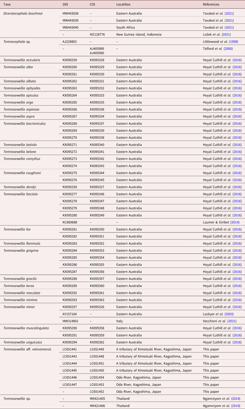
Using five specimens of one of the caligids fixed in 80% ethanol, DNA was extracted and sequenced for two genes, cytochrome c oxidase subunit I (COI) in mitochondria and 28S rDNA in nucleus. These methods are based on those we have published in the past (Tosuji et al., Reference Tosuji, Nishinosono, Hsieh, Glasby, Sakaguchi and Sato2019). The primer sets used were 425F (5′-GGNGCTAGNTCNATWTTAGGRGC-3′) + new 1200R (5′-CCCATTGAWAMNACATAATGAAAATG-3′) for COI, and Ltem180 (50-GAAGTTCGCACGATTGCGG-30) + Ltem1000R (50-CACAAGCATAGTTCACC-30) for 28S, according to Hoyal Cuthill et al. (Reference Hoyal Cuthill, Sewell, Cannon, Charleston, Lawler, Littlewood and Blair2016). The data have been submitted to the DNA Data Bank of Japan (DDBJ) database under accession numbers LC651441 and LC651443–LC651454. Using the nucleotide sequence data of these specimens with other temnocephalids obtained from GenBank (table 1), Bayesian inference (BI) trees were obtained. The sequences were aligned using the DNA alignment software MUSCLE ver. 3.8.31 (Edgar, Reference Edgar2004). MrBayes v. 3.1.6 (Ronquist et al., Reference Ronquist, Teslenko and van der Mark2012) was used to obtain BI tree on the basis of the substitution model GTR + G + I. As a result, 2,000,000 generations [Average Standard Deviation of Split Frequencies (ASDSF) = 0.005009] for COI and 500,000 generations (ASDSF = 0.006170) for 28S were obtained. The first 25% of the samples were discarded as burn-in samples.
List of temnocephalids using the phylogenetic analysis in this paper with GenBank accession numbers for 28S rDNA (28S) and cytochrome c oxidase subunit I (COI).

The taxonomy of temnocephalids follows Tyler et al. (Reference Tyler, Schilling, Hooge and Bush2006–2022), and the terminology basically follows Cannon (Reference Cannon1993) and Sewell et al. (Reference Sewell, Cannon and Blair2006). Measurements were reported in mm or μm as range followed by mean, and standard deviation in parentheses. Materials are deposited in the Platyhelminthes collection of the National Museum of Nature and Science, Tsukuba (NSMT), Japan, and the Kagoshima University Museum (KAUM), Kagoshima, Japan. Materials used by Okawachi et al. (Reference Okawachi, Uyeno, Miyazaki and Kamezaki2013) deposited in the Ryukyu University Museum, Fujukan (RUMF) were examined.
Results
Genus Temnosewellia Damborenea & Cannon, Reference Damborenea and Cannon2001
Japanese name: Yadori-itsu-tsuno-mushi-zoku
Temnosewellia aff. vietnamensis Damborenea & Brusa, Reference Damborenea and Brusa2009
Syn. Temnosewellia sp.: Okawachi et al., Reference Okawachi, Uyeno, Miyazaki and Kamezaki2013: 76–79, fig. 1c–h.
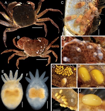
Live specimens of Temnosewellia aff. vietnamensis from host crabs, (a, c, e, g, h) specimens or eggs attached on Eriocheir japonica (De Haan, 1835) from Oda River, (b, d, f, i, j) specimens and eggs attached on Geothelphusa exigua Suzuki & Tsuda, 1994 from a tributary of Kimotsuki River. (a) crab with eggs of T. aff. vietnamensis on legs; (b) crab with specimens of T. aff. vietnamensis on carapace; (c, d) specimens on hosts; (e, f) habitus of specimens, dorsal view; and (g–j) specimens and eggs on meri of legs of crabs. Scale bars: 20 mm (a); 5 mm (b); 2 mm (c, d, g); 500 μm (e, f); 200 μm (h, j); and 1 mm (i).

New Japanese name: Yamataro-yadori-tsuno-mushi
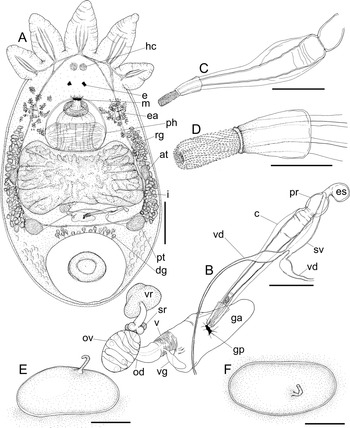
Temnosewellia aff. vietnamensis, (a, b) specimen (NSMT-Pl 6494), (c, d) specimen (NSMT-Pl 6494), (e, f) egg (NSMT-Pl 6495). (a) habitus, ventral, hc = Haswell's cell, e = eye, m = mouth, ph = pharynx, i = intestine, ea = excretory ampulla, rg = rhabdite glands, dg = disc glands, at = anterior testis, pt = posterior testis; (b) male and female reproductive systems, ventral, vr = vesicula resorben, sr = seminal receptacle, ov = ovary, od = oviduct, vg = vaginal gland, v = vagina, ga = genital atrium, gp = gonopore, c = cirrus, pr = prostate, es = ejaculatory sac, sv = seminal vesicle, vd = vas deferens; (c) cirrus, ventral; (d) distal portion of cirrus; (e) egg, lateral view; and (f) same, apical view. Scale bars: 400 μm (a); 100 μm (b, c); 40 μm (d); and 300 μm (e, f).

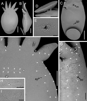
Temnosewellia aff. vietnamensis, scanning electron microscope micrographs of specimens, m = mouth, gp = gonopore, ep = excretory pore, white arrowheads indicating edges of syncytial plates, (a–h) specimens attached on Eriocheir japonica (De Haan, 1835) from Oda River, (i) specimen attached on Geothelphusa exigua Suzuki & Tsuda, 1994 from a tributary of Kimotsuki River. (a) habitus, dorsal view; (b) same, lateral view; (c) habitus without tentacles, ventral view; (d) posterior part of habitus, lateral view; (e) gonopore; (f) partial habitus, right side of anterior part, dorsal view; (g) same, left side of anterior part, lateral view; and (h, i) horizontal bar-shaped syncytium situated posterior to post-tentacular syncytium. Scale bars: 400 μm (a–c); 200 μm (d, f); 40 μm (e); 100 μm (g, h); and 50 μm (i).

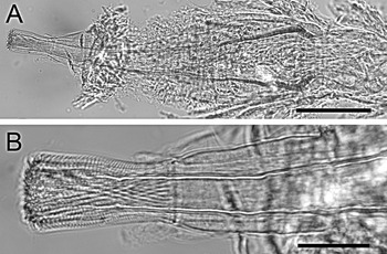
Temnosewellia aff. vietnamensis, specimen (NSMT-Pl 6495), cirrus, ventral. (a) General view. (b) Distal portion. Scale bars: 100 μm (a), 30 μm (b).

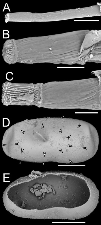
Temnosewellia aff. vietnamensis, scanning electron microscope micrographs of cirri and eggs. (a) distal portion of cirrus with spined region of introvert not exposed; (b) distal portion of cirrus with spined region of introvert slightly exposed; (c) distal portion of cirrus, spined region of introvert exposed; (d) egg attached on host, arrowheads indicating both outer and inner borders of opercular plates, apical view; and (e) egg shell hatched along outer margin of opercular plates, apical view. Scale bars: 40 μm (a); 20 μm (b, c); and 500 μm (d, e).

Materials examined
One specimen (NSMT-Pl 6494), one specimen (NSMT-Pl 6494), one specimen (NSMT-Pl 6494), two specimens (NSMT-Pl 6494) and one specimen (NSMT-Pl 6494) in whole mount slide, ex E. japonica (De Haan, 1835) (Brachyura: Varunidae) from middle reaches of Oda River (31°15′N, 131°3′E), Kimotsuki, Kagoshima, Japan, 4 November 2018; one specimen (NSMT-Pl 6495), one specimen (NSMT-Pl 6495), one specimen (NSMT-Pl 6495), one specimen (NSMT-Pl 6495), one specimen (NSMT-Pl 6495), one specimen (NSMT-Pl 6495), and one specimen (NSMT-Pl 6495) in whole mount slides, three specimens (NSMT-Pl 6495) in 10% formalin in distilled water, 65 specimens (NSMT-Pl 6495) in 99.5% ethanol, and 14 eggs (NSMT-Pl 6495) in 10% formalin in distilled water, ex E. japonica from middle reaches of Oda River (31°15′N, 131°3′E), Kimotsuki, Kagoshima, Japan, 20 September 2017; two specimens (NSMT-Pl 6496) in whole mount slides, five specimens (NSMT-Pl 6496) in 10% formalin in distilled water, 12 specimens (NSMT-Pl 6496) in 99.5% ethanol, and 13 eggs (NSMT-Pl 6496) in 10% formalin in distilled water, ex E. japonica from middle reaches of Oda River (31°15′N, 131°3′E), Kimotsuki, Kagoshima, Japan, 9 August 2021; three specimens (KAUM–PT–1) in 10% formalin in distilled water, collection data same as that of (NSMT-Pl 6496); nine specimens, prepared for observation by SEM, collection data same as that of (NSMT-Pl 6495); four specimens, prepared for observation by SEM, collection data same as that of (NSMT-Pl 6496); two specimens (NSMT-Pl 6497), two specimens (NSMT-Pl 6497), two specimens (NSMT-Pl 6497) in whole mount slides, four specimens (NSMT-Pl 6497) in 10% formalin in distilled water, six specimens (NSMT-Pl 6497) in 99.5% ethanol, and nine eggs (NSMT-Pl 6497) in 10% formalin in distilled water, ex Geothelphusa exigua Suzuki & Tsuda, 1994 (Potamidae) from upper reaches of a tributary of Kimotsuki River (31°18′N, 131°0′E), Kimotsuki, Kagoshima, Japan, 9 August 2021; 2 specimens (KAUM–PT–2) in 10% formalin in distilled water, collection data same as that of (NSMT-Pl 6497); two specimens, prepared for observation by SEM, collection data same as that of (NSMT-Pl 6497); five specimens (NSMT-Pl 6498) in 99.5% ethanol, ex G. exigua from upper reaches of Kushira River (31°31′N, 130°47′E), the Takakuma Experimental Forest, Kagoshima University, Tarumizu, Kagoshima, Japan, 20 October 2021; five specimens (RUMF-ZF-00006) in 2.5% lutaraldehyde in distilled water, ex G. exigua from upperstream of the Ogawa River, Osumi Peninsula, Kagoshima, Japan, 8 November 2012; five specimens (RUMF-ZF-00007) in 2.5% glutaraldehyde in distilled water, ex E. japonica from upperstream of Hitotsutani River, Osumi Peninsula, Kagoshima, Japan, 8 November 2012; and five specimens (RUMF-ZF-00008) in 2.5% glutaraldehyde in distilled water, ex E. japonica from upperstream of Honjo River, Osumi Peninsula, Kagoshima, Japan, October 2012.
Molecular sequence
The sequences of the following accession numbers of DDBJ: LC651446 and LC651447 (28S), LC651452–LC651454 (COI) were obtained from specimens collected from Oda River. LC651441 and LC651443–LC651445 (28S) and LC651448–LC651451 (COI) were obtained from specimens collected from another locality, a tributary of Kimotsuki River.
Description
General external characters: body (figs 1c–f, 2a and 3a, b) oval, dorsoventrally compressed bearing five tentacles, fresh colour white or brownish white, 1.21–5.56 (2.83 ± 0.97) mm long (n = 22) not including tentacles and 0.93–3.18 (1.75 ± 0.62) mm wide (n = 22); and tentacle elongate, gradually narrower to tip, 223–1154 (438 ± 195) 231–517 (336 ± 86) μm, length of tentacle/body ratio 0.10–0.29 (0.16 ± 0.04) (n = 21). Black pigments confined to fresh and fixed eyes (see fig. 1e, f). Posterior adhesive disc (figs 2a and 3b–d) pedunculate; disc 415–1331 (726 ± 222) μm in diameter (n = 22); and stalk 212–685 (397 ± 103) μm in diameter (n = 22). Epidermis thin, smooth; cilia entirely absent.
Syncytial plates: post-tentacular syncytium (fig. 3f, g) saddle shaped with constriction at adjacent areas of excretory pore. Horizontal bar-shaped syncytium situated posterior to post-tentacular syncytium (fig. 3f, h, i).
Alimentary system: mouth opening (figs 2a and 3c) situated posterior to level of eyes. Pharynx (fig. 2a) wider than long, 149–972 (417 ± 200) 302–1084 (596 ± 187) μm (n = 22), bearing large sphincter. Intestine (fig. 2a) saccular, rectangular with septa, 0.50–1.65 (0.97 ± 0.36) 0.87–2.70 (1.52 ± 0.57) mm (n = 21).
Excretory system: pair of excretory pores situated on dorsolateral surface of body syncytium not on post-tentacular syncytium (fig. 3f, g). Paired excretory ampullae (fig. 2a) represented by simple sac; major excretory duct conspicuous on dorsal surface.
Glands: numerous rhabdite glands (fig. 2a) forming bunches developed in lateral fields of body from posterior to excretory ampullae to posterior testis. Pair of Haswell's cells (fig. 2a) situated anterior to eyes. Clusters of disc glands (fig. 2a) surrounding basement of posterior sucker.
Reproductive system of female: gonopore (figs 2a, b and 3c, d) situated mid ventral, posterior third of body. Genital atrium (fig. 2b) commodious, elongate, connecting to oviduct via vagina. Vagina 18–92 (50 ± 22) μm long (n = 16), muscular with small sac-like vaginal gland. Oviduct long, curved, constricted at middle. Ovary elliptical, longer than wide, 38–172 (87 ± 33)26–107 (53 ± 19) μm (n = 20). Four spherical seminal receptacles present. Vitellaria developed on dorsal and ventral surfaces of intestine: length and width as in intestine (fig. 2a). Vesicula resorbens elliptic or mushroom shape.
Reproductive system of male: pair of anterior testes ellipsoid (fig. 2a), 88–806 (334 ± 185) μm long (n = 22), situated in lateral side of intestine, connecting to posterior testes via vasa deferentia. Pair of posterior testes (fig. 2a) globular, 108–992 (361 ± 206) μm long (n = 22), longer than anterior testes, situated posterior to intestine, connecting to seminal vesicle via vasa deferentia with swollen pyriform proximal parts; seminal vesicle slender, 68–220 (119 ± 40) (n = 20) 12–68 (24 ± 13) μm (n = 19). Ejaculatory sac (fig. 2b) spherical, 21–97 (50 ± 24) 13–69 (34 ± 16) μm (n = 21). Prostate (fig. 2b) spherical, 18–71 (39 ± 16; 30) 13–65 (32 ± 14) μm (n = 21), separated by constriction from cirrus. Cirrus (figs 2b, c, 4a and 5a) 137–426 (311 ± 88) μm long, 13–68 (51 ± 18) μm wide (n = 21); shaft cone-shaped, straight or slightly curved; introvert (figs 2b–d, 4b and 5a–c) cylindrical, 42–85 (65 ± 11) μm long (n = 21), with distal opening not oblique, eversible distal region covered with approximately 30 vertical rows of fine sharp spines connecting to unspined distal region via thick ring.
Egg: these (figs 1g–j, 2e, f and 5d, e) longer than wide, 570–866 (746 ± 99) 338–423 (394 ± 28) μm (n = 7), bean-shaped, fresh colour yellow to pale yellow, directly attached to host without peduncle. Polar filament short (figs 1h, j, 2e, f and 5d), situated on middle of operculum; opercular plates forming ring (fig. 5d, e).
Attachment site
Specimens attached on body surface of hosts. Eggs attached on lateral sides of carapaces and on meri of walking legs 2 to 4 (fig. 1a–d, g–j).
Remarks
Damborenea & Cannon (Reference Damborenea and Cannon2001) reviewed the species of the genus Temnocephala Blanchard, 1849 recorded from South to Central America and defined several synapomorphic characters. In their review, the genus Temnosewellia Damborenea & Cannon, Reference Damborenea and Cannon2001 was established based on T. minor, which was originally described from Australia, to accommodate 15 species of Temnocephala from Australia and Southeast Asia. Temnosewellia clearly differs from Temnocephala by having the single saddle-shaped post-tentacular syncytium without excretory pores (vs. a pair of ellipsoid post-tentacular syncytia with an excretory pore) (see Damborenea & Cannon, Reference Damborenea and Cannon2001). Subsequently, Cannon & Sewell (Reference Cannon and Sewell2001) reviewed Temnosewellia inhabiting the crayfish genus, Cherax Erichson, 1846 (Parastacidae), from Australia and described four species of the genus. Sewell et al. (Reference Sewell, Cannon and Blair2006) added 31 species in their review of Temnosewellia from another Australian crayfish genus, Euastacus Clark, 1936 (Parastacidae). So far, a total of 52 valid species are recognized in the genus with Temnosewellia vietnamensis recently added from Vietnam (Tyler et al., Reference Tyler, Schilling, Hooge and Bush2006–2020; Damborenea & Brusa, Reference Damborenea and Brusa2009). Since the specimens collected in this study are closely related to T. vietnamensis, the species is tentatively identified as T. aff. vietnamensis. Of those congeners, T. aff. vietnamensis share a cirrus composed of a cone-shaped shaft and cylindrical introvert covered with fine spinules on the eversible region and the distal opening not or slightly oblique with. Temnosewellia athertonensis (Cannon, Reference Cannon1993), Temnosewellia cita (Hickman, Reference Hickman1967), Temnosewellia maculata Sewell et al., Reference Sewell, Cannon and Blair2006, Temnosewellia rouxi, Temnosewellia semperi and T. vietnamensis. The species differ from T. athertonensis and T. maculata by lacking the body pigmentation and the length of introvert of cirrus (42–85 μm vs. 15 μm and 96–98 μm, respectively) (see Cannon, Reference Cannon1993; Sewell et al., Reference Sewell, Cannon and Blair2006). The species is differentiated from T. cita by having the longer introvert (7–27 μm, see Hickman, Reference Hickman1967). The general shape and measurements of the cirrus of T. aff. vietnamensis are very similar to that of T. vietnamensis (i.e. cirrus length: 137–426 μm vs. 309.4 μm; cirrus width 13–68 μm vs. 73.78 μm; introvert length 42–77 μm vs. 59.26 μm, see Damborenea & Brusa, Reference Damborenea and Brusa2009; this paper). While the spinules on the introvert of T. aff. vietnamensis are sharp, those of T. vietnamensis are relatively small and fine (see Damborenea & Brusa, Reference Damborenea and Brusa2009, fig. 9; this paper, fig. 6). In addition to these, T. aff. vietnamensis has several minor differences on the female reproductive system from T. vietnamensis: the four seminal receptacles present (vs. two); bearing a long, curved oviduct with vaginal gland (vs. a short oviduct without visible vaginal gland) (see Damborenea & Brusa, Reference Damborenea and Brusa2009, fig. 1b), but it is considered to be insufficient to separate those species clearly. The two congeners, T. rouxi and T. semperi, were regarded as similar species with T. vietnamensis by Damborenea & Brusa (Reference Damborenea and Brusa2009) but the length of cirrus and introvert of those were not shown in the previous descriptions (see Semper, Reference Semper1872; Weber, Reference Weber1889; Merton, Reference Merton1913, Reference Merton1914; Cannon, Reference Cannon1991). Temnosewellia aff. vietnamensis is also similar to T. semperi but is distinguished by having the cylindrical introvert (vs. slightly dilated, i.e. scoop-shaped, see Semper, Reference Semper1872; Weber, Reference Weber1889). It is difficult to compare the morphology of cirrus to that of T. rouxi because the previous descriptions are elementary. In the previous descriptions of T. rouxi, a post-tentacular arc composed of two paired large gland cells are present (Merton, Reference Merton1914; Cannon, Reference Cannon1991) while only one pair of the Haswell's cells are observed on T. aff. vietnamensis as well as T. vietnamensis. From Kagoshima, southern Japan, T. minor was recorded on the Australian crayfish C. tenuimanus from the culture ponds but its cirrus with a distinctly inflated introvert is clearly differentiated from that of T. aff. vietnamensis (see Oki et al., Reference Oki, Tamura, Takai and Kawakatsu1995; Cannon & Sewell, Reference Cannon and Sewell2001; this paper).
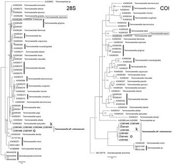
Bayesian inference trees for the genus Temnosewellia derived from the analysis of fragments of nuclear 28S rDNA (556–596 base pairs (bp)) and cytochrome c oxidase subunit I (COI) (526 bp) sequences. Diceratocephala boschmai (accession numbers MW443038–443040 and MZ128776) was used as an outgroup for rooting the trees. The posterior probability values are indicated on the branches. The scale bars correspond to the substitutions per nucleotide site. k = sequences of Temnosewellia aff. vietnamensis from a tributary of Kimotsuki River. o = sequences of T. aff. vietnamensis from Oda River. COI sequences, AJ05989 and AJ05990, were combined to be used for the analysis.

Molecular phylogeny
Monophyletic clades were formed by the phylogenetic analysis based on partial sequences of both 28S and COI of T. aff. vietnamensis (fig. 6). Small differences in nucleotide sequences were shown between specimens from Oda River (LC651446, LC651447 and LC651452–LC651454) and a tributary of Kimotsuki River (LC651441, LC651443–LC651445 and LC651448–LC651451). These localities are on adjacent but unconnected rivers, and the distance between them is approximately 7 km. The geographical variations among them were one base substituted within the 741 bases of 28S, and 24 bases substituted within the 569 bases of COI (amino acid substitution was observed in one residue between sites. One more residue was substituted in the single individual from Oda River). They constituted species-specific clades from other congeners.
Discussion
Phylogenetic position of T. aff. vietnamensis with other temnocephalids
Hoyal Cuthill et al. (Reference Hoyal Cuthill, Sewell, Cannon, Charleston, Lawler, Littlewood and Blair2016) conducted the phylogenetic analyses using 28S and COI with 23 species of Temnosewellia associated with the crayfishes of Euastacus Clark, 1936 from eastern Australia and T. minor and T. dendyi as outgroups. In our analyses using Diceratocephala boschmai Baer, 1953 as an outgroup, the clade of T. aff. vietnamensis was located between those of the 23 species and T. minor and T. dendyi in 28S (fig. 6). Two sequences registered as T. fasciata (KC869888) and T. minor (AY157164) in GenBank are doubted to possibly be T. minor and T. dendyi, respectively (see Vecchioni et al., Reference Vecchioni, Chirco, Bazan, Marrone, Arizza and Arculeo2021). In COI, the clade of T. aff. vietnamensis formed another clade with T. minor. While the monophyletic clades were formed by T. aff. vietnamensis both in the trees of 28S and COI (fig. 6), the almost sequences used in the phylogenetic analysis are of congeners from eastern Australia obtained by Hoyal Cuthill et al. (Reference Hoyal Cuthill, Sewell, Cannon, Charleston, Lawler, Littlewood and Blair2016). Although T. aff. vietnamensis is morphologically similar to Asian species, that is, T. semperi and T. vietnamensis, there is no genetic data published for these species. Further phylogenetic analysis is needed to reveal the phylogenetic relationship among Asian species. Along with this, as Vecchioni et al. (Reference Vecchioni, Chirco, Bazan, Marrone, Arizza and Arculeo2021) pointed out, sequences available in the public databases must correspond to the comprehensive and systematic revision of temnocephalans.
In this study, the specimens of T. aff. vietnamensis collected from two adjacent rivers which belong to different water systems show the geographical variations for both 28S and COI. These might indicate that the individuals do not migrate between rivers with hosts via land or sea while Eriocheir japonica is catadromous and migrate downstream into the sea to mate and lay eggs (e.g. Kobayashi, Reference Kobayashi1998). In the future study, it will be necessary to be confirmed with sequences of a larger number of individuals.
Records of Temnosewellia in Japan
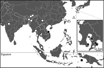
The previous records of the species which seems to be the members of the genus Temnosewellia from East to South Asia are plotted in fig. 7. In southern Japan, since the record of a species of Temnocephala from Okinawa Island by Okada (Reference Okada1938), its unidentified congeners have been reported several times in various localities. From Iriomote and Tanegashima Islands, the Ryukyu Islands, species of Temnocephala were recorded from Geothelphusa marginata fulva Naruse, Shokita & Shy, 2004 (=G. miyazakii) (Potamidae) and E. japonica, respectively (Suzuki et al., Reference Suzuki, Ninagawa and Kawakatsu1983). Although Kawakatsu et al. (Reference Kawakatsu, Sluys and Sasaki2004) listed the species as Temnosewellia semperi (=Temnocephala semperi), the authors also noted that it was not identified based on the specimens. Shimazu (Reference Shimazu, Otsuru, Kamegai and Hayashi2003) reported a finding of a species of temnocephalid that might be T. semperi associated with E. japonica from Tokushima Prefecture [listed as Temnosewellia semperi (Weber, Reference Weber1889)? of Shikoku by Kawakatsu et al. (Reference Kawakatsu, Gelder and Ponce de León2007a)]. Iwata et al. (Reference Iwata, Iino, Nakamura and Yasuda2004) recorded a species of Temnocephala associated with E. japonica from the northern part of Miyazaki Prefecture, and the authors implied the possibility of immigration of the species to Saga Prefecture. Further, Kawakatsu et al. (Reference Kawakatsu, Gelder and Ponce de León2007a) noted the findings of the congeners associated with G. minei Shy & Ng, 1998 and E. japonica from Ishigaki Island in the Ryukyu Islands and unspecified localities in Kagoshima Prefecture, respectively. Damborenea & Cannon (Reference Damborenea and Cannon2001) reviewed the species of Temnocephala from Central to South America, and transferred all species previously assigned to Temnocephala in Australia and Southeast Asia to Temnosewellia. Since then, the distribution of all valid species of Temnocephala is restricted from Central to South America, and thus it is reasonable that the species previously reported from Japan are considered to be members of Temnosewellia. For the species level identification, re-examinations of all the previous records from the Ryukyu Islands, Kyushu and Shikoku based on the specimens are required. Specimens found from E. japonica and G. exigua in Kagoshima were provisionally identified as Temnosewellia sp. (Okawachi et al., Reference Okawachi, Uyeno, Miyazaki and Kamezaki2013), but they are considered to be T. aff. vietnamensis. Additionally, an alien species T. minor has been recorded from the culture ponds of the crayfish C. tenuimanus in Ibusuki, Kagoshima (Tamura et al., Reference Tamura, Oki, Kawakatsu, Ninagawa, Matsusato and Suzuki1985; Oki et al., Reference Oki, Tamura, Takai and Kawakatsu1995; Kawakatsu et al., Reference Kawakatsu, Gelder and Ponce de León2007a, Reference Kawakatsu, Nishino and Ohtakab).
Map of East to South Asia showing the distributional records of the genus Temnosewellia Damborenea et Cannon, 2001. Black filled star = Temnosewellia aff. vietnamensis newly collected from the Osumi Peninsula, Kagoshima, Japan in this study; open star = same from the Osumi Peninsula, Kagoshima, Japan by Okawachi et al. (Reference Okawachi, Uyeno, Miyazaki and Kamezaki2013); black filled triangle = Temnosewellia minor from Ibusuki, Kagoshima, Japan (Tamura et al., Reference Tamura, Oki, Kawakatsu, Ninagawa, Matsusato and Suzuki1985; Oki et al., Reference Oki, Tamura, Takai and Kawakatsu1995); open circle = Temnosewellia semperi from Sumatra, Java, and various sites of Sulawesi, Indonesia (Weber, Reference Weber1889), Luzon and Mindanao, Philippines (Semper, Reference Semper1872), Malaysia (Rohde, Reference Rohde1966; Cannon, Reference Cannon1991), Dawna Hills and Dawei, Myanmar (Gravely, Reference Gravely1913), Narmada River in Madhya Pradesh, Jaintia Hills, and Manipur Hills, India (Gravely, Reference Gravely1913; Chauhan & Ramakrishna, Reference Chauhan and Ramakrishna1953), Yembung and Siyom Rivers in Abor Country (Wood-Mason, Reference Wood-Mason1875; Gravely, Reference Gravely1913; Chauhan & Ramakrishna, Reference Chauhan and Ramakrishna1953), and Yunnan and Foochow, southern China (Gravely, Reference Gravely1913; Lee, Reference Lee1936); black filled square = Temnosewellia rouxi from Aru Island, Indonesia (Merton, Reference Merton1914); black filled circle = Temnosewellia vietnamensis from Quangnam, Vietnam (Damborenea & Brusa, Reference Damborenea and Brusa2009); and open triangle = unidentified species of Temnosewellia from Tokushima, Saga, Miyazaki, Kagoshima, Tanegashima Island, Okinawa Island, Ishigaki Island, and Iriomote Island, Japan (Okada, Reference Okada1938; Suzuki et al., Reference Suzuki, Ninagawa and Kawakatsu1983; Shimazu, Reference Shimazu, Otsuru, Kamegai and Hayashi2003; Iwata et al., Reference Iwata, Iino, Nakamura and Yasuda2004; Kawakatsu et al., Reference Kawakatsu, Gelder and Ponce de León2007a), Taipei, Taiwan (Okada, Reference Okada1938), Lombok, Indonesia (Ng & Anker, Reference Ng and Anker2016), and eastern Thailand (Ngamniyom et al., Reference Ngamniyom, Sriyapai, Sriyapai and Panyarachun2019).

Species diversity of Temnosewellia from East to South Asia
Previous records of the species of Temnosewellia in East to South Asia are shown in fig. 7. Temnosewellia aff. vietnamensis is similar to T. semperi as well as T. vietnamensis, which are known from freshwater crabs in tropical Asian countries. Temnosewellia semperi was originally described from Indonesia (i.e. Sumatra, Java and various sites of Sulawesi) based on the specimens associated with freshwater crabs [reported as species of Telphusa by Weber (Reference Weber1889)]. The species is now known from various freshwater crabs in Luzon and Mindanao, Philippines (Semper, Reference Semper1872) and Malaysia including Stoliczia rafflesi (Roux, 1936) (=Potamon rafflesi) (Rohde, Reference Rohde1966; Cannon, Reference Cannon1991). Temnosewellia semperi has been found from Barytelphusa lugubris (Wood-Mason, 1871) (=Paratelphusa (Barytelphusa) lugubris), Tiwaripotamon adiatretum (Alcock, 1909) (=Po. adiatretum), Indochinamon andersonianum (Wood-Mason, 1871) (=Po. andersonianum), Po. (Potamon) manii (Rathbun, 1904) (=Po. manii), and Alcomon superciliosum (Kemp, 1913) (=Po. superciliosum) which were caught from Myanmar, India, southern China, and their border, Abor Country (Gravely, Reference Gravely1913; Lee, Reference Lee1936; Chauhan & Ramakrishna, Reference Chauhan and Ramakrishna1953). Furthermore, Gravely (Reference Gravely1913) noted that T. semperi is often found from the members of Geothelphusa Stimpson, 1858 (=Geotelphusa) and Potamon Savigny, 1816 in large numbers. He also mentioned that a specimen which was accidentally attached to fishes from Daphla Hill, Abor Country and was identified as Temnocephala chilensis (Moquin-Tandon, 1846) by Wood-Mason (Reference Wood-Mason1875) was T. semperi. Recently, Ng & Anker (Reference Ng and Anker2016) reported a species that was possibly a member of Temnosewellia when the authors described the freshwater crab Sundathelphusa tuerkayi Ng & Anker, Reference Ng and Anker2016 (Gecarcinucidae) from Lombok, Indonesia. Considering the locality and host, this species might be T. semperi or a closely related species. If all of those records are of true T. semperi, the facts mean that the species is widely distributed from isolated islands to Eurasian continent in tropical to temperate Asia. However, T. vietnamensis and T. aff. vietnamensis, which are similar to T. semperi, were found from Vietnam and Japan, respectively (Damborenea & Brusa, Reference Damborenea and Brusa2009; this paper). Furthermore, no species of the host that has such a wide distribution range is known. Based on these facts, it is doubtful that all of the records so far were conspecific. Re-examinations of previous records of temnocephalids identified as T. semperi are needed based on the specimens (see Kawakatsu et al., Reference Kawakatsu, Gelder and Ponce de León2007a), and redescription of the species is also strongly required. There are other records of the congener. From Taiwan, Okada (Reference Okada1938) found a congener from Yanmingshan, Taipei, and he mentioned that the species is conspecific to the unidentified congener from Okinawa Island, Japan. In eastern Thailand, an unidentified species of Temnosewellia was found from the red claw crayfish Cherax quadricarinatus von Martens, 1868 introduced from Australia (Ngamniyom et al., Reference Ngamniyom, Sriyapai, Sriyapai and Panyarachun2019). Together with the host, the species of Temnosewellia might be invaded. Just as Ngamniyom et al. (Reference Ngamniyom, Sriyapai, Sriyapai and Panyarachun2019) claimed that the species genetically similar to T. minor and T. fasciata, the species was formed a clade with T. minor by the partial sequences of COI in this study (fig. 6). Damborenea & Brusa (Reference Damborenea and Brusa2009) suggested that great numbers of potential hosts of undescribed temnocephalids are present in Southeast Asia. Nevertheless, the freshwater crabs in tropical to temperate regions of East to South Asia are actually highly diverse, only four native species including T. aff. vietnamensis so far being recognized. Discoveries of many undescribed species in the regions from future studies using morphology and molecular characterization are expected.
Acknowledgements
The authors thank Kenzaburo Arimura (Tarumizu City), Shuntaro Watanabe, Shuichiro Tagane, and Kenshiro Nagayoshi (Kagoshima University) for their help to collect materials; Seiichi Ashihara (Kagoshima University) for his kind guidance around the grounds of the Takakuma Experimental Forest, Kagoshima University; and Hiroshi Suzuki (Kagoshima University) and Masato Nitta (Setouchi Parasite Biodiversity Laboratory) for providing valuable information on sampling and preparing materials.
Financial support
This study was partially supported by the ‘Establishment of Glocal Research and Education Network in the Amami Islands’ project of Kagoshima University adopted by the Ministry of Education, Culture, Sports, Science and Technology, Japan.
Conflict of interest
None.
Ethical standards
The authors assert that all procedures contributing to this study comply with the ethical standards of the relevant national and institutional guides on the care and use of laboratory animals. This study is not related to human experimentation.