Introduction
Cerebrospinal fluid (CSF) leak after cerebellopontine angle surgery is not a rare occurrence, with a reported incidence of 2–21.9 per cent.Reference Becker, Jackler and Pitts1–Reference Rodgers and Luxford7 In a meta-analysis of 25 case series (which included a total of 5964 patients), Selesnick et al. reported an average incidence of 10 per cent.Reference Selesnick, Liu, Jen and Newman8 Numerous prophylactic measures have been employed; however, none significantly reduced the CSF leak rate below the average for the grouped data.Reference Selesnick, Liu, Jen and Newman8 The findings of comparative studies assessing CSF leak rates associated with various surgical procedures have remained remarkably similar. Leaks via the eustachian tube into the nasopharynx have been reported as slightly more common.Reference Cueva and Mastrodimos9
Usually these leaks develop in areas where dura cannot be sealed. If CSF leak dynamics are altered, they lead to increased intracranial pressure at sites where communication exists between the intracranial and extracranial compartments.Reference Kwartler, Schulder, Baredes and Chandrasekhar10
Different surgical and non-surgical techniques can be used to avert this problem. These techniques range from conservative treatment (e.g. bed rest management) to more invasive surgery involving skull re-exploration and dural defect obliteration.Reference Friedman, Cullen, Ulis and Brackmann11 In cases of refractory CSF rhinorrhoea where the above-mentioned measures are exhausted, endoscopic surgery should be considered.Reference Orlandi and Shelton12 Endoscopic surgery can be used to obliterate or close the eustachian tube endonasally.Reference Kwartler, Schulder, Baredes and Chandrasekhar10, Reference Orlandi and Shelton12, Reference Doherty and Slattery13
Case report
A 44-year-old fit and well woman with a more than 2-year history of right-sided facial pain was referred for a neurosurgical opinion. A diagnosis of trigeminal neuralgia was made.
The patient underwent glycerol injection into Meckel's cave, which failed to improve the facial pain symptoms. She subsequently underwent retromastoid suboccipital craniotomy and microvascular decompression of a vascular loop.
Post-operatively, the patient developed right-sided CSF rhinorrhoea. This was initially managed with bed rest, with the patient lying in the supine position at a 45-degree, head-up incline, and a lumbar drain. The leak persisted and subsequent posterior craniotomy with obliteration of the dural defect using strips of fat was undertaken. The leak continued and further surgery was necessary in the form of an obliterative right mastoidectomy through a middle fossa approach, in order to seal the dural defect and close the communication between the intracranial compartment and mastoid air cells (extracranial compartment). The above surgical procedures proved unsuccessful in abating the leak, and at this stage the patient opted to withdraw from any further surgical intervention.
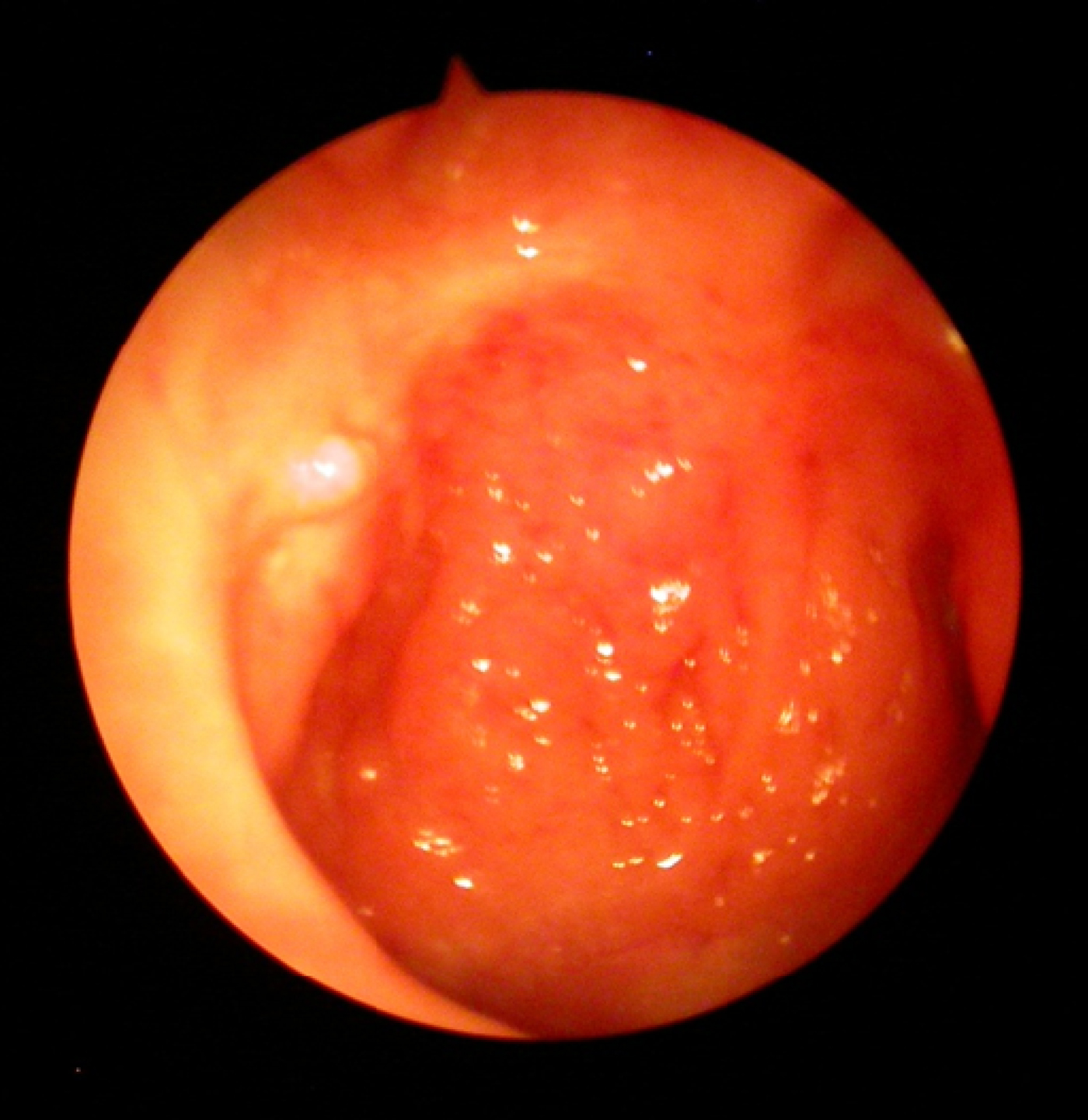
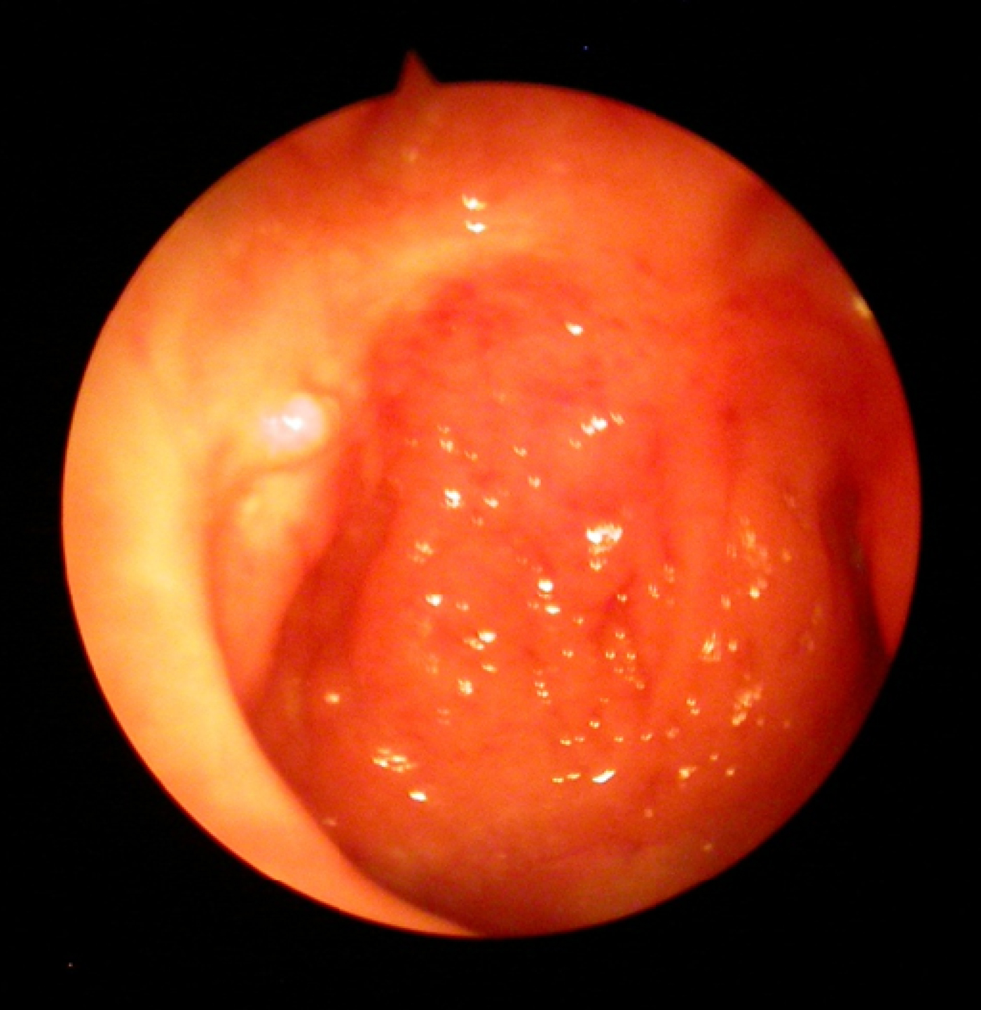
In light of the continuous leak and the several hospital admissions, the patient was referred to the ENT department. The option of eustachian tube obliteration using an endonasal endoscopic approach was discussed. Simple endoscopic unipolar diathermy of the eustachian tube orifice, performed under general anaesthetic, led to more than a 75 per cent reduction in CSF rhinorrhoea, but this was not sufficient to completely stop the leak. A second procedure was therefore carried out using the technique described below. Twenty-four months post-operatively, there remained no CSF leak (Figure 1).
Post-operative view of the eustachian tube.
Surgical technique
The surgery was performed under general anaesthetic, with topical adrenaline (1:1000) placed on neurosurgical pledgets in the nasal cavity for several minutes for vasoconstriction. A local anaesthetic containing 2 per cent lignocaine and noradrenaline (1:80 000; Lignospan®) was then injected using a spinal needle. Angulating the end of the needle helped to deliver the anaesthetic at the correct angle. Less than 1 ml was injected into the lateral eustachian tube wall and around the fossa of Rosenmüller. A 30-degree, 4 mm Karl Storz telescope was used to view the procedure.
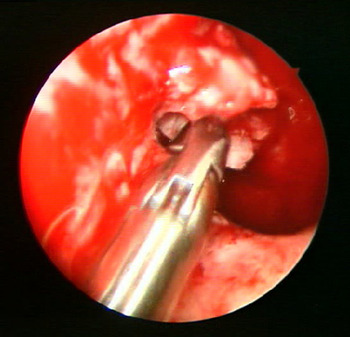
A piece of harvested septal cartilage was cut to size so that it filled the orifice. This was inserted after freshening of the mucosa. The graft was then impregnated with fibrinogen aprotinin solution (Tisseel® VH Baxter Fibrin Sealant Kit).
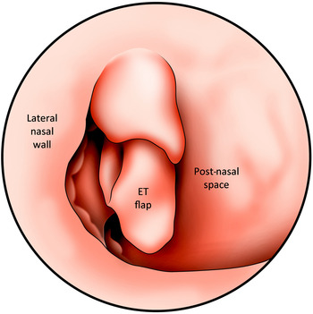
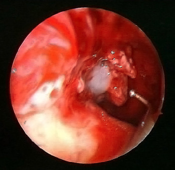
A circumferential incision was created at the torus tubarius, from 2 o'clock to 8 o'clock, and a mucosal flap was partially freed and elevated with endoscopic scissors. The flap was rotated cephalically to seal the eustachian tube orifice (Figures 2–4).
Flap design. ET = eustachian tube

Harvested septal cartilage in situ.

Infolding the eustachian tube flap inward.

Bio-resorbable nasal dressing (NasoPore®) was applied to aid fixation. No lumbar drain was required. The patient was discharged home the same day.
Discussion
A CSF leak may manifest after skull base or otological surgery as a watery discharge through the nostril on the side of surgery. In refractory cases, the patient may present with signs and symptoms of meningitis. A CSF leak can have serious consequences. More commonly, the consequences are less serious, but are prolonged; these hinder the recoveryReference Becker, Jackler and Pitts1 and prove more concerning to patients.
Management ranges from conservative to surgical approaches. Some studies have shown good improvement with a non-surgical approach.Reference Hoffman14 However, the management of some refractory rhinorrhoea cases remains very challenging, especially in patients who are reluctant to undergo further potentially debilitating and invasive neurosurgery, as in our case. In addition, the use of a lumbar drain, which is thought to be the main conservative method, is associated with unwanted complications.Reference Graf, Gross and Beck15, Reference Roland, Marple, Meyerhoff and Mickey16
Endoscopic obliteration of the eustachian tube to stop a CSF leak is technically simple and straightforward, and takes no more than 20 minutes to carry out. The rationale behind the technique is to create a complete seal of the eustachian tube and provide secondary obliteration of the middle-ear cavity. This should, in theory, allow time for the defect to heal completely. The resultant conductive hearing loss may be reversed in due time with a ventilation tube if the defect has healed.
The potential benefits of eustachian tube closure outweigh the risks of open craniotomy or mastoidectomy, which include facial nerve palsy, permanent hearing loss and other morbidities.
• Cerebrospinal fluid leak after cerebellopontine angle surgery is not uncommon, and recalcitrant cases could lead to meningitis
• Management ranges from conservative to surgical
• Management of some refractory rhinorrhoea cases remains challenging, especially in those reluctant to undergo surgery
• The condition can be treated successfully via transnasal endoscopic techniques that obliterate and close the eustachian tube
• Endoscopic obliteration of the eustachian tube orifice using a double seal technique is an easy procedure
• To our knowledge, we are the first to use this surgical technique to manage such rare and difficult cases
Eustachian tube closure for the treatment of patulous eustachian tube has been attempted previously. The methods used ranged from myringotomy with eustachian tube scarring to achieve closure, through stenosis, to complete closure using a fat plug.Reference Doherty and Slattery13 The latter technique was not applicable in our case as we needed to achieve complete obliteration of the tube rather than stenosis or even partial closure. Patulous eustachian tubes are frequently associated with depletion of the soft tissue surrounding the cartilaginous part of the eustachian tube,Reference Cox17–Reference Pulec and Simonton19 hence this technique was deemed unsuitable. All other techniques which might theoretically achieve closure via stenosis were shown to be incompletely effective.Reference O'Connor and Shea20, Reference Ogawa, Satoh and Tanaka21
Kwartler et al. employed a technique where autologous muscle was used to plug the eustachian tube; the 2 patients in whom this was carried out continued to be symptom-free after 12 months.Reference Kwartler, Schulder, Baredes and Chandrasekhar10 In our case, we opted to use cartilage as a plug, as the cartilage is more durable, less absorbable and more resilient to CSF flowReference Pappada, Formaggio, Regalia, Panzarasa and Geuna22 than a fat or muscle plug. Additionally, we feel that the use of a rotation flap is key to long-term closure.
We did not use posterior nasal packing, and the patient was able to go home the same day (with post-operative advice). The use of degradable packing (NasoPore) was very effective in stabilising the flap, as we found that suturing was not technically possible.Reference Orlandi and Shelton12
Conclusion
Endoscopic obliteration of the eustachian tube using a double seal technique is an easily performed procedure in cases of refractory CSF leak when other neurosurgical procedures have failed or are not possible.