Introduction
Radiation therapy (RT) plays an important role in the management of head and neck cancer (HNC). Most HNC patients have historically been treated with photon-based radiation techniques such as 3D conformal radiation therapy (3D-CRT), intensity-modulated radiation therapy (IMRT) and volumetric-modulated arc therapy (VMAT). RT has been well established as an essential tool to treat HNCs in various clinical settings. IMRT and VMAT (a form of IMRT utilising rotating gantry and dynamic dose rate) are advanced form of 3D-CRT that utilises non-uniform radiation beam intensities determined using various computer-based optimisation techniques incident on patients. The anatomically complex head and neck region is ideal for IMRT or VMAT because the technology allows for planning target volume (PTV) dose escalation, sharp dose gradients around target resulting in superior sparing of normal structures and organs at risk (OARs). However, 3D-CRT demonstrated limitations in these respects. Advancements in diagnostic imaging, RT planning and delivery have modernised the photon-based HNC treatments. IMRT and VMAT plans can yield highly conformal dose distributions with reduced toxicity, faster treatment times and less monitor unit (MU), though VMAT provides somewhat improved efficiency compared to IMRT. Reference Verbakel, Cuijpers and Hoffmans1–Reference Mann, Indira and David6
There are limited dosimetric comparisons available in literature among 3D-CRT, IMRT and VMAT for HNC. Larynx cancer is the most common head and neck malignancy and approximately half of all patients diagnosed at an early disease stage. Reference Gomez, Cahlon and Mechalakos7 Successful treatment options include surgery and RT, but no single modality has been proven superior. Reference Pfister, Laurie and Weinstein8 RT, however, is considered to be a mainstay treatment approach. Reference Teshima, Chatani and Inoue9 The traditional technique for early-stage Larynx RT included the use of small, parallel opposed lateral fields proven to be very effective but unfortunately did not allow dose sparing of the adjacent structures in the neck including the skin, bilateral carotid arteries and the thyroid gland. IMRT allowed superior dose conformality to PTV and provided steep dose gradients to nearby uninvolved structures. Reference Chera, Amdur and Morris10,Reference Rosenthal, Fuller and Barker11 VMAT with RapidArc allowed similar treatment dose conformality to PTV with the addition of reduced treatment and dose delivery times. Reference Dobbler, Weidner and Koelbl12 A recent study clearly indicated a potential clinical advantage for IMRT and VMAT compared to 3D-CRT, where VMAT planning was shown to be the most effective modality to maintain or improve PTV coverage with the most reduction in mean carotid and thyroid dose. IMRT and VMAT showed superiority with less variation among themselves compared with 3D-CRT plans, with exception of spinal cord dose. Reference Matthiesen, Herman and Singh13
Background
Majority of the patients with HNC treated with a curative intent receive a dose between 50 and 70 Gy. Radiation causes damage in normal tissue composed of several dissimilar structures that respond differently to radiation; mucosal lining, skin coverings, subcutaneous connective tissue, teeth and bone/cartilage. The oral complications that arise are primarily responsible for determining patient’s quality of life. The main causes of developing these cancers are consumptions of betel quid, betel nut, cigarettes, viral infection, poor oral hygiene, exposure to radiation, industrial fumes, alcohol, chemicals etc.
IMRT was first conceptualised in the 1960s. However, it was not implemented until the 1980–1990s when computing capability required for complex inverse planning algorithms became commercially available. In 1994, NOMOS Peacock system was introduced as the first commercial IMRT delivery unit. Reference Curran14 The unique feature of IMRT is the leaves known as MLC (multi-leaf collimator) that help create the complex shape of the beam. IMRT proposes many technical advantages Reference Hong, Ritter and Tomé15 over prior technologies, but it is not always the best technique for patients. Since the IMRT radiation beams are oriented usually up to nine beam angles surrounding the patient, a low-dose ‘bath’ of radiation may be created just outside the main PTV. This effect may also occur in 3D-CRT planning as well where only 2–4 beam angles are used and so the low-dose region is not spread out as widely. Sometimes, complex shapes of radiation dose with IMRT result in unwanted ‘hot spots’ or ‘cold spots’ of radiation within the patient. Hot spots located in important organs can put the patient at higher risk for side effects and cold spots within the target could mean that the tumour is not receiving enough radiation doses to control the cancer.
IMRT is a technique where hundreds of small radiation beams with different intensities are delivered with various beam angles providing precise radiation doses to a tumour while minimising the dose to adjacent normal tissues. It can generate a conformal dose distribution and has steep dose fall-off at the boundary between the tumour and the normal structures. IMRT enables dose escalation without increasing toxicity to the critical organs, potentially improving the therapeutic ratio. VMAT is a special type of IMRT technique where treatment is delivered with intensity-modulated beam that rotates around the patient. The beam is modulated by dynamic multi-leaf collimation, variable dose rate and variable gantry speed in optimised arc/arcs around the patient to generate dose distributions. VMAT treatments can significantly reduce the time and MU required to deliver a patient’s treatment. VMAT has emerged as a mainstream treatment option for HNC given the complexity of the anatomy in the head and neck region. A VMAT plan usually consists of 2–3 full or partial arcs. VMAT plans are capable of creating a more conformal dose distribution to PTV when compared with traditional IMRT. Both conventional IMRT and VMAT plans are based on the inverse planning system where expected dose distributions are given first then a mathematical equation is solved to find appropriate beam intensities needed to provide the dose.
Dose homogeneity index (HI) is usually measured as the standard deviation (SD) of the PTV dose. For a PTV, with a homogeneous dose, the dose fall-off is very steep and SD is very small. The conformity of dose to PTV is expressed by conformity index (CI) being the volume of the body receiving more than 95% of the prescribed dose, divided by the volume of the PTV. Reference Verbakel, Cuijpers and Hoffmans1 During radiation treatments, patients are required to stay comfortably immobilised for the entire time when the beam is on; small patient movements may offset the conformity. When a patient’s ability to maintain the treatment position is limited, VMAT is probably the best option to treat a complex shape tumour with a high conformity and lower treatment time. Reference Studenski, Bar-Ad and Siglin16,Reference Teoh, Clark and Wood17
Results
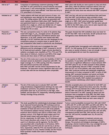
The inconsistency of results through several investigations in literature show differences in plan optimisation and delivery techniques. Thus, it is important to take into account all the results in terms of selection of beam parameters, treatment machine and dose calculation algorithm. The main characteristics for 3D-CRT and IMRT delivery methods are that the dose rate and gantry speed are constant. For VMAT delivery method, the dose rate and gantry speed are variable. For beam intensity, IMRT and VMAT are modulated and 3D-CRT is uniform. Based on the literature review, comparison of 3D-CRT, IMRT and VMAT are pointed out in Table 1.
Comparison of 3D-CRT, IMRT and VMAT advantages and disadvantages for HNC

Discussion and Conclusions
After investigating the literature, it is indeed difficult to indicate definitely the best technique in all cases of HNC. IMRT and 3D-CRT both techniques were largely similar in terms of OAR irradiation, when the only notable exception being the cerebellum, which received more radiation with IMRT than 3D-CRT. The HI and CI did not show any major statistical differences. In comparison of OAR sparing, the parotid glands had better sparing in VMAT than IMRT. In case of nasopharynx, hypopharynx, larynx, oral cavity etc., most of the papers showed that the IMRT provided better CI than 3D-CRT which insured more accurate dose distribution.
In comparison of IMRT and VMAT dose coverages, both plans have similar coverage. The volume of normal tissue dose received in VMAT was less than IMRT, which probably reduced the late effect of low-dose radiation (radiation-induced carcinomas). The number of MUs required to deliver a prescribed dose in one study was higher in VMAT (both single and dual arc) than IMRT. Other studies showed that the VMAT had less MU than IMRT. VMAT was found superior to IMRT because of its faster and efficient delivery than IMRT. Reference Nithya, Raj and Kumar24 Planning studies in different tumour sites comparing VMAT with IMRT have reported that the VMAT plans were comparable with a shorter delivery time, less MU in the arc delivery Reference Singh, Rawat and Bhushan25 .
Future studies are required to clarify the impact of reduced delivery time with cost utility factor on clinical outcomes and possible improvement on small OAR sparing. There are inherent limitations with these planning studies. Even if the same strict planning objectives and calculation algorithms were used, it is extremely difficult to completely eliminate planner bias, especially if multiple planners are involved in the process. Direct comparisons between different studies are not possible because of significant differences in target volume definitions, dose prescriptions and treatment schedules. Radiation techniques also vary among studies, for example, number of fields and arcs used in fixed field IMRT (sliding window or step and shoot) and VMAT plans, respectively.
Acknowledgements
None.