“Vascular sling” is the terminology used to describe the arrangement in which the left pulmonary artery arises anomalously from the right pulmonary artery and reaches the hilum of the left lung by coursing between the trachea and the oesophagus. Glaevecke and Doehl first described the entity in 1897, with Contro et al first using the term “sling” in 1958.Reference Marins, Anderson and Aiello 1 A rarer variant of the lesion has also been described as the partial pulmonary arterial sling, in which the pulmonary trunk bifurcates in a normal manner into its left and right branches, but in which a second pulmonary arterial branch supplying the left lung arises from the right pulmonary artery. In this report, we describe two additional cases of anomalous origin of a branch of the left pulmonary artery from the right pulmonary artery, which cannot strictly be termed partial pulmonary arterial slings, as there is neither tracheal nor oesophageal compression. We also discuss the potential developmental background for this rare malformation.
Case report
Case 1
The first patient was a male baby with normal arrangement of a left-sided heart, a large unrestrictive atrial septal defect, mitral atresia with a small left ventricle, no tricuspid regurgitation, a large inlet ventricular septal defect with muscular extension, a bicuspid aortic valve with diameter of 5.5 mm, and adequate antegrade flow into the ascending aorta. The segment of the aortic arch between the left common carotid and left subclavian arteries was hypoplastic, and the duct was large, with predominantly right-to-left flow.
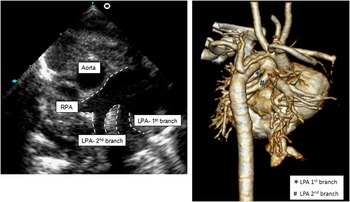
The pulmonary arteries were noted to have an unusual configuration. The pulmonary trunk divided normally into the right and left pulmonary arteries, with the right artery having a diameter of 4.3 mm, and the left of 2.5 mm. A second left-sided pulmonary artery, of diameter 2.5 mm, then arose anomalously from the proximal right pulmonary artery (Fig 1, Supplementary figure S1). The normally positioned branch supplied the left upper lobe, with normal relation to the airway, coursing superior to the left bronchus. The anomalous branch arose from the proximal right pulmonary artery below the tracheal bifurcation and passed posterior and inferior to the left main bronchus and anterior to the oesophagus to supply the left lower lobe. There was no tracheal, bronchial, or oesophageal compression noted on computerised tomographic imaging.
Parasternal short-axis view echocardiogram and CT angiogram 3D reconstruction – case 1. LPA=left pulmonary artery; RPA=right pulmonary artery.

Although the baby was appropriate for a staged single ventricle approach, he was also detected to have anal atresia and vertebral anomalies, specifically a third thoracic butterfly vertebra, and a left fourth thoracic hemivertebra. No genetic abnormality was detected on karyotyping and molecular cytogenetic analysis. In view of the comorbidities, his family elected palliative management with a colostomy and no other cardiac intervention. The baby died on his 40th day of life.
Case 2
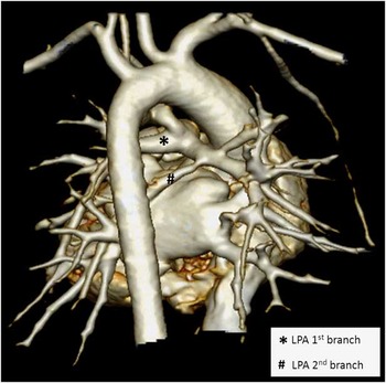
The second patient was a male baby with tetralogy of Fallot who was admitted on the 1st day of life. The right and left pulmonary arteries, with diameters of 3.5 and 3.8 mm, respectively, arose in a normal manner from the pulmonary trunk. In addition, a smaller left pulmonary arterial branch, of 2-mm diameter, was noted to originate from the right pulmonary artery (Fig 2, Supplementary figure S2). This anomalous left pulmonary arterial branch coursed between the left upper lobe bronchus anteriorly and the oesophagus posteriorly to supply the left lower lobe. There was no tracheal, bronchial, or oesophageal compression.
CT angiogram 3D reconstruction – case 2. LPA=left pulmonary artery.

He was also detected to have CHARGE association with coloboma, middle ear abnormalities, and retinal detachment. He underwent a left-sided modified Blalock–Taussig shunt on the 5th day of life, and subsequent total correction of his tetralogy of Fallot at 4 months of age. The anomalous left pulmonary branch supply was left unaltered. His ensuing cardiac and clinical status has been stable, with no stridor or wheezing reported.
Discussion
Anomalous origin of a branch of the left pulmonary artery from the right pulmonary artery in the setting of normal bifurcation of the pulmonary trunk is extremely rare. Our search of the literature revealed only four similar cases. In the case reported by Bamman et al,Reference Bamman, Ward and Woodrum 2 the anomalous left pulmonary artery circled over the bronchus to the right lower lobe and passed to the left hilum between the trachea and the oesophagus, forming a partial pulmonary arterial sling. In the case described by Erickson et al,Reference Erickson, Cocalis and George 3 the aberrant artery to the left lower lobe ran anterior to the trachea and caudal to the left lower lobe bronchus. As there was no encirclement of the trachea, this case cannot be considered to represent an arterial sling. In the example reported by Collins et al,Reference Collins, Weinberg, Goldmuntz and Harris 4 the anomalous artery coursed anterior to the left bronchus, producing no compression, and therefore again is not a sling. In the case of Ge et al,Reference Ge, De Groff, Knudson, Strain and Chan 5 the anomalous left lower pulmonary artery ran posterior to the left main bronchus. The case described by Marins et alReference Marins, Anderson and Aiello 1 is also of interest, as in their patient the entire left pulmonary artery had an extrapericardial origin, but again without producing a sling. Our two cases are most comparable to the arrangement described by Ge et al.Reference Ge, De Groff, Knudson, Strain and Chan 5
Such anomalous origin of the left lower lobe pulmonary artery from the right pulmonary artery has previously been described as a partial pulmonary arterial sling. Our review shows, however, that only the patient described by Bamman et alReference Bamman, Ward and Woodrum 2 had the anomalous artery coursing between the trachea and oesophagus, thus forming the so-called sling. In all other cases, including our own, the anomalous branch caused neither tracheal nor oesophageal compression. Therefore, all the cases reviewed, including the two from this report, except the one reported by Bamman and associates, are more accurately described as partial anomalous origin of the left pulmonary artery.
When first formed, the right and left pulmonary arteries arise within the pharyngeal mesenchyme and take their origin from the base of the aortic sac.Reference Anderson, Chaudhry and Mohun 6 When seeking to explain the development of the pulmonary arterial sling, Jue et alReference Jue, Raghib, Amplatz, Adams and Edwards 7 postulate that when the primitive lung tissue is still undivided into the left and right lungs, the pulmonary supply from the left sixth aortic arch either fails to form or, having formed, gets obliterated. Consequent to a lack of arterial supply, the right sixth aortic arch sends a collateral branch into the part of the primitive pulmonary mass, which will later become the left lung. This explanation, however, fails to stand rigorous examination, as the lung buds themselves arise separately from the tracheal groove, and the pulmonary arteries arise from the base of the aortic sac, rather than from the sixth pharyngeal arch arteries. Therefore, it remains difficult to provide a rational explanation for either the pulmonary arterial sling, or partial anomalous origin of the left pulmonary artery from the right pulmonary artery.
With regard to clinical implications, the partial anomalous origin of the left pulmonary artery would have affected flows of blood to the left lung had our first patient been selected for further surgery, and may ultimately have impacted on the suitability for a cavopulmonary circulation. The abnormal branching pattern had no clinical manifestations in our second case. Awareness of this rare variation is important, nonetheless, for formulating an appropriate therapeutic plan, as the finding of normal bifurcation of the pulmonary trunk does not exclude additional abnormal origin of part of the left pulmonary artery.
Acknowledgements
The authors thank Professors Robert H. Anderson, Richard Harvey, and Margaret Kirby for their advice regarding the development of the pulmonary arteries and assistance in revising the manuscript.
Financial Support
This research received no specific grant from any funding agency, commercial, or not-for-profit sectors.
Conflicts of Interest
None.
Supplementary material
To view supplementary material for this article, please visit http://dx.doi.org/10.1017/S1047951114001528