Introduction
Ptychadena is a true African frog and belongs to the anuran family Ptychadenidae, with 49 species currently recognized (Frost, Reference Frost2010). These species are widespread in savanna bushveld where they are especially common in low-lying areas, ranging south from Egypt through subSaharan Africa, excluding the south-western parts of Africa. They are also known from the Seychelles, the Mascarene Islands and Madagascar (Du Preez & Carruthers, Reference Du Preez and Carruthers2009). Du Preez & Kok (Reference Du Preez and Kok1992a) stated that Ptychadena species fulfil the requirements for the perfect host for a polystome in that they breed in stagnant or slow-flowing water and are opportunistic breeder with extended breeding seasons.
Polystomatids of anurans are represented in Africa by the genera Eupolystoma Kaw, 1950, Metapolystoma Yamaguti, 1963, Polystoma Zeder, 1800 and Protopolystoma Bychowsky, 1957. The genus Polystoma has a widespread occurrence in all zoogeographical realms except the Australian realm and 31 of the 64 currently known species are known from Africa. Of these, 13 are known from the grass frog genus Ptychadena. Polystomes known from Ptychadena include Polystoma aeschlimanni Bourgat & Murith 1980 from Pt. pumilio, P. assoulinei Bourgat 1975 from Pt. tellinii, P. baeri Maeder et al. 1970 from Pt. bibroni, P. dawiekoki Du Preez et al. 2002 from Pt. anchietae, P. ebriensis Maeder, 1973 from Pt. aequiplicata, P. lamottei Bourgat & Murith 1980 from Pt. pumilio, P. mangenoti Gallien 1956 from Pt. superciliaris, P. pricei Vercammen-Grandjean, Reference Vercammen-Grandjean1960 from Ptychadena sp., P. prudhoei Saoud 1967 from Pt. oxyrhynchus, P. sodwanensis Du Preez & Kok, Reference Du Preez and Kok1992b from Pt. porosissima and P. togoensis Bourgat, 1977 from Pt. hylaea. Furthermore Ptychadena also harbours Metapolystoma in that M. brygoonis (Euzet & Combes, 1964) is known from Pt. mascareniensis in Madagascar and M. cachani (Gallien, 1956) is known from Pt. longirostris. During further studies of African polystomes, Pt. oxyrhynchus has been found infected with an undescribed species of Polystoma in the KwaZulu Natal province of South Africa. This paper reports the second polystome species from Pt. oxyrhynchus, but the fifteenth record of a polystome from the anuran genus Ptychadena.
Materials and methods
Collection and examination of frogs
Adult frogs were collected in KwaZulu-Natal (South Africa) during December 1990, November 1991, May 1992, October 1992, March 1993 and October 2007. Prior to dissection, frogs were anaesthetized with benzocaine. They were dissected and the complete urinary bladder of infected frogs was removed and transferred to 0.3% saline solution. Following fixation for 2 h in 10% neutral buffered formalin (NBF) while under coverslip pressure, parasites were removed and preserved in 10% NBF. Prior to staining, parasites were rinsed in tap water for 1 h, agitating the Petri dish every 10 min and replacing the water after 30 min, stained overnight in a weak solution of acetocarmine, dehydrated, cleared in xylene and mounted using Canada balsam. Two of the specimens were fixed in 96% ethanol (EtOH) for future molecular studies. Parasite eggs were harvested from infected hosts prior to dissection. Eggs were collected by sieving water through plankton netting with a mesh size of 112 μm and incubated in 10 ml of aged tap water in glass Petri dishes. Oncomiracidia were mounted in ammonium picrate glycerine. Marginal hooklet pairs were numbered one to eight, with pair one being the posteriormost pair closest to the median (see Murith, 1981a). All measurements are in micrometres and are given with the mean followed by the range in parentheses.
Results
Levels of infection
The single host specimen collected during December 1990 was infected with a single parasite and the specimen collected during November 1991 with two parasites. The two immature specimens collected during May 1992 were infected with one and two immature parasites, respectively. The four specimens collected during October 1992 were not infected. One of the two immature specimens collected during March 1993 was infected with 18 immature parasites. Of the three frogs collected in October 2007 two were infected with respectively six and four mature parasites, which were identified as Polystoma vernoni n. sp., with a prevalence and mean intensity of 53.8% and 4.9, respectively.
Polystoma vernoni n. sp.
Thirteen sexually mature worms, 21 immature worms. Holotype (NMB P312) and five paratypes (NMB P313–317) deposited in the Parasitic Worm Collection, National Museum, Aliwal Street, 9300 Bloemfontein, South Africa; two paratypes (2010.8.27.1-2) in the Parasitic Worms Collection, Natural History Museum, Cromwell Road, London; remaining specimens in the collection of the author.
Type host. Ptychadena oxyrhynchus, sexually mature male (AACRG 695) deposited in the Amphibian Collection, African Amphibian Conservation Research Group, North West University, Potchefstroom, South Africa.
Site. Urinary bladder.
Type locality. Vernon Crookes Game Reserve, KwaZulu-Natal, South Africa (30°16′39″S, 30°36′33″E).
Etymology. The specific name refers to the Vernon Crookes Nature Reserve which was named after naturalist Vernon Crookes.
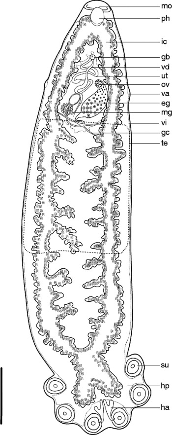
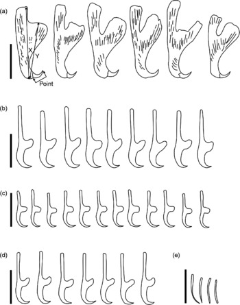
Morphological characteristics. General characteristics of mature, egg-producing parasite (fig. 1) typical of Polystoma. Body elongate; total body length 7244 (5067–9628); greatest width 1816 (956–2578); haptor length 1407 (1008–2291); haptor width 1796 (1119–2404); haptor length to body length ratio 0.19; six haptoral suckers, mean diameter 356 (255–444); hamulus length to tip of handle 395 (332–459); hamulus length to tip of guard 338 (291–381), handle longer than guard and mean X/Y ratio 1.16 (fig. 2); hamulus hook length 73 (70–77). Mouth subterminal, ventral. False oral sucker 422 (303–531) wide; pharynx length 279 (207–312); pharynx width 223 (182–249). Intestine bifurcate, caeca confluent posteriorly extending into haptor; caeca with 16 (13–23) medial diverticula, 42 (30–54) small lateral diverticula; no pre-haptoral anastomoses.
Polystoma vernoni n. sp. Ventral view of holotype; the dotted line indicates the outline of the testis and the dashed line indicates the outline of the vitelline system. Abbreviations: eg, egg; gc, genito-intestinal canal; gb, genital bulb; ha, hamulus; hp, haptor; ic, intestinal caecum; mg, Mehlis gland; mo, mouth; ov, ovary; ph, pharynx; su, sucker; te, testis distribution; ut, uterus; va, vagina; vd, vas deferens; vi, vitelline distribution. Scale bar: 1 mm.

Polystoma vernoni n. sp. (a) Hamuli from holotype and paratypes; (b) marginal hooklets 1 from oncomiracidia hatched from eggs laid by holotype and paratypes; (c) marginal hooklet 2–7; (d) marginal hooklet 8; (e) genital spines. Abbreviations: X, distance from hook to tip of handle; Y, distance from hook to tip of guard. Scale bars: 200 μm (a), 20 μm (b)–(e).

Testis single and follicular, well developed and extensive, situated post-ovarian, ventral, extending full width of body and halfway towards the haptor (fig. 1). Seminal vesicle prominent. Genital atrium median, ventral; genital bulb 84 (71–90) posterior to intestinal bifurcation, 8–9 genital spines 29 (28–29) long. Ovary sinistral, submedian; anterior in body, ovary length 719 (487–959); ovary width 254 (146–360). Ootype well developed. Genito-intestinal canal present on same side as ovary, joining intestinal caecum posterior to ovary. While observing a live parasite under coverslip pressure it was observed how waste products were rhythmically pumped via the genito-intestinal canal to the intestine. Uterus tubiform and, judging from the length, it may hold about ten eggs; specimens studied had a single egg in utero and one specimen had two. Eggs operculate. Egg length 201 (180–219), egg width 127 (116–140). No intrauterine development of eggs observed. Vitellarium distributed throughout the body excluding oral region, region around ovary and uterus, and haptoral suckers (fig. 1).
Oncomiracidium. Ciliated oncomiracidium has narrow cylindrical body with circular cup-shaped opisthaptor and resembles a typical polystomatid oncomiracidium. Opisthaptor bears 16 marginal hooklets, which are retained in adult parasites and do not increase in size. Marginal hooklet 1 (postero-medial), 36 (34–38) in length; hooklets 2–7, 21 (20–23) in length and hooklet 8, 31 (30–31) in length (fig. 2). Hamulus primordia 13 (13–14) in length.
Diagnosis
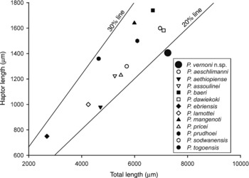
Polystoma vernoni n. sp. differs from all 11 other members of the genus Polystoma that infect Ptychadena by a combination of characteristics. Whereas most other polystomes have a spindle-shaped body with a haptor length–body length ratio of between 0.2 and 0.3, P. vernoni n. sp. has a long cylindrical shape with a mean haptor length–body length ratio of 0.19 (fig. 3). The only other African polystome for which this ratio is below 0.2 is P. chiromantis Dupouy & Knoepffler, 1978 from the tree frog Chiromantis rufescens (Günther, 1868) with a ratio of 0.18. Although the body measurements for P. chiromantis do overlap with that of P. vernoni n. sp., their hosts belong to two totally different anuran families, namely Rhacophoridae and Pyxicephalidae and, based on the high degree of host specificity that has been documented for African polystomes (Combes, Reference Combes1966, Reference Combes1968; Tinsley, Reference Tinsley1973, Reference Tinsley1974a; Euzet et al., Reference Euzet, Bourgat and Salami-Cadoux1974a, Reference Euzet, Combes and Knoepfflerb; Bourgat & Salami-Cadoux, Reference Bourgat and Salami-Cadoux1976; Combes & Channing, Reference Combes and Channing1979; Murith, Reference Murith1981a, Reference Murithb, Reference Murith1982; Kok & Van Wyk, Reference Kok and Van Wyk1986; Kok & Du Preez, Reference Kok and Du Preez1987; Du Preez & Kok, Reference Du Preez and Kok1992b, Reference Du Preez and Kok1993, Reference Du Preez and Kok1997), it is highly unlikely that the two would be related.
Scatter diagram of haptor length relative to the total length for Polystoma vernoni n. sp. and other Polystoma species known from Ptychadena. The 20% and 30% lines are indicated to show the relative position for the various species.

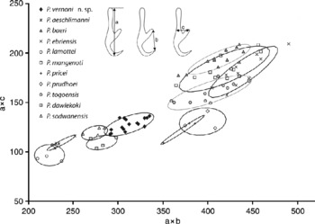
A plot of the products of the total length (a in fig. 4) and the width at the level if the guard (c in fig. 4) versus the product of the total length and the length of a tangent between the tip of the blade to the guard (b in fig. 4) of marginal hooklet 1 (Du Preez & Maritz, Reference Du Preez and Maritz2006) reveals that P. vernoni n. sp. does not overlap with any other Polystoma species. It borders the plots for two other polystomes known from South Africa, namely P. sodwanensis and P. dawiekoki. Polystoma vernoni n. sp. differs from both of these regarding the shape and number of intestinal diverticulae as well as the length of marginal hooklet 1.
Scatter diagram of the product of the total length (a) and the width at the level if the guard (c) versus the product of the total length and the length of a tangent between the tip of the blade to the guard (b) of marginal hooklet 1 for Polystoma vernoni n. sp. and other Polystoma species known from Ptychadena.

Discussion
Ptychadena is one of the most successful anuran genera in Africa and, according to Perret (1979), it is currently undergoing an explosive radiation. Du Preez & Kok (Reference Du Preez and Kok1992a) referred to Ptychadena as an extremely suitable host for polystomes as it fulfils ecological and physiological requirements for the completion of the polystome life cycle. With the high degree of host specificity within the genus, Polystoma host–parasite co-evolution is a reality, implying that diversification of hosts might be mirrored by diversification of polystome diversity. A study of the origin and evolution of African Polystoma conducted by Bentz et al. (Reference Bentz, Leroy, Du Preez, Mariaux, Vaucher and Verneau2001) included five specimens retrieved from Ptychadena. Within the robust African clade, two subgroups were identified, among which polystomes of Ptychadena fall in a tight group supported with high bootstrap values in both neighbour-joining and maximum-parsimony analyses. Based on experimental infection and cross-infection studies Du Preez & Kok (Reference Du Preez and Kok1997) suggested that not all polystomes exhibit the same degree of host specificity. Bentz et al. (Reference Bentz, Leroy, Du Preez, Mariaux, Vaucher and Verneau2001) proposed an alternative hypothesis, namely that some polystomes may be generalists rather than specialists and that differences in host ecology and physiology might result in either temporal or behavioural isolation precluding polystome speciation, and that the isolation within non-closely related hosts would generate speciation.
An in-depth study of the polystomes infecting Pt. mascareniensis might shed further light on host specificity and speciation among polystomes infecting Ptychadena. This frog is known to be widespread in Africa and also occurs on Madagascar, in the Seychelles and the Mascarene Islands, including the type locality Reunion. Metapolystoma brygoonis has originally been described as Polystoma brygoonis parasitizing Pt. mascareniensis from Madagascar and was later placed in a separate genus based on internal arrangement of the reproductive organs and the extent of the uterus. Ptychadena mascareniensis furthermore has been reported as host for Polystoma africanum from Zaire (Vercammen-Grandjean, Reference Vercammen-Grandjean1960), Uganda and Kenya (Tinsley, Reference Tinsley1974a) and Polystoma aethiopiense from Ethiopia (Meskal, Reference Meskal1970). On the basis of this, Bentz et al. (Reference Bentz, Leroy, Du Preez, Mariaux, Vaucher and Verneau2001) postulated that specificity in this case was operating below the species level of the host, which might reveal that the host itself was undergoing speciation. Aisien & Du Preez (Reference Aisien and Du Preez2009) redescribed P. africanum and concluded that the Ugandan material from Pt. mascareniensis was not P. africanum but most likely P. pricei. After re-examining the types from P. pricei Vercammen-Grandjean and P. aethiopiense Meskal, Tinsley (Reference Tinsley1974b) concluded that these two species were conspecific but that the name P. pricei takes precedence. This implies that polystomes known from Pt. mascareniensis from Africa and the Mascarene Islands include M. brygoonis and P. pricei. Genetic studies, however, revealed that mitochondrial haplotypes from Madagascar are similar to those from the Seychelles and Mascarene Islands but very different to those in Africa. In addition, those from Africa were very divergent from each other, indicating that up to six different species might be involved (Vences et al., Reference Vences, Kossuch, Rödel, Lötters, Channing, Glaw and Böhme2004). This could imply that the high degree of host specificity that has been reported for Polystoma is still being upheld. However, only an in-depth study of Pt. mascareniensis and its polystomes conducted throughout its known range of extension will shed light on this question.
Acknowledgements
Thanks to Ché Weldon, Leon Meyer and Donnavan Kruger from the North-West University, Potchefstroom for collecting the infected specimens in 2007; and Mrs Cynthia Kemp for proofreading the manuscript.