Ventilator-associated pneumonia (VAP) is the second most common hospital-acquired infection in pediatric and neonatal intensive care units (ICUs).Reference Foglia, Meier and Edward1 However, the diagnosis of neonatal pneumonia remains clinically problematic because there is no diagnostic test for pneumonia. In neonates, it can be difficult to discern pneumonia from exacerbations of primary lung disease. In cases of suspected pneumonia, providers often rely upon a combination of clinical symptoms and laboratory and radiographic studies that have been associated with neonatal infection.Reference Apisarnthanarak, Holzmann-Pazgal, Hamvas, Olsen and Fraser2–Reference Garland4 Practitioners variably employ noninvasive endotracheal aspirates to aid in the diagnosis of suspected pneumonia. Tracheal aspirates are obtained by collecting sputum through suctioning of the distal endotracheal tube and are typically sent for Gram stain and culture. A positive tracheal aspirate culture may prompt clinicians to administer an extended course of antibiotics or tailor antibiotic choice for the organism isolated.Reference Classen and Keenan5 However, bacterial growth from a tracheal aspirate may reflect a pathogenic organism causing true infection or may represent microbial contamination or colonization. This is especially problematic because tracheal aspirate cultures often grow antibiotic-resistant organisms, necessitating the use of broad-spectrum antibiotics to “treat” the isolate.
A number of studies have tried to establish an appropriate set of diagnostic criteria for pediatric ventilator-associated pneumonia (VAP), and many algorithms have been extrapolated from adults.Reference Cocoros, Priebe and Logan6 In adults, microbiologic criteria for VAP assume that pulmonary specimens are obtained through invasive testing such as bronchoalveolar lavage. Studies have demonstrated that tracheal aspirates have no correlation to samples obtained through mini-bronchoalveolar lavage,Reference Artuk, Gul, Mert, Karakas, Bedir and Eyigun7 and they pose a high contamination risk. In neonates, obtaining a noncontaminated sample requires the use of a bronchoscope, which is invasive and can present risk in this population. As a proxy, clinicians obtain endotracheal aspirates, despite existing data suggesting the variable diagnostic reliability of cultures obtained using this method.
To our knowledge, no studies in neonates have defined the utility of tracheal aspirates in pneumonia. In our study, we measured the association of positive tracheal aspirate cultures with clinical and laboratory and radiographic findings associated with infection that are commonly used in clinical practice. Given that antimicrobial stewardship is necessary to reduce antimicrobial resistance,Reference Schulman, Dimand, Lee, Duenas, Bennett and Gould8 we also sought to determine whether positive tracheal aspirate cultures were associated with increased antibiotic use.
Methods
This study was a retrospective chart review of patients hospitalized between January 2016 and December 2017 at a single level IV neonatal intensive care unit in Los Angeles, California, at the University of California–Los Angeles.
Participants
All infants hospitalized in the neonatal intensive care unit (NICU) who required intubation and had a tracheal aspirate sent for culture for suspected infection were eligible. Exclusion criteria included infants with ureaplasma and mycoplasma infections (because these infants receive treatment with a macrolide) and infants with a tracheostomy. The institutional review board at UCLA approved the research protocol.
Study protocol
Using electronic medical record review, the UCLA Microbiology Lab identified infants who had a tracheal aspirate culture. At the time the culture was sent and within 24 hours, clinical findings, laboratory and radiographic study results had been recorded. Patient demographic data, including gestational age, sex, birth weight, route of delivery, Apgar scores at 1 and 5 minutes, diagnosis of chronic lung disease, and death before hospital discharge were collected. Chronic lung disease was defined as the need for supplemental oxygen at 36 weeks corrected gestational age.
Outcome measures
The primary outcome variable was the presence or absence of a positive tracheal aspirate culture. If a repeat positive tracheal aspirate culture was found in a single patient, only the first culture event was considered. A positive tracheal aspirate was defined as any culture result with a potential pathogenic species. Cultures were grown on a blood agar plate composed of tryptic soy agar with 5% sheep's blood at 37°C at the UCLA Clinical Laboratory. If an organism grew, it was identified by matrix-assisted laser desorption ionization time-of-flight mass spectrometry (MALDI-TOF) on the Vitek mass spectrometer (bioMérieux, Marcy-l’Étoile, France).
We gathered data for several measures in our retrospective chart review. Demographic data included gestational age at birth, birth weight, corrected gestational age at the time of the “event,” weight at the time of the “event,” intubation >7 days or ≤7 days. Comorbidities included the presence of a patent ductus arteriosus as visualized on echocardiography, diagnosis of chronic lung disease, and death. Clinical events included an increase in the fraction of inspired oxygen (FiO2) by >0.05 and increased ventilator settings (ie, an increase in the mean airway pressure >5% or 10%). Laboratory testing included Gram stain quantitation of white blood cells (WBCs) from tracheal aspirates, serum WBC count and immature to total neutrophil ratio (I/T ratio), C-reactive protein (CRP), blood, urine, and cerebrospinal fluid cultures. Radiographic findings included chest radiograph notable for change from baseline, as determined by a radiologist. Finally, the antibiotic course was evaluated as ≤72 hours or >72 hours. Expanded broad-spectrum antibiotic administration was also determined. Broad-spectrum antibiotics were defined as third- or fourth-generation cephalosporins (cefotaxime or cefepime), carbapenems (meropenem), extended penicillins (piperacillin-tazobactam), oxazolidinones (linezolid), and aminoglycosides (tobramycin).
Analyses
Continuous variables were compared using the 2-sample t test. Association and trend were analyzed for categorical variables using the χ2 test and the Cochran Armitage trend test, respectively. For continuous variables, the normality of distribution was checked using the D’Agostino test. Multivariable logistic regression model was used to identify factors associated with culture positivity. Variables that showed some differences (P < .20) between positive and negative tracheal aspirate cultures in the univariable analyses were entered into the initial multivariable model, and the backward elimination method was used to construct a final multivariable model. A separate logistic regression model was constructed following forward selection method to confirm the model selected by backward elimination. P < .05 was considered statistically significant. All analyses were performed using SAS version 9.4 software (SAS Institute, Cary, NC).
Results
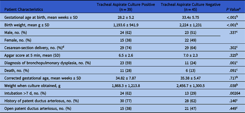
In total, 86 infants were identified as having at least 1 tracheal aspirate culture, but 1 infant was excluded for a positive ureaplasma culture and 1 was excluded for having a tracheostomy. Overall, 84 infants were included in our analyses. Only the first event concerning sepsis was included in the final analysis (ie, subsequent events were not included), yielding a final analysis of 84 tracheal aspirate cultures. We chose to conservatively include only the first event to avoid skewing our results toward falsely positive associations: 10 infants had multiple tracheal aspirates sent and 92% of repeat tracheal aspirate cultures were positive, which could have led to a biologic replicate–like phenomenon. Among 84 infants, 39 (46%) had a positive tracheal aspirate culture. In those infants with a positive tracheal aspirate culture, certain findings were apparent. They had lower birth weights (P < .0001) and lower gestational ages at birth (P < .0001) than their negative-culture counterparts. Additionally, they were more likely to require mechanical ventilation for >7 days (P = .002) and to carry a diagnosis of chronic lung disease by time of discharge from the NICU. Infants with positive tracheal aspirate cultures were also smaller than their negative counterparts at the time of tracheal aspirate collection (P = .038) (Table 1).
Comparison of Patient Characteristics Between Positive Tracheal Aspirate Cultures and Negative Respiratory Cultures

Note. SD, standard deviation.
a Data per univariable analysis.
b Student t test.
c χ2 test.
d Delivery details for 2 patients not recorded.
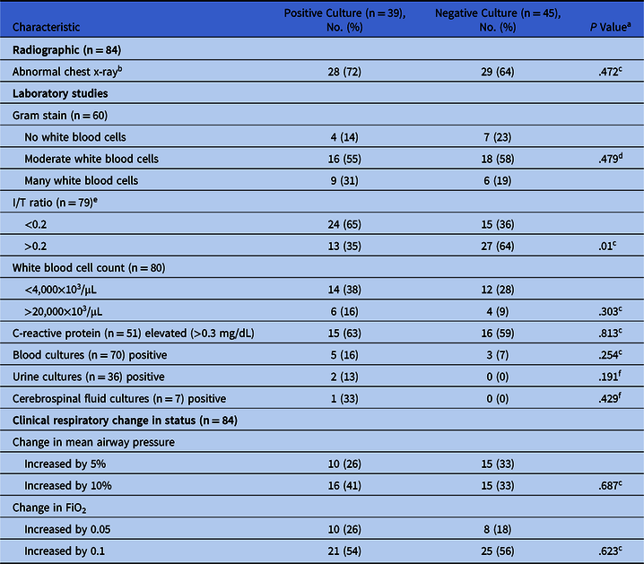
When evaluating the diagnostic evaluation of positive versus negative tracheal aspirate cultures, none of the considered factors (presence of WBCs on Gram stain, findings on chest radiograph, serum WBC, or CRP) were associated with having a positive tracheal aspirate culture. Interestingly, having an I/T ratio <0.2 was more likely to be associated with a positive tracheal aspirate culture (P = .005). Increased ventilator requirements (increased mean airway pressure and increased FiO2) in the setting of tracheal aspirate acquisition were nonsignificant (P = .687; P = .623) (Table 2). Also, we detected no statistical significance between blood cultures, urine cultures, or cerebrospinal fluid cultures compared with positive tracheal aspirate cultures.
Comparison of Diagnostic Evaluation Between Positive and Negative Tracheal Aspirate Cultures

Note: I/T, immature to total neutrophil ratio. Percentages may not add up to 100% due to rounding.
a Data per univariable analysis.
b Abnormal chest radiograph defined as any increase in infiltrate or new opacities from baseline film.
c χ2 test.
d Cochran Armitage trend test.
e The I/T ratio was calculated by dividing immature neutrophils by sum of immature and mature neutrophils.
f Fisher exact test.
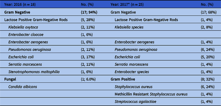
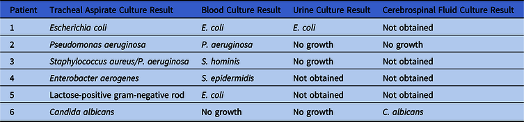
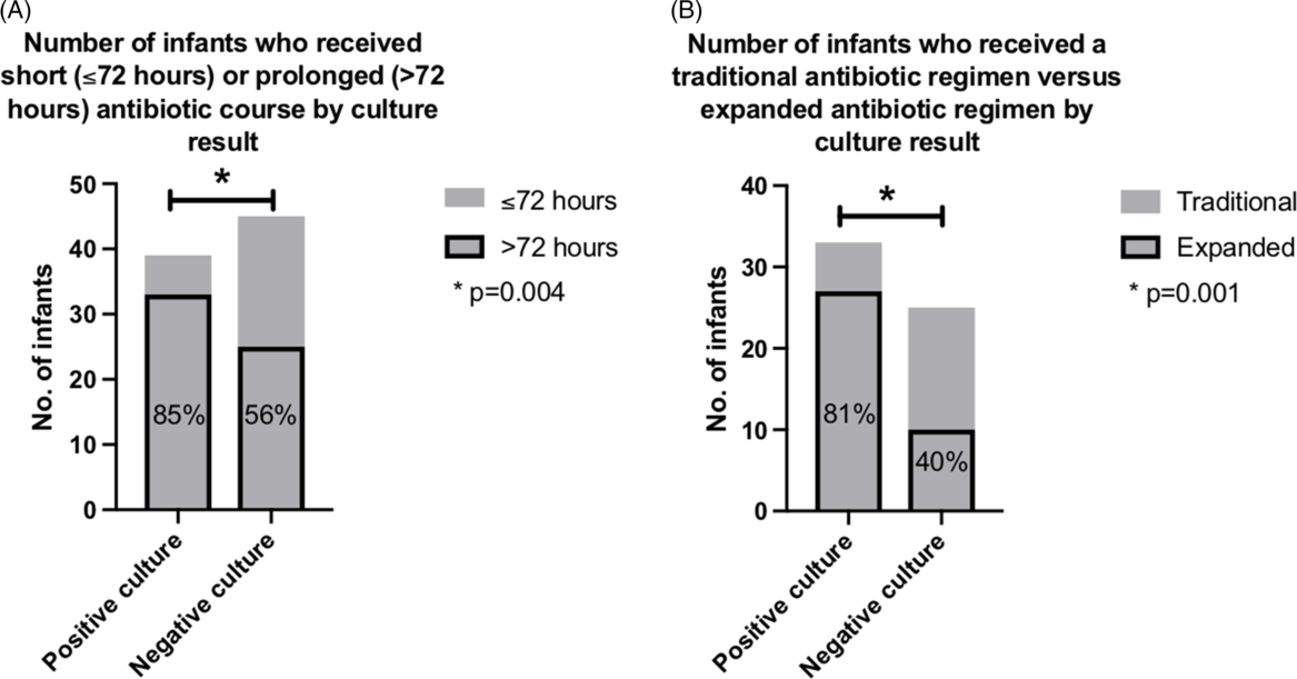
Most speciated pathogens were gram-negative organisms (Table 3). Also, 6 patients had concurrent infections with a positive tracheal aspirate culture (Table 4). Among patients with positive tracheal aspirate cultures, 85% received prolonged antibiotic courses compared to 56% of patients with negative tracheal aspirate cultures (P = .004). Among patients with prolonged antibiotic courses, 81% of patients with a positive tracheal aspirate culture received broad-spectrum antibiotics versus 40% who had a negative tracheal aspirate culture (Fig. 1). When those subjects with positive tracheal aspirate cultures and a concurrent systemic infection (n = 6) were excluded from the data analysis, there continued to be a statistically significant association of positive tracheal aspirate cultures with increased antibiotic days (P = .015) and exposure to broad-spectrum antibiotics (P = .024).
Identity and Distribution of Organisms Identified on Positive Respiratory Cultures

Note: Percentages may not add up to 100% due to rounding.
* 5 cultures with 2 organisms.
Concurrent Infections with Positive Tracheal Aspirate Cultures

Antibiotic duration and type by positive or negative tracheal aspirate culture. (A) Infants with a positive tracheal aspirate culture (n = 39; left column) were more likely than infants with a negative culture (n = 45; right column) to receive a prolonged course (>72 hours) of antibiotics (black outlined portion of columns) (P = .004 by univariate analysis, χ2 testing). (B) Among the infants who received a prolonged course of antibiotics, infants with a positive culture (n = 33, left column) were more likely than infants with a negative culture (n = 25, right column) to receive an expanded antibiotic regimens: broad-spectrum antibiotics including third- or fourth-generation cephalosporins (cefotaxime or cefepime), carbapenems (meropenem), extended penicillins (piperacillin-tazobactam), oxazolidinones (linezolid), and aminoglycosides (tobramycin) versus traditional empiric antibiotics defined as ampicillin or vancomycin and gentamicin (black outlined portion of columns) (P = .001 by univariate analysis, χ2 testing).

The final model based on multivariable analysis revealed statistically significant associations between positive tracheal aspirate cultures and low birth weight (P = .001), normal I/T ratio (P = .027), and increased number of antibiotic days (P < .001).
Discussion
Diagnosing neonatal pneumonia in intubated infants remains a challenge for many reasons, including the lack of a feasible gold standard for diagnosis and a population with comorbidities that make it uniquely difficult to distinguish primary respiratory etiologies from true respiratory infection. We sought to determine whether a positive tracheal aspirate culture correlates with routinely used clinical, laboratory, and radiographic tests. We identified factors that correlate with a positive tracheal aspirate culture as birth weight, a normal I/T ratio, and increased number of antibiotic days per multivariable analyses. Significant findings of our univariable analysis included gestational age of the neonate, the weight at time of tracheal aspirate collection, whether the infant had been intubated for >7 days, a diagnosis of chronic lung disease, and broad-spectrum antibiotic exposure. Low birth weight and earlier gestational age are already known to be associated with increased risk of infection.Reference Shane, Sánchez and Stoll9 Whether the positive tracheal aspirate culture group received longer courses of antibiotics due to the positive culture or due to the surrounding events at time of concern for sepsis remains unclear. However, the lack of association of positive tracheal aspirate cultures with clinically significant changes in respiratory status or infection-associated markers compared to negative cultures suggests that the positive tracheal aspirate cultures may represent non–clinically significant bacterial growth such as biofilm colonizing the endotracheal tube.Reference Leroue, Harris and Burgess10 However, in this study, we did not distinguish whether this means that those clinical or laboratory biomarker measures have poor diagnostic accuracy or whether tracheal aspirate cultures are diagnostic.
The literature continues to be conflicted regarding the utility of endotracheal aspirates. In a small study looking at the pediatric ICU population, tracheal aspirates were sensitive (90%) but only had a specificity of 40% compared to a reference standard of ventilator-associated pneumonia based on clinical, microbiological, and radiologic data.Reference Gauvin, Dassa, Chaibou, Proulx, Farrell and Lacroix11 In neonates, the use of TAs to diagnose pneumonia may be useful when aspirates are collected in the immediate neonatal period, with an overall positive culture incidence of 6.5% in tracheal aspirate cultures obtained 12 hours after birth.Reference Booth, Al-Hosni, Ali and Keenan12 Interestingly, the Gram stain results in this study demonstrated low specificity and sensitivity for pathogen growth, consistent with our results, in which we found no statistical association with the number of WBCs on the Gram stain.Reference Booth, Al-Hosni, Ali and Keenan12 In contrast, in neonates who have been chronically intubated, the risk of colonization increases, therefore increasing the chances of obtaining a positive tracheal aspirate culture.Reference Harris, Wirtschafter and Cassady13,Reference Gibbs and Holzman14 One study in ventilated preterm infants using molecular identification of bacteria in tracheal aspirates found that in the first 72 hours after birth, most tracheal aspirate cultures had undetectable bacterial loads. However, within 7 days, there is sustained diverse bacterial colonization.Reference Mourani, Harris, Sontag, Robertson and Abman15 A more recent study using 16S ribosomal RNA sequencing on tracheal aspirates found that a microbial community is present in the airway even at birth and that this community changes over the postnatal period.Reference Lal, Travers and Aghai16 These studies collectively suggest that bacterial growth in tracheal aspirate cultures in chronically intubated neonates may be misleading in the diagnosis of pneumonia.
Murila et alReference Murila, Francis, Bland, Kumbla, Doherty and Sehgal17 demonstrated that very-low-birth-weight neonates with positive tracheal aspirate cultures do indeed have increased ventilator requirements 3 days prior to sepsis event compared to settings during and at conclusion of treatment and abnormal inflammatory markers (absolute neutrophil count <1,100/mm3 or >6,600/mm3; I/T ratio >0.2; elevated CRP >0.5 mg/dL). However, Murila et al did not have a control group of negative respiratory cultures; they evaluated very-low-birth-weight infants (n = 38) exclusively, and they did not factor in duration of mechanical ventilation. In contrast, within the pediatric population (n = 61), Wilson et alReference Wilson, Conaway, Kelly and Hendley18 showed that tracheal aspirates can have pathogenic bacteria and inflammatory cells in patients that are not clinically symptomatic.
Our study yielded a concerning finding. Although positive tracheal aspirate cultures had no association with any of the traditional “markers” of infection, the presence of a positive tracheal aspirate was associated with prolonged antibiotic use. Prolonged antibiotic use has been associated with the development of multidrug-resistant organisms,Reference Tsai, Chu and Hsu19 increased risk of late-onset sepsis, necrotizing enterocolitis, increased number of hospital days, and increased ventilator days.Reference Tripathi, Cotton and Smith20 Although our retrospective study could not definitely prove that a positive tracheal aspirate alone caused prolongation of antibiotic courses, we did demonstrate that many of the other clinical features that are typically considered concerning as part of a pneumonia work-up (eg, increased respiratory support requirement, complete blood count [CBC], CRP abnormalities, chest radiograph abnormalities, etc) were not different between the positive and negative tracheal-aspirate groups. Thus, the only differentiating factor identified in these cohorts was a positive tracheal aspirate.
We also observed marked differences among providers pertaining to which laboratory analysis was performed at the time of concern for infection. A screening CBC was the most frequently ordered laboratory test when sepsis was a concern (n = 80), but indices on the CBC (eg, WBC and I/T ratio) do not have good sensitivity, and only extremely low or high WBCs exhibit good specificity for identifying sepsis.Reference Hornik, Benjamin and Becker21 Meanwhile, neonatologists continue to debate the validity of CRP values in neonatal sepsis because it has variable sensitivity and specificity and should be combined with other indicators of sepsis to increase its validity.Reference Xu, Li, Mo and You22 Despite the lack of evidence to suggest that these laboratory values are sensitive or specific for neonatal infection, or for pneumonia specifically, they continue to be used routinely. Procalcitonin has demonstrated promise as a marker for the diagnosis and management of suspected pneumoniaReference Christ-Crain, Jaccard-Stolz and Blingisser23; however, its utility in NICU patients with suspected pneumonia is unclear. Strategies to improve antibiotic use in the NICU should include diagnostic stewardship, in other words, appropriate ordering and interpreting of diagnostic tests.Reference Willis and de St Maurice24 On a population level, we found no significant worsening of respiratory status in our infants with positive tracheal aspirates, but infants with an isolated positive tracheal-aspirate culture were more likely to receive an extended course of antibiotics.
In conclusion, pneumonia in the intubated neonate remains difficult to diagnose, given the lack of sensitive or specific clinical criteria or laboratory testing. Especially in the vulnerable population of premature infants with primary lung disease, the fear of missing ventilator-associated pneumonia and the inability to distinguish infection from other causes of respiratory distress often prompts the liberal administration of antibiotics. Our study provides important information for the clinician; we have clearly shown that a positive tracheal aspirate culture is not associated with commonly used biomarkers associated with infection or with an increased need for respiratory support. These findings suggest that routine use of an endotracheal aspirate culture in the diagnosis of sepsis, especially in infants without clinical deterioration, should be questioned. Future studies should aim (1) to address the development of guidelines to standardize the evaluation of a patient with sepsis to reduce inappropriate antibiotic exposure, (2) to determine whether timing of tracheal aspirate acquisition affects utility of the tracheal aspirate in pneumonia evaluation, and (3) to evaluate whether our findings can be validated in a larger multicenter cohort using databases such as the California Perinatal Quality Care Collaborative, where our ventilator-associated pneumonia or ventilator-associated events are reported annually.
Acknowledgments
None.
Financial support
No financial support was provided relevant to this article. A.C. is supported by National Institutes of Health (NIH grant no. K08-HD093874).
Conflicts of interest
All authors report no conflicts of interest relevant to this article.