INTRODUCTION
Trypanosoma brucei lives in the blood and tissue fluids of mammalian hosts and an infection can last several years. This lifestyle requires invariant cell surface proteins for nutrient uptake, signaling and structural integrity. However, the invariant surface proteins need to be shielded from the immune system to establish a long-lasting infection.
African trypanosomes avoid clearance by the immune system in several ways. First, they modulate the innate and adaptive immune response by an incompletely understood mechanism dependent on trypanosome-derived factors (Mansfield and Paulnock, Reference Mansfield and Paulnock2005; Leppert et al. Reference Leppert, Mansfield and Paulnock2007). Second, trypanosomes have a dense surface coat mainly composed of a single polypeptide, the variant surface glycoprotein (VSG), that shields the invariant surface proteins from effectors of the immune system and prevents complement activation (Ferrante and Allison, Reference Ferrante and Allison1983). Third, antibody binding to VSG leads to rapid internalization of the antibody-VSG complex, subsequent proteolysis of the antibody and recycling of the VSG back to the surface (Engstler et al. Reference Engstler, Pfohl, Herminghaus, Boshart, Wiegertjes, Heddergott and Overath2007). This process prolongs the survival of a trypanosome at low antibody titres. Finally, the survival of the population is dependent on antigenic variation that allows the infection to stay ahead of the host immune system.
As an infection progresses, a high antibody titre against a particular VSG leads to the clearance of trypanosomes expressing that VSG, trypanosomes that have switched VSG expand as a new population until they are recognized in turn – these cycles can go on for years. The rate of VSG switching varies between isolates but models of long-term infections are consistent with estimated rates and temporal expression of VSG genes is dependent only on parasite-intrinsic factors and a density-dependent differentiation to the non-dividing trypanosomes (Lythgoe et al. Reference Lythgoe, Morrison, Read and Barry2007).
This review describes some components of the trypanosome plasma membrane, the endocytosis and recycling of VSGs back to the surface and finally summarizes what is known about antigenic variation.
THE TRYPANOSOME CELL SURFACE
An unusual feature of the external face of the plasma membrane of trypanosomes is the predominance of a glycosylphosphatidylinositol (GPI)-anchored protein, the VSG (Ferguson et al. Reference Ferguson, Homans, Dwek and Rademacher1988; Melhert et al. Reference Mehlert, Bond and Ferguson2002). The numbers are staggering; the VSG is over 95% of externally disposed cell surface proteins and ∼15% of the total cell protein of a trypanosome cell with ∼5×10⁶ VSG dimers per cell (Jackson et al. Reference Jackson, Owen and Voorheis1985).
Why has there been selection for GPI-anchoring of the superabundant VSG? There are several possible advantages of a GPI-anchor over the single transmembrane helix present in type 1 membrane proteins that are probably only significant at high packing densities. First, a higher packing density on the cell surface could be achieved using GPI-anchors as an equivalent packing density of type 1 membrane proteins might congest the cytoplasmic face of the plasma membrane. This is important as it is probable that the VSG density on the surface approaches the maximum possible. Secondly, GPI-anchoring may play a role in trafficking and recycling of the VSG. In the absence of a GPI-anchor, VSG is mislocalised to the lysosome (Triggs and Bangs, Reference Triggs and Bangs2003) or to non-lysosomal compartments close to the flagellar pocket (Bohme and Cross, Reference Bohme and Cross2002). In trypanosomes GPI-valence seem to play a role in the trafficking of GPI-anchored proteins. The dimeric VSG, with two GPI anchors, mainly localises to the cell surface whereas the transferrin receptor, with a single GPI-anchor, and reporter proteins, with a single GPI anchor, cycle through the flagellar pocket and endosome, and are delivered to the lysosome (Schwartz et al. Reference Schwartz, Peck, Tazeh and Bangs2005; Schwartz and Bangs, Reference Schwartz and Bangs2007). Thirdly, during differentiation from the bloodstream to the procyclic form the VSG coat is rapidly shed from the surface by the activities of the GPI-specific phospholipase C and the zinc metalloprotease TbMSP-B (Grandgenett et al. Reference Grandgenett, Otsu, Wilson, Wilson and Donelson2007).
In addition to a GPI-anchor, VSGs are modified with N-linked oligosaccharides that are either high-mannose oligosaccharides or complex oligosaccharides (Bangs et al. Reference Bangs, Doering, Englund and Hart1988; Zamze et al. Reference Zamze, Wooten, Ashford, Ferguson, Dwek and Rademacher1990; Zitzmann et al. Reference Zitzmann, Mehlert, Carrouee, Rudd and Ferguson2000). It has been suggested that VSG glycosylation contributes to the volume of VSGs and thus its ability to form a protective surface barrier (Blum et al. Reference Blum, Down, Gurnett, Carrington, Turner and Wiley1993). The model of the N-terminal domain of the MITat1.5 glycoprotein shows that the N-linked oligosaccharides occupy a significant amount of the inter-VSG space (Mehlert et al. Reference Mehlert, Bond and Ferguson2002).
There are three types of N-terminal VSG domains (A, B and C) and six types of C-terminal domains (type1–6) (Carrington et al. Reference Carrington, Miller, Blum, Roditi, Wiley and Turner1991; Berriman et al. Reference Berriman, Ghedin, Hertz-Fowler, Blandin, Renauld, Bartholomeu, Lennard, Caler, Hamlin, Haas, Bohme, Hannick, Aslett, Shallom, Marcello, Hou, Wickstead, Alsmark, Arrowsmith, Atkin, Barron, Bringaud, Brooks, Carrington, Cherevach, Chillingworth, Churcher, Clark, Corton, Cronin, Davies, Doggett, Djikeng, Feldblyum, Field, Fraser, Goodhead, Hance, Harper, Harris, Hauser, Hostetler, Ivens, Jagels, Johnson, Johnson, Jones, Kerhornou, Koo, Larke, Landfear, Larkin, Leech, Line, Lord, Macleod, Mooney, Moule, Martin, Morgan, Mungall, Norbertczak, Ormond, Pai, Peacock, Peterson, Quail, Rabbinowitsch, Rajandream, Reitter, Salzberg, Sanders, Schobel, Sharp, Simmonds, Simpson, Tallon, Turner, Tait, Tivey, Van Aken, Walker, Wanless, Wang, White, White, Whitehead, Woodward, Wortman, Adams, Embley, Gull, Ullu, Barry, Fairlamb, Opperdoes, Barrell, Donelson, Hall, Fraser, Melville and El-Sayed2005). The different VSG types are classified according to their pattern of conserved cysteine residues. All combinations of the N-terminal domains with the C-terminal domains appear possible (Hutchinson et al. Reference Hutchinson, Smith, Jones, Chattopadhyay, Welburn and Carrington2003). The structures of the N-terminal domain of MITat1.2 and ILTat1.24, both type A, have been determined by X-ray crystallography (Freymann et al. Reference Freymann, Down, Carrington, Roditi, Turner and Wiley1990; Blum et al. Reference Blum, Down, Gurnett, Carrington, Turner and Wiley1993). Despite having only 16% sequence identity the proteins were folded in the same way (see Fig. 1). Disulphide mapping experiments showed that type B domains have a similar tertiary structure (Bussler et al. Reference Bussler, Linder, Linder and Reinwald1998). Type 2, 4 and 5 C-terminal domains are single domains, type 1, 3 and 6 di-domains. The structure of the type 2 domain of MITat1.2 (Chattopadhyay et al. Reference Chattopadhyay, Jones, Nietlispach, Nielsen, Voorheis, Mott and Carrington2005) and the type 1 di-domain of ILTat1.24 (Jones et al. Reference Jones, Nietlispach, Sharma, Burke, Eyres, Mues, Mott and Carrington2008) has been solved. The C-terminal domain of MITat1.2 and the two parts of the type1 domain of ILTat1.24 adopt similar tertiary structures. The structural conservation of the N- and C-terminal VSG domains is probably due to a selection pressure to ensure a protective cell surface barrier, rapid VSG internalization and recycling despite the need for antigenic variation. The proper VSG fold seems to be required for high protein expression as mutating several structural features of VSGs resulted in reduced protein expression compared to wildtype VSGs (Wang et al. Reference Wang, Bohme and Cross2003). Several other cell surface proteins have a similar structure to the N-terminal domain of VSGs; amongst these are the invariant surface glycoproteins ISG65 and ISG75 (Carrington and Boothroyd, Reference Carrington and Boothroyd1996), the transferrin receptor (Salmon et al. Reference Salmon, Hanocq-Quertier, Paturiaux-Hanocq, Pays, Tebabi, Nolan, Michel and Pays1997) and the SRA protein which renders T. brucei rhodesiense resistant to lysis by human serum (Campillo and Carrington, Reference Campillo and Carrington2003). The evolutionary relationship between VSGs and VSG-like proteins is unclear. They could have evolved in parallel from a common ancestor or the VSGs evolved from a VSG-like protein or the other way round. The VSG MITat1.5 of T. brucei shares extensive homology with the transferrin subunits ESAG6 and ESAG7. Interestingly, it was possible to make a chimeric transferrin receptor of VSG/ESAG6 and VSG/ESAG7 that was still able to heterodimerise and bind transferrin (Salmon et al. Reference Salmon, Hanocq-Quertier, Paturiaux-Hanocq, Pays, Tebabi, Nolan, Michel and Pays1997). SRA looks like a truncated VSG which lacks the surface loops and again a VSG exists, WATat1.2, which has extensive homology to SRA (Campillo and Carrington, Reference Campillo and Carrington2003). Why is the VSG fold conserved in non-VSG proteins? In the case of the transferrin receptor it is possible that the elongated VSG-like shape is needed to allow access to free transferrin in the flagellar pocket, but it is less clear why the ISGs have the VSG-like fold.
VSG structure: A. The superimposed structures of the N-terminal domains of MITat1.2 and ILTat1.24 demonstrate the conservation of the tertiary structure of VSGs. The disulphide bridges are marked in yellow. B. Representation of a VSG dimer and linear representation of a VSG MITat1.2 monomer. In the structural cartoon, one monomer is shown in grey, the other is shown in different colours that correspond to regions indicated in the primary structure. The white boxes are regions for which the structure has not been solved. The vertical lines represent cysteines, the horizontal lines disulphide bonds.

How does the VSG shield other cell surface proteins? The simplest explanation is that the VSG physically prevents immunoglobulin binding to other cell surface proteins. The VSG layer has a thickness of 12 to 15 nm (Vickerman, Reference Vickerman1969; Blum et al. Reference Blum, Down, Gurnett, Carrington, Turner and Wiley1993) and there is evidence that antibodies are able to recognize VSG epitopes up to 6 to 8 nm into this layer (Hsia et al. Reference Hsia, Beals and Boothroyd1996). Many proteins, such as the hexose transporters, have only small extracellular domains (Borst and Fairlamb, Reference Borst and Fairlamb1998) that could be physically protected from host antibodies by the larger VSG. However, ISG65 and 75 are type 1 transmembrane proteins and have an extracellular domain similar in the size to the VSGs and are distributed over the entire surface of the trypanosome (Ziegelbauer and Overath, Reference Ziegelbauer and Overath1993). The low abundance of ISG65 and ISG75 is only relative to the VSG, there are 7×10⁴ copies of ISG65 and 5×10⁴ copies of ISG75 per cell (Ziegelbauer et al. Reference Ziegelbauer, Multhaup and Overath1992), but in any other cell they would be moderately abundant cell surface proteins. Infection serum both from humans and mice contains antibodies that recognize invariant cell surface proteins but the trypanosome is not susceptible to a killing response mediated by these immunoglobulins (Ziegelbauer and Overath, Reference Ziegelbauer and Overath1993). The extracellular domains ISG65 and 75 are similar in size to VSGs, but it remains unclear if and how the VSG coat prevents or negates the effect of antibody binding. The problem is perhaps exemplified by comparison with the transferrin receptor which is a similar size to the ISGs but is localised to the flagellar pocket, an invagination of the plasma membrane where the flagellum exits the cell, which renders them inaccessible to effectors of the immune system. Whether the transferrin receptor is restricted to the flagellar pocket or not is unknown. Overexpression of the transferrin receptor by serum starvation or by introduction of extra copies of the transferrin receptor subunit genes resulted in localisation over the entire cell surface (Mussmann et al. Reference Mussmann, Janssen, Calafat, Engstler, Ansorge, Clayton and Borst2003; Schwartz et al. Reference Schwartz, Peck, Tazeh and Bangs2005). Several explanations are possible: (1) The transferrin receptor is localised to the entire cell surface but due to the limit of detection it is only detected in the flagellar pocket. (2) There is an easily saturable transferrin receptor retention mechanism in the flagellar pocket (Mussmann et al. Reference Mussmann, Janssen, Calafat, Engstler, Ansorge, Clayton and Borst2003). (3) As overexpressed transferrin receptor on the surface was not able to bind transferrin and was not shed into the medium at a higher rate, the single GPI-anchor on the transferrin receptor does not seem sufficient for passive plasma membrane retention, and the detected cell surface transferrin receptor could be in reality a homodimer of the GPI-anchored ESAG6 (Schwartz et al. Reference Schwartz, Peck, Tazeh and Bangs2005). The biological significance of the finding that serum starvation leads to overexpression of the transferrin receptor also remains questionable. Trypanosomes encode in their bloodstream form expression sites different transferrin receptors that have different affinities for the diverse mammalian transferrins. It was suggested that overexpression of the transferrin receptor takes place at the beginning of an infection when a low-affinity receptor is expressed until the trypanosomes have switched to a bloodstream form expression site encoding for a high-affinity receptor. Calculations based on the Kd values show that trypanosomes expressing a low-affinity receptor should nevertheless be able to grow in any mammalian host without the need to switch to another bloodstream form expression site (Steverding, Reference Steverding2003).
THE ENDOCYTOSIS OF SURFACE PROTEINS AND REMOVAL OF BOUND ANTIBODIES TO VSG
The endocytic system in bloodstream form trypanosomes is exceptionally active with the equivalent of the entire cell surface being recycled every 12 minutes (Engstler et al. Reference Engstler, Thilo, Weise, Grunfelder, Schwarz, Boshart and Overath2004). For comparison, cell surface turnover in macrophages is 2.6 times slower and in fibroblasts 5–9 times slower than in Trypanosoma brucei (Thilo, Reference Thilo1985; Engstler et al. Reference Engstler, Thilo, Weise, Grunfelder, Schwarz, Boshart and Overath2004; Overath and Engstler, Reference Overath and Engstler2004). There is evidence that this high rate of endocytosis is restricted to mammal-infective forms with much lower rates in insect forms (Morgan et al. Reference Morgan, Allen, Jeffries, Hollinshead and Field2001; Natesan et al. Reference Natesan, Peacock, Matthews, Gibson and Field2007). It has been suggested that the up-regulation in bloodstream form cells contributes to the evasion of the immune system (Morgan et al. Reference Morgan, Hall, Denny, Carrington and Field2002). The VSG half-life is 32 hours, representing ∼5 cell doublings, with VSG being shed to the medium rather than degraded by internal turnover (Bulow et al. Reference Bulow, Nonnengasser and Overath1989; Seyfang et al. Reference Seyfang, Mecke and Duszenko1990). Hence, VSGs are able to remain functional through more than 100 cycles of traversing acidic endosomal compartments.
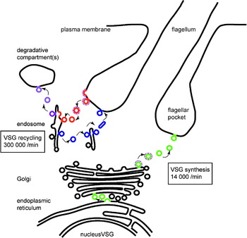
All endocytosis and exocytosis occurs via the flagellar pocket and VSG is taken up from the plasma membrane by large clathrin-coated vesicles and transported to the early endosome, which is marked by TbRAB5 (Fig. 2). Small clathrin-coated vesicles not containing VSG bud from the early endosomes resulting in an indirect concentration of the VSG. These vesicles presumably contain cargo for late endosomes and lysosomes. The mechanism of the VSG exclusion from these clathrin-coated vesicles is not known. From the early endosomes the VSGs move to recycling endosomes which are TbRAB11-positive structures and, from there, to the flagellar pocket via TbRAB11-positive exocytic carriers (Jeffries et al. Reference Jeffries, Morgan and Field2001; Grunfelder et al. Reference Grunfelder, Engstler, Weise, Schwarz, Stierhof, Morgan, Field and Overath2003). The volume of vesicular transport involved in VSG recycling far exceeds the volume of vesicular transport from the Golgi apparatus delivering newly synthesized VSG to the flagellar pocket (Fig. 2).
Diagram of a simplified model of VSG transport in the trypanosome. VSG is synthesised on the rough endoplasmatic reticulum and is exported to the flagellar pocket plasma membrane in vesicles (green) via the Golgi apparatus. VSG is internalised from the plasma membrane by the removal of vesicles (red) from the flagellar pocket. The VSG and receptors are separated from bound immunoglobulin and cargo in the endosome and the VSG is returned to the cell surface in elongated vesicles (blue). Immunoglobulin and possibly ligands are transported to a degradative compartment(s) via vesicles (purple). The rate of VSG recycling far exceeds that of new synthesis. Clathrin-mediated steps are indicated by the purple shell around some vesicles, although a complete description of clathrin-dependent transport remains an aspiration. The values for rates of VSG movement are estimates based on published measurements (Grunfelder et al. Reference Grunfelder, Engstler, Weise, Schwarz, Stierhof, Morgan, Field and Overath2003) and a cell doubling time of 6 hours.

VSGs diffuse freely in the surface coat but movement is under some restriction due to the dense VSG packing in the plasma membrane. The diffusion coefficient for a VSG in the plane of a membrane is 1×10⁻¹⁰ cm2/s, similar to other plasma membrane glycoproteins (Bulow et al. Reference Bulow, Overath and Davoust1988). VSG molecules form a complex with anti-VSG immunoglobulins and these complexes move more rapidly in the plane of the membrane than free VSG. The movement of the complexes is directional, towards the posterior pole, the location of the flagellar pocket. This directional movement is dependent on trypanosome cell motility and is explained by hydrodynamic flow forces dragging the antibody VSG complex as the cell swims forward (Engstler et al. Reference Engstler, Pfohl, Herminghaus, Boshart, Wiegertjes, Heddergott and Overath2007). Endocytosed immunoglobulins are present in TbRAB5A-positive sorting endosomes (early endosomes) and in the recycling endosome. The antibodies are degraded to short peptides and then secreted showing that a proteoloytic system is operating within the endosome (Pal et al. Reference Pal, Hall, Jeffries and Field2003).
ANTIGENIC VARIATION
African trypanosomes express a surface coat composed of a single VSG. There are two interconnected processes important for antigenic variation; first, the genetic or epigenetic changes that lead to a change in the identity of the expressed VSG, and second the mechanisms that ensure monoallelic expression of the active VSG gene so that any one cell presents only a single VSG.
A change in VSG being expressed occurs stochastically and independently of any host factors, the trypanosome population generates cells expressing different VSGs and each variant will expand unless or until killed by the host immune system. The rate of switching varies from once per 1×10² to 1×10⁶ cell doublings depending on the origin and history of the isolate investigated (Turner and Barry, Reference Turner and Barry1989; Turner, Reference Turner1997). Up to 20% of the protein coding capacity of the genome is dedicated to VSGs, a huge repertoire of at least 1700 potential VSGs per diploid genome (Horn and Barry, Reference Horn and Barry2005). Nearly all VSG sequences are located in tandem arrays in subtelomeric regions, a location that favours mitotic recombination (Linardopoulou et al. Reference Linardopoulou, Williams, Fan, Friedman, Young and Trask2005). The central ‘housekeeping’ parts of homologous chromosomes have synteny but the VSG arrays do not (Melville et al. Reference Melville, Leech, Gerrard, Tait and Blackwell1998; Berriman et al. Reference Berriman, Ghedin, Hertz-Fowler, Blandin, Renauld, Bartholomeu, Lennard, Caler, Hamlin, Haas, Bohme, Hannick, Aslett, Shallom, Marcello, Hou, Wickstead, Alsmark, Arrowsmith, Atkin, Barron, Bringaud, Brooks, Carrington, Cherevach, Chillingworth, Churcher, Clark, Corton, Cronin, Davies, Doggett, Djikeng, Feldblyum, Field, Fraser, Goodhead, Hance, Harper, Harris, Hauser, Hostetler, Ivens, Jagels, Johnson, Johnson, Jones, Kerhornou, Koo, Larke, Landfear, Larkin, Leech, Line, Lord, Macleod, Mooney, Moule, Martin, Morgan, Mungall, Norbertczak, Ormond, Pai, Peacock, Peterson, Quail, Rabbinowitsch, Rajandream, Reitter, Salzberg, Sanders, Schobel, Sharp, Simmonds, Simpson, Tallon, Turner, Tait, Tivey, Van Aken, Walker, Wanless, Wang, White, White, Whitehead, Woodward, Wortman, Adams, Embley, Gull, Ullu, Barry, Fairlamb, Opperdoes, Barrell, Donelson, Hall, Fraser, Melville and El-Sayed2005), the effect is to increase the VSG repertoire but it also implies that the arrays are excluded from meiotic recombination (Hutchinson et al. Reference Hutchinson, Picozzi, Jones, Mott, Sharma, Welburn and Carrington2007).
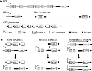
The VSG genes are located in four different types of loci (see Fig. 3A). The first is the bloodstream expression site (BES). There are at least 10 BESs in the diploid genome and only one is active per time (Becker et al. Reference Becker, Aitcheson, Byles, Wickstead, Louis and Rudenko2004; Hertz-Fowler et al. Reference Hertz-Fowler, Figueiredo, Quail, Becker, Jackson, Bason, Brooks, Churcher, Fahkro, Goodhead, Heath, Kartvelishvili, Mungall, Harris, Hauser, Sanders, Saunders, Seeger, Sharp, Taylor, Walker, White, Young, Cross, Rudenko, Barry, Louis and Berriman2008; Young et al. Reference Young, Taylor, Kurioka, Becker, Louis and Rudenko2008). Each BES has a VSG promoter and contains several expression site-associated genes (ESAGs). The gene content is variable but, in addition to the VSG, most contain ESAG6 and 7 which encode the two subunits of the transferrin receptor (Hertz-Fowler et al. Reference Hertz-Fowler, Figueiredo, Quail, Becker, Jackson, Bason, Brooks, Churcher, Fahkro, Goodhead, Heath, Kartvelishvili, Mungall, Harris, Hauser, Sanders, Saunders, Seeger, Sharp, Taylor, Walker, White, Young, Cross, Rudenko, Barry, Louis and Berriman2008). The VSG gene is always located at the very end of each BES that is closest to the telomere. In T. brucei rhodesiense one or two BESs contain the SRA gene. When T. brucei rhodesiense infects a human, this specific BES needs to be active to prevent lysis by human serum (Xong et al. Reference Xong, Vanhamme, Chamekh, Chimfwembe, Van Den Abbeele, Pays, Van Meirvenne, Hamers, De Baetselier and Pays1998). Later in the infection, switching is only possible by recombination of the VSG to a BES containing a SRA gene. Transcriptional switching, activation of another BES not containing SRA, presumably leads to cell death. The BESs are polycistronically transcribed (Johnson et al. Reference Johnson, Kooter and Borst1987; Kooter et al. Reference Kooter, van der Spek, Wagter, d'Oliveira, van der Hoeven, Johnson and Borst1987) by RNA polymerase I (Gunzl et al. Reference Gunzl, Bruderer, Laufer, Schimanski, Tu, Chung, Lee and Lee2003). Single mRNAs are produced by trans-splicing and simultaneous polyadenylation. In the ‘inactive’ BESs transcription is initiated but transcriptional elongation and processing is inhibited so that no VSG mRNA is made from these sites (Vanhamme et al. Reference Vanhamme, Poelvoorde, Pays, Tebabi, Van Xong and Pays2000). Only the active BES is located in an expression site body (ESB) that is an extranucleolar structure in the nucleus where transcription of the BES takes place (Navarro and Gull, Reference Navarro and Gull2001). VSG double expressors utilising two BES are unstable, thus it seems that the ESB favours the expression of a single BES (Chaves et al. Reference Chaves, Rudenko, Dirks-Mulder, Cross and Borst1999). A BES can be driven by an rDNA promoter (Rudenko et al. Reference Rudenko, Blundell, Dirks-Mulder, Kieft and Borst1995) and such a BES is also located in an extranucleolar structure, presumably in an ESB. This suggests that sequences other than the promoter sequence are responsible for ESB recruitment of a BES (Chaves et al. Reference Chaves, Zomerdijk, Dirks-Mulder, Dirks, Raap and Borst1998). Early in the differentiation to procyclic cells the active BES is repositioned from the ESB to the nuclear envelope. In this location, the VSG promoter is inactivated and undergoes chromatin condensation (Landeira and Navarro, Reference Landeira and Navarro2007).
A. VSG loci: There are around 10 or more VSG genes located in bloodstream form expression sites, 25 in metacyclic expression sites, around 1500 in subtelomeric arrays and around 200 on minichromosomes in the diploid genome. B. VSG switching mechanisms: When gene conversion occurs, the VSG gene in the bloodstream form expression site is replaced by a copy of another VSG gene, often located in subtelomeric arrays. Telomere exchange involves the crossover of the active bloodstream form expression site telomere with another telomere from minichromosomes. The active VSG gene is transferred to a ‘silent’ location on the other telomere and the formerly silent VSG gene is transferred into the active bloodstream form expression site. Transcriptional switching involves no recombination event unlike gene conversion and telomere exchange. Here the active bloodstream form expression site is down-regulated by an epigenetic mechanism and another bloodstream form expression site is activated.

VSG genes are also located in less than 25 metacyclic expression sites (MES). MESs are only active in metacyclic trypanosomes, the life stage that differentiates in the tsetse fly salivary gland and infects a mammalian host when the fly feeds. These expression sites have also a promoter but are monocistronic and are not expressed once differentiation to bloodstream forms has occurred (Graham et al. Reference Graham, Terry and Barry1999).
The silent repertoire of VSG sequences is present in subtelomeric arrays and minichromosomes. At subtelomeres there are around 1500 silent VSG genes located in arrays ranging from 5 to more than 150 genes. From these genes, only around 4% are intact genes, 65% are pseudogenes, 21% gene fragments and 9% encode for not-functional whole-length VSGs which lack the required modification signals. In addition, there are around 200 VSG genes located on minichromosomes within 5 kbp of the telomere tract. These are less well characterized but it is believed that there is only one and sometimes no VSG gene per subtelomere (Weiden et al. Reference Weiden, Osheim, Beyer and Van der Ploeg1991; Wickstead et al. Reference Wickstead, Ersfeld and Gull2004).
Antigenic variation occurs when the identity of the active VSG gene switches. There are three different switching mechanisms called duplicative gene conversion, telomere exchange and transcriptional switching (see Fig. 3B). Gene conversion is the predominant switching mechanism in pleomorphic bloodstream form cells (Robinson et al. Reference Robinson, Burman, Melville and Barry1999). Monomorphic bloodstream form cells that have been cultivated for a long time in the laboratory without selective pressure to undergo antigenic variation have a 10,000 fold reduced switching rate compared to pleomorphic bloodstream form cells (Lamont et al. Reference Lamont, Tucker and Cross1986) and more often use transcriptional switching rather than gene conversion (Liu et al. Reference Liu, Michels, Bernards and Borst1985).
VSG switching is significantly more frequent than the rates of background implying that it is initiated in a locus-specific manner, possibly by an endonuclease. The rate of switching can be increased by more than two orders of magnitude by engineering an endonucleolytic cleavage (Boothroyd et al. Reference Boothroyd, Dreesen, Leonova, Ly, Figueiredo, Cross and Papavasiliou2009). The 5′ endpoint of gene conversion lies in the tandem array 70 bp repeats upstream of the VSG (Boothroyd et al. Reference Boothroyd, Dreesen, Leonova, Ly, Figueiredo, Cross and Papavasiliou2009), and the 3′ endpoint occurs within a region spanning the 3′ end of the VSG coding sequence to the subtelomeric repeats (Bernards et al. Reference Bernards, Van der Ploeg, Frasch, Borst, Boothroyd, Coleman and Cross1981; Campbell et al. Reference Campbell, van Bree and Boothroyd1984; Matthews et al. Reference Matthews, Shiels, Graham, Cowan and Barry1990). There are some unusual features to the recombination mechanisms involved in antigenic variation. RAD51 is one of the key proteins for homologous recombination and enables a DNA single strand to pair with a homologous region in a DNA double strand. The switching rate was reduced in RAD51 knock-out cells but nevertheless the cells were able to use recombination for switching to some degree (McCulloch and Barry, Reference McCulloch and Barry1999). Mutating RAD51-3, a RAD51-related protein, (Proudfoot and McCulloch, Reference Proudfoot and McCulloch2005) and BRCA2, one of the regulators of RAD51 function, (Hartley and McCulloch, Reference Hartley and McCulloch2008) also impaired antigenic variation. There are two homologous recombination pathways in trypanosomes (Conway et al. Reference Conway, Proudfoot, Burton, Barry and McCulloch2002), one is RAD51-dependent pathway and the other is RAD51-independent that uses DNA substrates with very short lengths (minimal substrate length 24 bp). It has been proposed that the latter is perhaps responsible for the microhomology-based recombination that is observed in segmental gene conversion events. In addition to gene conversion, VSG switching can result from telomere exchange, the crossover of the chromosome end carrying the active BES with the chromosome end of another chromosome. This leads to the location of the former inactive VSG gene into the active BES and the formerly expressed gene is inactivated by recombination into the silent chromosome end (Pays et al. Reference Pays, Guyaux, Aerts, Van Meirvenne and Steinert1985).
When the epigenetic change that leads to VSG switching occurs, the active BES is inactivated and a different BES is activated. The process must involve two linked events: (1) the loss of the ESB from the silenced BES and the appearance of a new ESB on the activated BES, and (2) the appearance of silencing modifications on the chromatin of the old BES and the removal of such modification on the newly activated BES. The difficulty is to separate cause and effect. Transcriptional switching is slowed in cells deleted of the histone methyltransferase DOT1B so that switching intermediates, cells expressing two VSGs, are detectable for weeks and silent BESs become partially derepressed in DOT1B deleted cells (Figueiredo et al. Reference Figueiredo, Janzen and Cross2008). TbISWI, a SWI2/SNF2-related chromatin-remodeling protein, is also required for the maintenance of BES down-regulation (Hughes et al. Reference Hughes, Wand, Foulston, Young, Harley, Terry, Ersfeld and Rudenko2007). The DNA of silent BESs and telomeres contains base J (β-glucosylhydroxymethyluracil) but it is not known if this base has an active role in BES silencing or if base J modification is just a consequence of BES silencing (van Leeuwen et al. Reference van Leeuwen, Wijsman, Kieft, van der Marel, van Boom and Borst1997). Recently, the accuracy of the inheritance of the ESB during the cell cycle has been investigated. An experimentally induced decrease in the amount of cohesin was used to cause a degree of premature separation of sister chromatids. The effect was to disrupt the inheritance of the ESB, probably due to one daughter cell inheriting the ESB and thus maintaining expression of the same VSG, the other daughter cell activated a BES de novo. The implications from these results are that the ESB replicates late in mitosis and that a new ESB can form spontaneously (Landeira et al. Reference Landeira, Bart, Van Tyne and Navarro2009).
The genomic structure of the VSG repertoire provides an explanation for the stochastic nature of antigenic variation. Intact VSG genes, which tend to be telomeric and have greater numbers of 5′ repeats, are activated early during infection. Later in infection, mosaic VSG genes assembled from pseudogenes from subtelomeric arrays are expressed (Morrison et al. Reference Morrison, Majiwa, Read and Barry2005; Marcello and Barry, Reference Marcello and Barry2007). Thus, expression of VSGs is constrained by the likelihood of recombination to an active BES.
FUTURE CHALLENGES
The study of the trypanosome surface and antigenic variation has led to the discoveries ranging from the first description of the GPI-anchor in eukaryotes, protein-sorting by hydrodynamic flow forces, polycistronic transcription of protein-coding genes by RNA polymerase I in an expression site body, the extreme efficient rate of VSG uptake and recycling outstanding in eukaryotes, accurate mathematical modeling of an infection and so on. Nevertheless, there is still much work to do. It is not understood how the VSG proteins are able to protect the invariant surface proteins from the adaptive immune system. Nothing is known about the surface coat of trypanosomes like Trypanosoma theileri which also live in the mammalian bloodstream but do not use classical antigenic variation for protection. Why is the VSG genetic repertoire used for antigenic variation so large? An infection with Anaplasma marginale, causative agent of bovine anaplasmosis, can persist for years although this parasite uses only 5–7 functional pseudogenes and a single expression site for antigenic variation (Brayton et al. Reference Brayton, Kappmeyer, Herndon, Dark, Tibbals, Palmer, McGuire and Knowles2005; Palmer et al. Reference Palmer, Futse, Knowles and Brayton2006). Is T. brucei really able to use the entire VSG repertoire? Is the enormous amount of VSG genes required to allow superinfection of an animal by a second strain (Hutchinson et al. Reference Hutchinson, Picozzi, Jones, Mott, Sharma, Welburn and Carrington2007) or is there another reason for the huge number of VSG genes?
ACKNOWLEDGEMENTS
Work in the authors' laboratory is funded by the Wellcome Trust.