Introduction
Metallothioneins (MT) are low-molecular-weight (2–7 kDa) and cysteine-rich (20 to 30%) metal-binding proteins (Hamer, Reference Hamer1986; Robinson et al., Reference Robinson, Tommey and Kuske1993; Kägi and Schaffer, Reference Kägi and Schaffer1988); they are widespread, found in microorganisms, plants and all invertebrate and vertebrate animals. MTs were first isolated as a cadmium-binding protein from horse kidneys (Margoshes and Vallee, Reference Margoshes and Vallee1957). Mammalian MTs are composed of four isoforms: MT-I, MT-II, MT-III and MT-IV. MT-I and MT-II exist in all tissues (Suzuki et al., Reference Suzuki, Kodama, Molotkov, Aoki and Tohyama1998); MT-III is found only in the brain (Uchida and Takio, Reference Uchida and Takio1991); MT-IV mainly exists in stratified squamous epithelia (Quaife et al., Reference Quaife, Findley and Erickson1994). Amino-acid sequences of mammalian MTs are all highly conserved, being composed of between 61 and 68 amino-acid residues, including 20 cysteines, which have the same basic location in the amino-acid sequence, and a characteristic sequence of Cys–X–Cys with the number of seven and the same degree of metal binding (Uchida et al., Reference Uchida, Gomi, Masumizu and Miura2002). There are no aromatic amino acids (Hamer, Reference Hamer1986; Kägi and Kojima, Reference Kägi and Kojima1991). Because it has a similar structure to MT-I and MT-II, MT-III was designated similarly, but is also named for its neuronal growth inhibitory factor (GIF) role. Human MT-III is composed of 68 amino acids, seven more than MT-I and MT-II (Uchida and Yasuo, Reference Uchida and Yasuo1995). MTs are involved in the storage, transport and metabolism of living organisms' microelements, detoxification of heavy metals, attenuation of ionizing radiation and elimination of free radicals, as well as growth, development, reproduction, aging, occurrence of tumours, immunization and emergency response, and so on (Breathnach and Chambon, Reference Breathnach and Chambon1981; Vallee, Reference Vallee1995). Research into the development and application of MTs is one of today's hot topics. In recent years, MTs have been extensively and deeply studied in the fields of agriculture, medicine, health, biotechnology, environmental protection and other fields (Vallee, Reference Vallee1991). There are two main facets to the research: the first is to make a model system to study genetic expression, regulation and engineering methods to explore the function of the MTs themselves as well as their physiology; the second is to explore clinical applications including heavy metal detoxification, detection of metal pollution in the environment, physiology research, functional foods and cosmetics, and so on (Vallee, Reference Vallee1991; An et al., Reference An, Wang and Han2005).
The yak is a unique bovine species living at altitudes of over 3000 m above sea level in the Qinghai-Tibet Plateau. They have a series of unique biological characteristics, including cold tolerance, hypoxia tolerance, anti-ultraviolet features and others suited to their particular ecological environment, resulting from long-term evolution (Zhang et al., Reference Zhang, Wu and Han1994). The objective of this research was to obtain the basic data for further study of the nature and function of MTs, and then to study the biological characteristics adapted to the plateau and the hypoxic environment there from the perspective of the molecular biology of the yak.
Materials and methods
Samples
Liver and brain tissue of the domestic yak (Bos grunniens) were obtained from Gannan, Gansu, China, for cloning MT genes. Samples were obtained from liver and brain tissue at 5 and 10 min intervals after slaughtering, respectively, and were gauzed and flash-frozen in liquid nitrogen and stored at −80°C until being thawed for RNA extraction.
Plasmids and strains
Plasmid carrier PZeroT-Vector for cloning the MT-I and MT-III gene was provided by the Gene Research Centre for Human Diseases of Peking University. Escherichia coli strains were cultured and prepared by the State Key Laboratory of Protein Engineering of Beijing University.
Reagents
The following materials were used: TRIzol® reagent (GIBCO, BRL, Gaithersburg, Maryland, USA), RNA PCR kit Version 3.0 (TaKaRa, Dalian, Japan), DNA Rapid-Purification/Recycling Kit (Dingguo, Beijing, China), TaqDNA Polymerase (Invitrogen, California, USA), T4 DNA Ligase, HindIII and XbaI (Promega, Madison, Wisconsin, USA), Lambda-pUC Mix Marker, 4 (MBI Fermentas), ribonuclease inhibitor (Human Placenta source; TaKaRa, Japan) and AMV Reverse Transcriptase (Promega, USA).
Primers for cloning MT-I and MT-III
Upstream primers:
YMT-ISP (17 bp): 5′-TGGACCCGAACTGCTCC-3′
YMT-IIISP (19 bp): 5′-ATGGACCCTGAGACCTGCC-3′
Downstream primers M13 Primers M4 (17 bp):
5′-GTTTTCCCAGTCA-CGAC-3′
Reverse transcription primers for the above (Oligo dT-Adaptor Primer) were provided with the Kit Ver. 3.0.
Equipment
The following equipment was used: Gene Amp PCR System 9700 (PE Biosystems, Foster City, California, USA), desktop high-speed centrifuges (Heraeus, Hanau, Germany), gel imaging system (SynGene, Cambridge, UK), Hettich high-speed refrigerated centrifuge (Heraeus) and DSHZ-300 heated water bath shaker (Botonyc, Shangai, China).
Methods
Total RNA extraction from liver and brain tissue of yak
Total RNA from the liver and brain tissue of the yak was extracted with TRIzol (GIBCO, BRL) as described in its protocol and stored at −70°C until being thawed for RNA extraction.
PCR primer design
Specific upstream primers YMT-ISP and YMT-IIISP were designed based on the cDNA coding region sequences (GenBank accession nos: MT-I, AY513744; MT-III, DQ323545) of MT-I and MT-III in the yak, respectively, and were synthesized by Osel, Inc., Beijing, China, and purified by polyacrylamide gel electrophoresis (PAGE). Downstream primers, M13 Primer M4, were provided using the RNA PCR Kit Ver. 3.0 (TaKaRa).
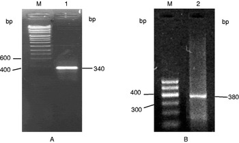
Electrophoresis of the amplified products of MT-I (A) and MT-III (B) genes by RT-PCR. Lanes: M, DNA marker; 1, MT-I cDNA fragment; 2, MT-III cDNA fragment.

3′-RACE
Reverse transcription polymerase chain reaction (RT-PCR) was carried out using RNA PCR kit Version 3.0 (TaKaRa). The total RNA of liver and brain tissue of the yak were used as a template, and Oligo dT-Adaptor Primer and avian myeloblastosis virus reverse transcriptase (AMV RT) was used for the first-strand DNA synthesis. YMT-ISP and M13 Primer M4 were then used for combination I, YMT-IIISP and M13 Primer M4 for combination II, and Taq DNA polymerase was used for PCR amplification. Response conditions and cycling parameters were as follows: denaturation at 94°C for 4 min; PCR amplification for 40 cycles of 94°C for 30 s, 42°C for 1 min (combination II: 46°C for 1 min) and 72°C for 2 min. The products (5–10 μl) were separated by electrophoresis on a 1.5% agarose gel. Finally, we identified and preserved the products, for further experimentation.
Cloning and sequencing of the objective gene
After the PCR products were recovered, the plasmid library was constructed with T4 DNA polymerase by ligating the DNA fragments into the cloning site of the plasmid pZeroT-Vector. The following steps were applied: isolation of library DNA, transfection of competent E. coli DH5α, extraction of DNA from transfected cells and so on, then electrophoresis, and identification by PCR in bacterium with enzyme digestion. Products were sequenced by Shanghai Sangon Biological Engineering Technology & Services Co., Ltd., China.
Sequence analyses
Sequence analyses of DNA were performed using BioEdit (http://www.mbio.ncsu.edu/) and the pDRAW32 program (http://www.bioon.com/soft/), and sequence analyses of the amino acids were performed using BioEdit and DNAMAN software (www.bioon.com). At the same time, the above sequences were subjected to BLASTN searching at the National Center for Biotechnology Information (NCBI).
Results and analyses
3′-RACE
Fragments (340 and 380 bp) (Fig. 2) with the expected size were amplified by 3′-RACE using the specific primers YMT-ISP (17 bp) and YMT-IIISP (19 bp), which were designed based on the cDNA coding region sequences of MT-I and MT-III in yak and M13 primers M4. A preliminary determination suggested that the two fragments might be Yak MT-I and MT-III cDNA 3′-end full-length sequences.
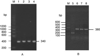
Electrophoresis of the amplified products of MT-I (A) and MT-III (B) genes by PCR in bacterium. Lanes: M, DNA marker; 1–4, MT-I cDNA fragments; 5–8, MT-III cDNA fragments.

Cloning and identification of PCR products
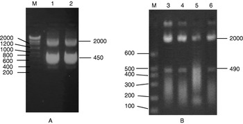
Following recovery of the fragments, they were ligated with pZeroT-Vector in vitro, transformed into efficient competent cells of E. coli DH5α. There was a sufficient quantity of white bacteria in the LB culture medium, and we selected a typical colony to carry out strain PCR (Fig. 2) and monoclonal strains to extract plasmids according to this. Electrophoresis analysis was then performed, with dual identification from enzyme digestion using HindIII and XbaI. The results showed that the size of the fragments inserted in the pZeroT-Vector carrier were similar to that of one of the objective DNAs (Fig. 3).
Electrophoresis of enzyme digestion of recombinant plasmids of MT-I (A) and MT-III (B) genes. Lanes: M, DNA marker; 1 and 2, MT-I cDNA fragments; 3–6, MT-III cDNA fragments.

Sequence analyses
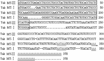
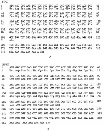
Sequence analysis was carried out using BioEdit software and pDRAW32, which showed that the 3′-ends of MT-I and MT-III were at 331 bp with a coding region sequence length of 183 bp, and at 378 bp with a length of 207 bp, respectively. There were tailing signals AATAAA and poly(A) at the 3′-ends of MT-I and MT-III (Fig. 4). The sequences were submitted to GenBank (GenBank accession nos: MT-I, AY758557; MT-III, DQ492300).
Analysis of MT-I and MT-III cDNA 3′-end full-length sequences. ATG…GCC/TGA, sequences of MT-I and MT-III gene cds; AATAAA, signal of Poly (A); AAAAAA, Poly (A).

Sequence analyses of amino acids
Sequence analyses of the yak MT-I and MT-III encoding amino acid
Analysis of the nucleotide sequences of MT-I and MT-III showed that they contained complete coding regions, and had specific start and termination codons. The lengths of MT-I and MT-III coding regions were 183 bp and 207 bp, respectively. MT-I encoded 61 amino acids, including 20 cysteines (32.79%) such as Phe (phenylalanine), Trp (tryptophan) and Tyr (Tyrosine); there were no aromatic amino acids. MT-III encoded 68 amino acids, including 19 cysteines (27.94%). These results showed that yak MT-I and MT-III were low-molecular-weight, cysteine-rich, non-aromatic amino-acid proteins (Fig. 5).
The amino-acid sequences encoded by MT-I (A) and MT-III (B) genes.

Homology and evolution analysis of amino-acid sequences based on mammalian MTs
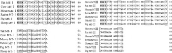
Comparisons of the amino-acid sequences of MT-I and MT-III in the yak with comparative sequences in cow, sheep, mouse, rabbit, pig and so on showed that they were all conserved tripeptides, such as Cys–X–Cys (Fig. 6) and the location of C–X–C is fixed. All amino acids except Cys had the following motif: MDPN/E, CXC——CXC—CXC–CXC–C—CCXC C—CXXC—CXC——CXCC,X, where X are aminoacids (including Ser, Thr, Ala, Pro, Val, and so on), excluding cysteines.
Conservation analysis of amino-acid sequences of MT-I and MT-III in the yak and other mammals. Shadows and boxes show specific conserved amino-acid sequences in MT proteins.

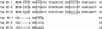
Analysis of the amino-acid sequences of MT-I, MT-II and MT-III in the yak (Fig. 7) indicated that sequences of MT-III had T, CPCP, GEGAEA different from MT-I and MT-II.
Analysis of amino-acid sequences of MT-I, MT-II and MT-III proteins. Boxes show special conserved amino-acid sequences.

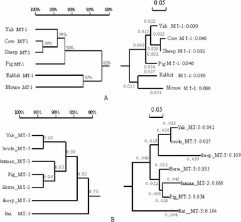
Molecular evolution analysis of MT-I and MT-III proteins in the yak, cow, sheep, mouse, rabbit and pig indicated that conservation of MT-I and MT-III proteins in yak, cow, sheep and pig were above 94% and 85%, respectively, and comparatively conservative in their evolution (Fig. 8).
Phylogenetic (right) and homology (left) trees of MT-I (A) and MT-III (B) protein sequences in the yak and other mammals.

Prediction of MT protein structure in the yak
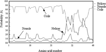
Software was used to predict the secondary structure of MT-III protein in the yak (Fig. 9). The results showed that MT-III has a similar secondary structure as MT-I and MT-II (Ma et al., Reference Ma, Ren and Wu2005), and amino acids 6–14 and 47–52 have irregular coils without B folds and have obvious strands and helices, respectively.
Secondary structure prediction of MT-III protein.

Discussion
In most cases, a new cDNA full-length sequence is selected by the cDNA library or RACE method. In this study, specific primers were designed using cDNA coding region sequences for MT-I and MT-III in the yak, and cDNA 3′-end sequences for MT-I and MT-III were cloned by RT-PCR and 3′-RACE. The cDNA coding sequences were exactly the same, and were obtained for both MT-I and MT-III in the yak. Analysis showed that the homology of the cDNA coding region sequences of yak MT-I and MT-III and protein sequences were consistent. Although the 3′-end non-translation regions have a short homology sequence, their non-encoding regions are lacking homology. The results confirmed that the conclusion that the cDNA homology of MT is mainly manifested in its coding region (Breathnach and Chambon, Reference Breathnach and Chambon1981) is correct. MTs are low-molecular-weight (~7 kDa) and cysteine-rich metal-binding proteins. MTs are believed to play a central role in the regulation of tissue concentrations of essential metals (such as zinc and copper) and to be involved in detoxification of non-essential highly toxic metals (such as cadmium and mercury) (Kägi and Kojima, Reference Kägi and Kojima1991; Roesijadi, Reference Roesijadi1992, Reference Roesijadi1996). The MT family in the liver is composed of two isoforms, MT-I and MT-II, which can be induced by metal ions, dexamethasone, chemical reagents and so on (Breathnach and Chambon, Reference Breathnach and Chambon1981; Robinson et al., Reference Robinson, Tommey and Kuske1993; Vallee, Reference Vallee1995). The mammalian brain has a particular kind of MT (MT-III), which can only be expressed in the brain. MT-III is a member of the MT family, and has a similar primary structure, more complex structure and metal-binding characteristics as other MTs of mammalian tissues, but induction of MT-I or MT-II does not affect MT-III. The normal brain is rich in MT-III, but in the brain of an Alzheimer's patient it is expressed at a reduced level. MT-III can inhibit the death of bud neurons, so has been termed a growth inhibitory factor (GIF) (Uchida and Takio, Reference Uchida and Takio1991). MT-I and MT-II do not have the same function, the functional difference probably arising from the unique structure of MT-III (Uchida et al., Reference Uchida, Gomi, Masumizu and Miura2002). Figure 7 shows that yak MT-III has Thr inserted at 5, CPCP at 6→9, and Glu–Ala–Ala–Glu–Ala–Glu at 54→59 (when compared with MT-I and MT-II). MT-III is a strong acid because its 8 Glu; the arrangement of 19 Cys according to the kind of mode: C–X–C, C–C–X–C–C or C–X–X–C, the location and quantity to which are comparatively conserved in MTs; these structures probably determine the conservative structure of the MT-III function. Thus MT-III not only has the biological functions of other MTs, but also one that is unique to it (Uchida and Takio, Reference Uchida and Takio1991; Vallee, Reference Vallee1995; Uchida et al., Reference Uchida, Gomi, Masumizu and Miura2002). It is therefore important to research the structure and function of MT-III, comparing it with MT-I and MT-II, and then clarify the space structure of the protein for further molecular biology research.
In general, MT amino-acid peptide chains contain 20 Cys in the human, pig and mouse (Hamer, Reference Hamer1986; Kägi and Kojima, Reference Kägi and Kojima1991). The results of this study have shown that yak MT-III contains 19 Cys, with a T mutation into A on the 88 bp of its base sequence, leading to a mutation of Cys into Ser on the 30 bp of the coding amino-acid sequences. Sheep also exhibit a missense mutation in this location that has an important effect on the structure and function of the MT-III protein. Following comprehensive analysis, sequences in yak and cattle have strong conservation, but have weak conservation when compared to sheep, which matches their evolutionary relationship. However, conservation between yak and sheep is higher than that between cattle and sheep. We see from the quantity and location of missense mutations that sheep and yak exhibit missense mutations leading to a change in a coding amino acid, but sheep have two mutations. Because the location is specific to metal binding and free radicals, which means less than one binding metal, this showed that the conservation region of MT-III for yak and sheep exhibits a mutation. So, further research is required regarding the cause of the mutation, and the way in which the mutations have an effect on the characteristics, structure and function of yak MT-III protein.
Acknowledgements
This work was supported by the University Doctors Subject Funds.