Introduction
Fine needle aspiration (FNA) and core biopsy are important diagnostic techniques routinely used alongside clinical examination and imaging in the investigation of neck lumps. Fine needle aspiration and core biopsy are widely accepted investigations, as they are a quick and relatively non-invasive way of acquiring pathology samples in the out-patient setting. In current literature, diagnostic accuracy rates are reported as 73–98 per cent for FNA and 83–100 per cent for core biopsy.Reference Tandon, Shahab, Benton, Ghosh, Sheard and Jones1,Reference Novoa, Gürtler, Arnoux and Kraft2
Despite few published reports of its incidence, the theoretical risk of tumour cell seeding, namely the spread of malignant cells along the needle tract, following core biopsy and FNA, remains a concern.Reference Yamaguchi, Strong, Shapshay and Soto3 Risk factors for seeding include technique, needle size, number of passes and biopsy site.Reference Robertson and Baxter5–Reference Imamura, Tateishi, Shiina, Goto, Sato and Ohki13 The risk of seeding following core biopsy is, understandably, higher than that for FNA.
Here we report a case of a 75-year-old patient where seeding to the skin occurred following first FNA, then core biopsy, of a metastatic lymph node secondary to a thyroid neoplasm.
Search strategy
A literature search was performed using PubMed, Cochrane Library and Ovid databases. The following search criteria were used: ‘neck lump’, ‘core biopsy’, ‘fine needle aspiration’ and ‘tumour/neoplasm seeding’. These searches were performed with the following limits applied: English language, involving humans and published within the last 50 years.
Case report
A 75-year-old woman was referred to the head and neck clinic with a 2-week history of a left-sided neck lump. She had no associated upper aerodigestive tract symptoms and was systemically well. Her only past medical history was of Ménière's disease.
On examination, there was a firm lesion in the left level IV region, adjacent to the left lobe of the thyroid extending into the root of the neck. The lesion was noted to be pulsatile. Initial ultrasound demonstrated a 5.3 cm soft tissue mass displacing the common carotid. Computed tomography (CT) of the neck suggested that the mass may have been a paraganglioma; however, appearances were atypical and lymph node metastases could not be excluded.
Cautious FNA of the left-sided level IV mass was carried out under ultrasound guidance. Histological analysis was suggestive of a thyroid neoplasm. There was a small number of follicular epithelial cell groups; however, moderate nuclear grooves and pseudo-inclusions could not completely exclude a diagnosis of papillary thyroid carcinoma. The sample, initially thought to be of a thyroid nodule, was therefore graded Thy3F (possible follicular neoplasm). The patient was subsequently referred to a consultant colleague with a special interest in thyroid pathology.
The decision was made to proceed with ultrasound-guided core biopsy, which was performed using an 18-gauge needle. This specimen demonstrated features of a poorly differentiated carcinoma; however, abundant necrosis and a limited number of lesional cells made the specimen, again, difficult to interpret. The immunoprofile of the sample was in keeping with a thyroid origin.
On clinical review, the patient complained of new-onset left arm pain. A repeat CT scan of the neck and thorax demonstrated enlargement of the mass with involvement and occlusion of the left internal jugular vein. There was new associated lymphadenopathy on the left at levels III and IV, and new pulmonary nodules.
The patient agreed to intervention for symptom palliation. After discussion with the patient and the multidisciplinary team, a total thyroidectomy and left-sided selective neck dissection was agreed upon, with the primary intention of relieving pressure on the affected nerve roots. In addition, excision of the specimen would confirm the histological diagnosis.
On the day of surgery, a superficial nodule was noted in the left supraclavicular region (Figure 1). This was separate to both the neck lump the patient initially presented with and the thyroid gland. This was the first occasion that the nodule had been noted. The patient confirmed that this had been close to the site where her core biopsy had been taken. This lesion was excised, along with a 1 cm margin of overlying skin, as part of the neck dissection specimen.
Cutaneous deposit noted at time of surgery.

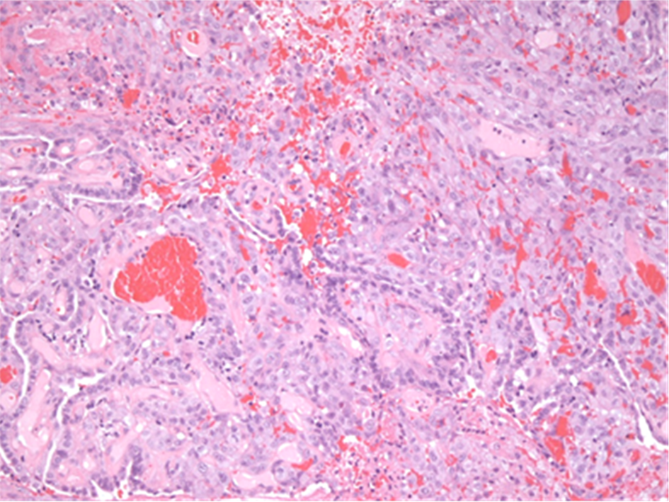
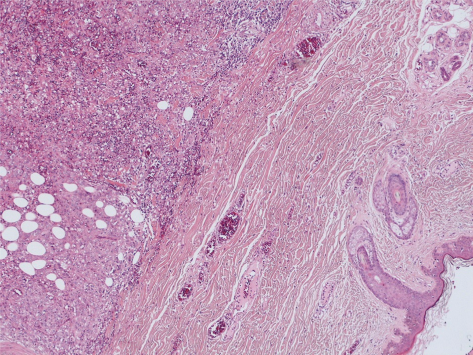
Histopathology confirmed metastatic, poorly differentiated papillary thyroid carcinoma in 8 of the 25 lymph nodes excised (Figures 2 and 3). This lymph node count included the initial presenting 5.3 cm mass in level IV of the neck. This was a separate entity from the thyroid, confirmed both radiologically and histologically.
Medium power view of the tumour. The tumour has a range of appearances, with conventional papillary thyroid carcinoma seen on the left of the image, and a more solid, poorly differentiated carcinoma seen on the right of the image. (H&E; ×??).

Low power view demonstrating the epidermis and dermis on the right of the image, with poorly differentiated carcinoma within the subcutaneous tissues on the left of the image. (H&E; ×??).

Examination of the superficial nodule demonstrated cutaneous deposits of poorly differentiated papillary carcinoma within the dermis and subcutis, but not extending to the epidermis. These deposits were identical to metastases present in the other lymph nodes. Within the thyroid, which was processed in its entirety, there were papillary microcarcinomas without evidence of poorly differentiated carcinoma. The patient went on to die at one month post-procedure, reflecting the aggressiveness of her disease.
Discussion
There is a theoretical risk of seeding with any form of biopsy. The estimated seeding risk is 0.003–0.009 per cent for FNA4 and 0–22 per cent for core biopsy.Reference Robertson and Baxter5 However, the dissemination of tumour cells following biopsy may be higher than indicated by the reported literature; animal studies have shown evidence of tumour seeding following FNA in 65–83 per cent of subjects implanted with solid tumours, with 1000 to 10 000 cells seeded along the needle tract.Reference Smith6,Reference Mighell and High7 In addition, in freshly excised human lung cancer specimens, 89 per cent of FNA needle tracts contained tumour cells.Reference Smith6,Reference Mighell and High7
However, these seeded cells rarely translate into clinically identifiable metastases. This may be the result of a combination of destruction by the host's immune system, physical damage sustained during the procedure and some cells being simply non-viable.Reference Mukunyadzi, Bardales, Palmer and Stanley8 The risk appears to vary widely depending on the site of the biopsy, as shown in Table 1. Other factors that appear to influence seeding rates include technique (aspiration vs capillary technique), needle gauge size and number of passes.Reference Robertson and Baxter5–Reference Imamura, Tateishi, Shiina, Goto, Sato and Ohki13 It is proposed that the increased risk of seeding with large gauge needles is secondary to the aspiration of sufficient stroma necessary for tumour survival.Reference Mukunyadzi, Bardales, Palmer and Stanley8,Reference Roussel and Dalion12 Evidence of vascular spread to distant sites from seeded cells has not been identified either clinically or experimentally; this is likely because of the absence of required essential molecular processes.Reference Yamaguchi, Strong, Shapshay and Soto3
Comparison of seeding rates according to siteReference Robertson and Baxter5–Reference Imamura, Tateishi, Shiina, Goto, Sato and Ohki13

FNAC = fine needle aspiration cytology
The relationship between seeding risk and tumour grade has also been studied, and poorly differentiated disease has been found to be an independent risk factor for seeding in hepatocellular carcinoma.Reference Imamura, Tateishi, Shiina, Goto, Sato and Ohki13 Comparatively limited information is available with regard to thyroid disease. However, a recent review of isolated case reports of cutaneous tumour seeding following FNA of follicular carcinoma demonstrated that those with earlier cutaneous presentation had biologically more aggressive tumours, as in this case.Reference Panunzi, Paliotta, Papini, Petrucci, Rinaldi and Nardi14,Reference Uchida, Suda, Inoue, Fujiwara and Ishiguro15
• Fine needle aspiration (FNA) and core biopsy are common percutaneous sampling methods with high diagnostic accuracy
• There are few reports on the incidence of tumour seeding following percutaneous sampling of malignancies
• Reports of skin seeding following sampling of malignancies of a thyroid origin are particularly scarce
• Skin seeding remains a rare but important risk following percutaneous sampling of neck lumps
• The decision regarding which sampling method to use should be based on the clinical situation rather than the tumour seeding risk
• If a patient progresses to surgery following FNA and core biopsy of an aggressive tumour, the biopsy site should be marked and the needle tract included in excision
To date, there are only isolated case reports of cutaneous seeding following the sampling of head and neck lumps (excluding thyroid nodule sampling). A recent systematic review focusing on the exact incidence of tumour seeding following FNA and core biopsy of head and neck lumps, produced crude estimated figures of incidence: 0.00012 per cent with FNA and 0.0011 per cent with core biopsy. These are the lowest reported rates of seeding associated with any form of biopsy from any biopsy site within the human body.Reference Shah and Ethunandan16 There is almost a 10-fold increased risk of seeding with core biopsy; therefore, some clinicians only resort to this sampling method following an inconclusive FNA, such as in the case discussed above. However, many clinicians maintain that core biopsy is their first choice of investigation in the examination of head and neck lumps because of the increased diagnostic accuracy.
Conclusion
This case highlights an extremely rare but important risk associated with the sampling of neck lumps, and radiologists should inform patients of this risk during the consent process. Nevertheless, we feel that ultrasound-guided sampling is still an essential part of investigation, as it provides a high degree of diagnostic accuracy and patient acceptability. Decisions regarding choice of sampling method should be made based on the individual clinical situation, rather than the risk of tumour seeding. Particular consideration should be given to the increased risk of seeding with core biopsy in suspected aggressive disease, as in this case. In those patients undergoing surgery for poorly differentiated carcinoma who have previously undergone diagnostic core biopsy, we recommend marking the biopsy site and including the sample tract with the excision specimen.
Acknowledgement
The authors would like to thank the subject's family for their permission to submit this case report.
Competing interests
None declared