




No CrossRef data available.
Article contents
Differences in olfactory and visual memory in patients with pathologically confirmed Alzheimer's disease and the Lewy body variant of Alzheimer's disease
Published online by Cambridge University Press: 01 October 2004
Abstract
Recognition and remote memory for odors, faces, and symbols were assessed in patients with pathologically confirmed Lewy body variant of Alzheimer's disease (LBV), patients with pathologically confirmed Alzheimer's disease (AD), and healthy elderly controls. On recognition memory tasks, LBV and AD patients showed significantly lower discriminability (d′) than controls, particularly for olfactory stimuli. However no significant differences were found in the bias measure (c). When participants rated familiarity (a proposed measure of remote memory) of olfactory stimuli LBV and AD patients reported significantly lower familiarity than controls. Familiarity ratings were significantly lower in LBV patients than in AD patients for olfactory, but not for visual stimuli. Consistent with prior reports, the LBV patients showed significantly poorer odor thresholds than AD patients. The results suggest that recognition memory for olfactory stimuli is impaired in LBV and AD. However, patients with LBV are more impaired than patients with AD on tasks requiring remote memory for olfactory but not visual stimuli. The findings suggest that odor memory tasks may be useful in the assessment of LBV and AD. (JINS, 2004, 10, 835–842.)
Information
- Type
- Research Article
- Information
- Journal of the International Neuropsychological Society , Volume 10 , Issue 6 , October 2004 , pp. 835 - 842
- Copyright
- © 2004 The International Neuropsychological Society
INTRODUCTION
Two of the most commons forms of dementia that affect the elderly are sporadic Alzheimer's disease (AD) and the Lewy body variant of Alzheimer's disease (LBV). Both AD and LBV are behaviorally characterized by gradual and progressive impairment in memory and cognition; however, patients with LBV also show extrapyrimadal symptoms (bradykinesia, rigidity, masked faces), visual hallucinations, and attentional fluctuations (Galasko et al., 1996; Hansen et al., 1990; Perry et al., 1990). Studies also have shown that patients with LBV show a more rapid rate of cognitive decline than patients with AD (Olichney et al., 1998). Pathological examinations of patients with LBV typically reveal Lewy body formations in brainstem nuclei, such as the substantia nigra and locus ceruleus, and various neocortical areas (Hansen et al., 1990). In addition, these patients also meet the National Institute on Aging (NIA) criterion (Khachaturian, 1985) and Consortium to Establish a Registry for Alzheimer's Disease (CERAD) neuropathological criteria for AD (Mirra et al., 1991). Although patients with LBV typically show senile plaque distribution that is similar to AD patients, neocortical tangles often are rare in the brains of patients with LBV (Hansen et al., 1990; Ince et al., 1991; SantaCruz et al., 2002). The pathology associated with LBV is different from that associated with diffuse Lewy body disease which is a neurological condition in patients with dementia who show subcortical and diffusely distributed cortical Lewy bodies without concomitant AD pathology (Galasko et al., 1996; Perry et al., 1990; Salmon et al., 1996).
A definite diagnosis of either AD or LBV is possible only following autopsy when the neuropathological changes in the brain are examined. As a result, the diagnosis of AD and LBV remains very challenging in the living patient. The diagnosis of LBV is particularly difficult in the living patient. Studies have reported that the diagnostic criteria for LBV have very low sensitivity (44%) in cases when the presence of Lewy body disease is superimposed with AD pathology (Verghese et al., 1999). However, other studies have reported different cognitive profiles in AD and LBV patients using subscales of a standard dementia screening measure (Connor et al., 1998). Therefore, there is a need for further research to examine behavioral differences between AD and LBV patients on cognitive and sensory measures.
It has been documented that AD affects the regions of the brain involved in processing olfactory information (Braak & Braak, 1997; Esiri & Wilcox, 1984; Hyman, 1997; Price et al., 1991). In support of this finding, a large number of studies have shown that patients with AD show deficits in short-term recognition memory for odors (Gilbert & Murphy, in press; Kesslak et al., 1988; Knupfer & Spiegel, 1986; Moberg et al., 1987; Nordin & Murphy, 1996), remote memory for odors (Niccoli-Waller et al., 1999), odor fluency (Bacon-Moore et al., 1999), and odor identification (Doty, 1991, 2003; Koss et al., 1987; Morgan et al., 1995; Serby et al., 1985). In addition, recent reports have shown that olfactory function also may be impaired in patients with LBV. McShane and colleagues reported that patients with LBV were more likely to show anosmia than patients with AD (McShane et al., 2001). In support of this finding, recent studies have shown that patients with LBV show poorer odor thresholds compared to patients with AD (Bailie et al., 2003; Olichney et al., 2002, 2004). Patients with LBV (Bailie et al., 2003) and diffuse Lewy body disease (Westervelt et al., 2003) also have been reported to show impairments in odor identification. Therefore, there is evidence that LBV may impair processing of olfactory information. However, no studies have been conducted to examine odor memory in patients with LBV.
The present study investigated whether recognition and remote memory for olfactory and visual stimuli were differentially affected by the type of stimulus to be remembered or by the pathology associated with AD and LBV.
METHODS
Research Participants
A retrospective study was conducted on participants (n = 36) who were volunteers in a longitudinal study conducted at the Alzheimer's Disease Research Center (ADRC) at the University of California, San Diego (UCSD). Data were included from patients with postmortem, pathologically confirmed AD (n = 12), pathologically confirmed LBV (n = 12), and healthy elderly controls (n = 12). All groups were matched on age, gender, and education. Table 1 provides a summary (M ± SE) of the demographic information, performance on the Dementia Rating Scale (DRS; Mattis, 1976), Mini-Mental State Exam (MMSE; Folstein et al., 1975) for LBV patients, AD patients, and controls. A multivariate analysis of variance (MANOVA) with group (control, AD, LBV) as a between group factor revealed no significant effects of age [F(2,33) = .05, p = .95] or education [F(2,33) = .28, p = .76]. However, the analysis revealed a significant effect of group on DRS scores [F(2,33) = 17.23, p < .0001], and on MMSE scores [F(2,33) = 19.02, p < .0001]. A Tukey's HSD test for multiple comparisons revealed that DRS and MMSE scores from both AD and LBV patients were significantly different from controls (p < .05). However, there were no significant differences between the AD and LBV patients on either the DRS or MMSE suggesting that there were no statistically significant differences in the level of dementia at the time of testing.
Mean (±SE) demographics, odor thresholds, and scores on the Mini-Mental Status Examination and Dementia Rating Scale from patients with pathologically confirmed AD, LBV, and healthy elderly controls

The neuropathological diagnosis of AD required sufficient senile plaques to meet National Institute on Aging (NIA) criterion (Khachaturian, 1985) and Consortium to Establish a Registry for Alzheimer's Disease (CERAD) neuropathological criteria for AD (Mirra et al., 1991). Neuritic plaques were assessed using Thioflavin-S or Bielchowsky silver stains of cerebral neocortex. In addition, all patients with LBV demonstrated brain stem and neocortical Lewy bodies (Hansen et al., 1990). Lewy bodies were detected either by anti-ubiquitin or hematoxylin and eosin staining.
Each participant received extensive laboratory, medical, and neuropsychological testing to rule out other possible causes of dementia. When deemed appropriate, blood tests, urinalysis, cerebrospinal fluid analysis, magnetic resonance imaging, computed tomography, electrocardiography, and electroencephalography were conducted. Participants with a history of alcoholism, psychiatric illness, cerebrovascular insult, head trauma, or any other neurological or psychiatric condition were excluded from the study.
Stimuli and Procedures
Odor threshold
A two alternative (odorant and blank), forced choice ascending method of limits test was administered to obtain an odor threshold for each nostril (Cain, 1989; Murphy et al., 1990). A series of 10 (aqueous solution) dilution steps of butanol in distilled water was used as stimuli. The solutions ranged from 349 ppb (Dilution Step 9) to 3055 ppm (Dilution Step zero). Each successive dilution step was one-third the concentration of the next highest concentrated dilution. The stimuli, vapor phase from 60-ml solutions, were presented in 250-ml squeezable polyethylene bottles with pop-up spouts that were inserted into the nostril for monorhinic testing. On each trial, the participant was presented with a bottle containing distilled water (blank) and a bottle containing a dilution step of butanol. The participant was instructed to indicate which of the two bottles contained the odorant. Beginning with Dilution Step 9, an incorrect choice (selecting the blank) led to the presentation of a dilution with a one-step increase in concentration. A correct choice (selecting the odorant) led to the presentation of the same concentration to a criterion of five consecutive correct choices. The order presentation of the odorant and the blank was randomized across trials. A 45-s intertrial interval was implemented to avoid adaptation (Ekman et al., 1967). A threshold was obtained for each nostril and testing order was randomized. The two threshold scores were averaged for final analysis.
Recognition memory task
Recognition memory was assessed using a task developed by Murphy et al. (1991). The stimulus sets consisted of (1) 15 household odors concealed in amber-colored glass jars, (2) 50 black and white, bust pictures of the faces of United States presidents and vice presidents (Clements, 1975), and (3) 50 abstract engineering symbols (see Murphy et al., 1991, for examples). Each visual stimulus was presented to the participant on an 8 × 9-cm laminated card. For olfactory stimuli, the participant was instructed to close the eyes and the jar containing the odorant was presented directly beneath the nose of the participant. Prior to testing, 10 stimuli were randomly selected from each of the three stimulus sets for each participant. The stimuli were presented to the participant one at a time in the order of odor, face, symbol. The participant was instructed to visually examine or smell each stimulus but was not informed that a recognition memory test would follow the presentation of the stimuli. Each stimulus was presented for 5 s with a 10-s interstimulus interval so that a 45-s interstimulus interval existed between each olfactory stimulus.
Immediately following the presentation of the three sets of stimuli, each participant was presented with 10 odors, 10 faces, and 10 symbols. Five stimuli in each set were randomly selected from the previously presented stimuli (old) and the remaining five stimuli were distractors randomly selected from the stimuli not previously presented (new). The participant was instructed to respond as to whether each stimulus was “old” or “new.” The interval between the initial presentation of the stimuli in each stimulus set and the recognition memory task was approximately 15 min. The task was structured so that the retention delay was approximately equivalent for each type of stimulus. The number of hits (response of “old” to an old stimulus) and false positive errors (response of “old” to a new stimulus) was recorded for each participant. For analysis, the data were grouped into one block of 10 trials for each of the three types of stimuli.
The recognition memory data were analyzed using standard signal detection theory procedures to measure discriminability and bias (Macmillan & Creelman, 1991). The raw number of hits and false positive errors were transformed into conditionalized rates. Since measures of performance using signal detection theory are undefined for hit rates equal to one or false positive rates equal to zero, a linear transformation was applied to the calculation of hit rates and false positive error rates as suggested by Upton (1978) for log-linear models using the following equations: Hit Rate (HR) = [(number of hits + .05)/(number of targets + 1)] and False Positive Error Rate (FR) = [(number of false positive errors + .05)/(number of distractors + 1)]. The z transformation was used to convert the hit rates and false positive error rates into a z score. The discriminability measure of signal detection theory (d′) was then calculated using the formula d′ = z(HR) − z(FR). The discriminability measure characterizes the ability of a participant to discriminate old stimuli from new stimuli. The hit and false positive error rates also were used to calculate the bias measure of signal detection theory (c, criterion) defined as c = −.05[z(HR) + z(FR)]. The bias measure (c) characterizes the tendency of a participant to favor one response over another.
Remote memory
To assess remote memory, each participant was asked to give a familiarity rating for each of the stimuli used during the sample (encoding) phase of the recognition memory task. When each stimulus was presented to the participant during the sample phase, the participant was asked to rate the familiarity of the stimulus. Familiarity ratings were made by making a pencil mark along a 160-mm bipolar visual analog scale ranging from “not familiar” to “very familiar.” The participants were instructed that they did not need to be able to identify the stimulus, but only rate how familiar the stimulus was to them. Mean familiarity ratings were calculated for each participant for each of the stimulus sets. The ratings were measured in millimeters and were linearly transformed into percentages by dividing the rating measurement by 160, which was the total length of the visual analog scale. Therefore, zero percent would correspond to a rating of zero mm on the visual analog scale (not familiar), whereas 100% would correspond to a rating of 160 mm (very familiar).
RESULTS
Odor Threshold
Mean (± SE) odor thresholds (Threshold) in dilution steps are shown in Table 1 for AD patients, LBV patients, and healthy controls. Higher threshold scores on the test indicate better odor detection. A one-way ANOVA of the threshold scores revealed a significant effect of threshold for group [F(2,33) = 13.62, p < .001]. A Tukey's test showed the threshold scores for controls to be significantly higher, indicating better odor detection, than for AD and LBV patients (p < .05). In addition, LBV patients had significantly lower (p < .05) odor threshold scores than AD patients, suggesting poorer odor detection.
Recognition Memory
Figure 1 shows mean discriminability (d′) measures for elderly controls compared to confirmed AD and LBV patients. AD and LBV patients had poorer d′ scores than controls for odors, faces, and symbols. However, AD and particularly LBV patients showed very poor discriminability for olfactory stimuli.

Mean (± SE) discriminability (d′) measures of recognition memory from patients with pathologically-confirmed AD, LBV, and healthy elderly controls.
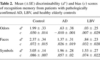
A Group (control, AD, LBV) × Measure (d′ odors, c odors, d′ faces, c faces, d′ symbols, c symbols) multivariate analysis of variance revealed a significant effect of group on the discriminability measure (d′) for odors [F(2,33) = 9.31, p < .001], faces [F(2,33) = 6.98, p < .01], and symbols [F(2,33) = 11.03, p < .001]. No significant differences were detected among the groups on the bias measure (c) for odors [F(2,33) = .89, p = .42], faces [F(2,33) = 1.82, p = .18], or symbols [F(2,33) = .72, p = .50]. Since both the AD and LBV groups showed significantly poorer thresholds than controls, a multivariate analysis of covariance with odor threshold as a covariate also was used to analyze the data. The analysis revealed a significant effect of group on the discriminability measure (d′) for odors [F(2,32) = 7.06, p < .01] and symbols [F(2,32) = 8.58, p < .01]. No significant differences were detected on d′ for faces [F(2,32) = 2.32, p = .11] or among the groups on the bias measure (c) for odors [F(2,33) = .89, p = .42], faces [F(2,33) = 1.82, p = .18], or symbols [F(2,33) = .72, p = .50]. A Tukey's HSD test for multiple comparisons on the effect of group on d′ measures revealed that both AD and LBV patients showed significantly lower (p < .05) measures of discriminability compared to controls for olfactory and visual stimuli. The lower d′ scores for the AD and LBV groups indicate that the ability of these participants to discriminate old stimuli from new stimuli was diminished compared to controls. The mean (±SE) d′ and c scores for each group are shown in Table 2.
Mean (±SE) discriminability (d′) and bias (c) scores of recognition memory from patients with pathologically confirmed AD, LBV, and healthy elderly controls

Remote Memory
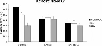
The data in Figure 2 show that there were no significant differences in mean familiarity ratings for visual stimuli among controls, AD patients, and LBV patients. However, both AD and LBV patients showed lower familiarity ratings compared to controls for olfactory stimuli. In addition, the familiarity ratings of the LBV patients were significantly lower than those of AD patients.

Remote memory measured by mean (±SE) familiarity ratings (expressed in percentage of a 160-mm scale) from patients with pathologically confirmed AD, LBV, and healthy elderly control.
For analysis, the data were grouped into one block of 10 trials for each of the three types of stimuli. A group (control, AD, LBV) by stimuli (odors, faces, and symbols) multivariate analyses of variance revealed that there were no significant differences between the groups on remote memory tasks involving faces [F(2,33) = .09, p = .92] or symbols [F(2,33) = .40, p = .68]. However, the groups differed on remote memory for odors [F(2,33) = 17.34, p < .001]. Since both the AD and LBV groups showed significantly poorer thresholds than controls, a multivariate analysis of covariance with odor threshold as a covariate also was used to analyze the data. The analysis revealed a significant effect of group on remote memory for odors [F(2,32) = 11.23, p < .001] but no effect of group on remote memory for faces [F(2,32) = .10, p = .90] or symbols [F(2,32) = .42, p = .66]. A Tukey's test revealed that both AD and LBV patients performed significantly worse (p < .05) than controls on the remote memory for odors task. In addition, LBV patients performed significantly worse (p < .05) than AD patients on the task.
DISCUSSION
The diagnosis of AD and LBV remains particularly challenging in the living patient. As a result, studies that involve living patients diagnosed with either AD or LBV are at risk for diagnostic error resulting in inclusion of patients who fit diagnostic criteria for a disease but may not fit the pathological criteria. One strength of the present study is that all patients with AD or LBV were pathologically confirmed and therefore the findings are not confounded by the potential for diagnostic error. The present manuscript offers the first study to compare memory for olfactory stimuli in patients with pathologically confirmed LBV and AD.
The results of the present study demonstrate that on tasks requiring recognition memory, LBV and AD patients showed significantly poorer discriminability (d′) than controls, particularly for olfactory stimuli. This finding illustrates that recognition memory for olfactory and visual stimuli is impaired in patients with LBV and AD. The data reveal that patients with LBV showed poorer d′ scores than AD patients; however, the difference did not reach statistical significance. The results also revealed no significant differences among the groups on the bias measure (c) suggesting that differences in task performance are not the result of a response bias. Prior experiments have reported similar impairments in recognition memory for olfactory stimuli in AD patients (Gilbert & Murphy, in press; Kesslak et al., 1988; Knupfer & Spiegel, 1986; Moberg et al., 1987; Nordin & Murphy, 1996). However, this is the first study to our knowledge to report deficits in recognition memory for olfactory stimuli in patients with pathologically confirmed LBV.
Recognition memory may be impaired in LBV and AD patients due to damage to mesial temporal lobe structures such as the hippocampus which support the encoding of episodic or declarative memories (Schacter & Wagner, 1999; Squire & Zola, 1996). Hippocampal degeneration has been well documented in patients with AD (Braak & Braak, 1992, 1997; Braak et al., 1996; Morris et al., 1996; Price et al., 1991). In addition, AD has been shown to affect neurons in the entorhinal cortex that form the perforant path input into the hippocampus (Braak & Braak, 1997; Braak et al., 1996; Morris et al., 1996; Price et al., 1991). Studies also have shown increased pathology in the entorhinal cortex, perforant path, and hippocampus in patients with LBV (Iseki et al., 2002; Thorns et al., 1997; Wakabayashi et al., 1994, 1997). A study by Hansen et al. (1990) showed that 85% of LBV patients showed spongiform vacuolizations of various size in entorhinal cortex; however, vacuoles were reported in only 17% of AD patients. Neocortical Lewy bodies also have been found in the deep layers of entorhinal cortex (Galasko et al., 1996). The entorhinal cortex has been shown to receive direct and indirect input from the olfactory bulb in rodents, nonhuman primates, and humans (Insausti et al., 2002). Braak and Braak (1992) have suggested that lesion in the entorhinal cortex, which is the primary input into the hippocampus, may disrupt the transfer of information into the hippocampus essential to memory function. Therefore, in addition to memory problems resulting from direct damage to the hippocampus in patients with AD and LBV, damage to the entorhinal cortex also may contribute to deficits in odor memory. However, the unique contributions of the entorhinal cortex and hippocampus to olfactory memory remain uncertain. Based on the aforementioned research, damage to the hippocampus and entorhinal cortex in LBV and AD patients may result in particularly poor encoding of olfactory stimuli and may account for the low d′ scores for odors in LBV and AD patients in the present study.
When participants were asked to rate the familiarity of olfactory stimuli (a proposed measure of remote memory) LBV and AD patients reported significantly lower familiarity ratings compared to controls, suggesting that regions of the brain involved in the retrieval and/or storage of remote memories for olfactory stimuli are affected by LBV and AD pathology. Since the task requires the participant to rate their subjective feeling of familiarity, performance of the task also may depend on regions of the brain involved in implicit memory. Using the same paradigm for measuring remote memory used in the present study, Niccoli-Waller et al. (1999) showed that remote memory for odors but not faces was impaired in patients with AD relative to controls. In addition, prior studies have reported that both AD patients (Doty, 1991; Koss et al., 1987; Morgan et al., 1995; Serby et al., 1985) and LBV patients (Bailie et al., 2003) show impairments in odor identification, a task that has been suggested to require remote memory. In contrast, the present study demonstrated that familiarity ratings for olfactory stimuli were significantly lower in patients with LBV compared to patients with AD. This finding suggests that patients with LBV are more impaired than AD patients on tasks requiring remote memory for olfactory stimuli. The present study did not detect any significant differences among the groups on familiarity ratings for visual stimuli. Similar findings also were reported by Niccoli-Waller et al. (1999) using the same paradigm to assess remote memory in patients with probable AD. Based on these findings, the regions of the neocortex that support remote memory for olfactory stimuli, measured by familiarity ratings, may be particularly affected in LBV patients and to a lesser extent in patients with AD.
Consistent with prior reports, the LBV patients showed significantly lower odor thresholds compared to AD patients (Bailie et al., 2003; Olichney et al., 2002, 2004) and both groups were significantly different from controls. This finding suggests that hyposmia in dementia is associated with both Lewy body and AD pathology. Since both the AD and LBV groups showed significantly poorer odor thresholds than controls, one may argue that the memory impairments in the AD and LBV group may be the result of impaired detection of the odors rather than impaired mnemonic encoding or retrieval. To statistically control for impairments in olfactory detection, butanol odor threshold scores were entered as a covariate in the MANCOVAs conducted on the recognition and remote memory data. The results of the analyses with odor threshold as a covariate were very consistent with the results of the MANOVAs suggesting that the reported deficits are not only the result of impaired sensory registration.
In conclusion, the results of the present study suggest that recognition memory for olfactory stimuli is impaired in both patients with LBV and AD. However, patients with LBV are more impaired than AD patients on tasks requiring remote memory for olfactory but not visual stimuli. The results suggest that tasks requiring odor memory may be potentially useful in discriminating LBV and AD patients.
ACKNOWLEDGMENTS
This research was supported by NIH grant #AG04085 from the National Institute on Aging to Claire Murphy, and Training Grant #DC00032 to Terence M. Davidson. We gratefully acknowledge Drs. Robert Katzman, Leon Thal, David Salmon as well as the staff, patients, and volunteers at the Alzheimer's Disease Research Center, UCSD (P50AG05131). We also thank Sally Ferdon, Jason Bailie, and Violeta Canton for their advice and assistance.
References
REFERENCES
Bacon-Moore, A.S.,
Paulsen, J.S., &
Murphy, C.
(1999).
A test of odor fluency in patients with Alzheimer's and
Huntington's disease.
Journal of Clinical and Experimental Neuropsychology,
21,
341–51.CrossRefGoogle Scholar
Bailie, J.M.,
Gilbert, P.E., &
Murphy, C.
(2003).
Odor identification deficits in Lewy body variant of
Alzheimer's disease.
Journal of the International Neuropsychological Society,
9,
164–165.Google Scholar
Braak, H. &
Braak, E.
(1992).
The human entorhinal cortex: Normal morphology and lamina-specific
pathology in various diseases.
Neuroscience Research,
15,
6–31.CrossRefGoogle Scholar
Braak, H. &
Braak, E.
(1997).
Frequency of stages of Alzheimer's-related lesion in different
age categories.
Neurobiology of Aging,
18,
351–357.CrossRefGoogle Scholar
Braak, H.,
Braak, E.,
Yilmazer, D.,
de vos, R.A.I.,
Jansen, E.N.H., &
Bohl, J.
(1996).
Pattern of brain destruction in Parkinson's and
Alzheimer's diseases.
Journal of Neural Transmission,
103,
455–490.CrossRefGoogle Scholar
Cain, W.S.
(1989).
Testing olfaction in a clinical setting.
Ear, Nose, and Throat Journal,
68,
78–86.Google Scholar
Clements, J.
(1975).
Chronology of the United States.
New York:
McGraw-Hill.
Connor, D.J.,
Salmon, D.P.,
Sandy, T.J.,
Galasko, D.,
Hansen, L.A., &
Thal, L.J.
(1998).
Cognitive Profiles of Autopsy-Confirmed Lewy body variant vs Pure
Alzheimer disease.
Archives of Neurology,
55,
994–1000.CrossRefGoogle Scholar
Doty, R.L.
(1991).
Olfactory dysfunction in neurodegenerative disorders. In
T.V. Getchell,
R.L. Doty,
L.M. Bartoshuk, &
J.B Snow, Jr. (Eds.),
Smell and taste in health and disease (pp.
735–751).
New York:
Raven Press.
Doty, R.L.
(2003).
Odor perception in neurodegenerative diseases. In
R.L. Doty (Ed.),
Handbook of olfaction and gustation (pp.
479–502).
New York:
Marcel Dekker.
Ekman, G.,
Berglund, B.,
Berglund, U., &
Lindvall, T.
(1967).
Perceived intensity of odor as a function of time of adaptation.
Scandinavian Journal of Psychology,
8,
177–186.CrossRefGoogle Scholar
Esiri, M.M. &
Wilcox, G.K.
(1984).
The olfactory bulbs in Alzheimer's disease.
Journal of Neurology, Neurosurgery, and Psychiatry,
47,
56–60.CrossRefGoogle Scholar
Folstein, M.F.,
Folstein, S.E., &
McHugh, P.R.
(1975).
“Mini-Mental State”: A practical method for grading the
cognitive state of patients for the clinician.
Journal of Psychiatric Research,
12,
189–198.CrossRefGoogle Scholar
Galasko, D.,
Katzman, R.,
Salmon, D.P., &
Hansen, L.
(1996).
Clinical and neuropathological findings in Lewy body dementias.
Brain and Cognition,
31,
166–175.CrossRefGoogle Scholar
Gilbert, P.E. &
Murphy, C.
(in press).
The effect of the ApoE ε4 allele on recognition memory for
olfactory and visual stimuli in patients with pathologically confirmed
Alzheimer's disease, probable Alzheimer's disease, and healthy
elderly controls.
Journal of Clinical and Experimental Neuropsychology.
Hansen, L.,
Salmon, D.,
Galasko, D.,
Masliah, E.,
Katzman, R.,
DeTeresa, R.,
Thal, L.,
Pay, M.,
Hofstetter, R.,
Klauber, M.,
Rice, V.,
Butters, N., &
Alford, M.
(1990).
The Lewy body variant of Alzheimer's disease: A clinical and
pathological entity.
Neurology,
40,
1–8.CrossRefGoogle Scholar
Hyman, B.T.
(1997).
The neuropathological diagnosis of Alzheimer's disease:
Clinical-pathological studies.
Neurobiology of Aging,
18,
S27–32.CrossRefGoogle Scholar
Ince, P.,
Irving, D.,
MacArthur, F., &
Perry, R.H.
(1991).
Quantitative neuropathological study of Alzheimer-type pathology in
the hippocampus: comparison of senile dementia of Alzheimer type,
senile dementia of Lewy body type, Parkinson's disease and
non-demented elderly control patients.
Journal of Neuroscience,
106,
142–152.Google Scholar
Insausti, R.,
Marcos, P.,
Arroyo-Jimenez, M.M.,
Blaizot, X., &
Martinez-Marcos, A.
(2002).
Comparative aspects of the olfactory portion of the entorhinal
cortex and its projection to the hippocampus in rodents, nonhuman
primates, and the human brain.
Brain Research Bulletin,
57,
557–560.CrossRefGoogle Scholar
Iseki, E.,
Takayama, N.,
Marui, W.,
Ueda, K., &
Kosaka, K.
(2002).
Relationship in the formation process between neurofibrillary
tangles and Lewy bodies in the hippocampus of dementia with Lewy bodies
brains.
Journal of Neuroscience,
195,
85–91.Google Scholar
Kesslak, J.P.,
Cotman, C.W.,
Chui, H.C.,
van Dan Noort, S.,
Fang, H.,
Pfeffer, R., &
Lynch, G.
(1988).
Olfactory tests as possible probes for detecting and monitoring
Alzheimer's disease.
Neurobiology of Aging,
9,
399–403.CrossRefGoogle Scholar
Khachaturian, Z.S.
(1985).
Diagnosis of Alzheimer's disease.
Archives of Neurology,
42,
1097–1105.CrossRefGoogle Scholar
Knupfer, L. &
Spiegel, R.
(1986).
Differences in olfactory test performance between normal aged,
Alzheimer, and vascular type dementia individuals.
International Journal of Geriatric Psychiatry,
1,
3–14.CrossRefGoogle Scholar
Koss, E.,
Weiffenbach, J.,
Haxby, J., &
Friedland, R.
(1987).
Olfactory detection and recognition in Alzheimer's disease.
Lancet,
1,
622.CrossRefGoogle Scholar
Macmillan, N.A. &
Creelman, D.C.
(1991).
Signal detection theory: A user's guide.
New York:
Cambridge University Press.
Mattis, S.
(1976).
Mental status examination for organic mental syndrome in the
elderly patient. In
L. Bellack &
T.B. Katsau (Eds.),
Geriatric psychiatry: A handbook for psychiatrists and primary care
physicians (pp.
77–121).
New York:
Grune & Stratton.
McShane, R.H.,
Nagy, Z.,
Esiri, M.M.,
King, E.,
Joachim, C.,
Sullivan, N., &
Smith, A.D.
(2001).
Anosmia in dementia is associated with Lewy bodies rather than
Alzheimer's pathology.
Neurology, Neurosurgery, and Psychiatry,
70,
739–743.CrossRefGoogle Scholar
Mirra, S.S.,
Heyam, A.,
McKeel, D.,
Sumi, S.M.,
Crain, B.J.,
Brownlee, L.M.,
Vogel, F.S.,
Hughes, J.P.,
van Belle, G., &
Berg, L.
(1991).
The Consortium to Establish a Registry for Alzheimer's Disease
(CERAD). Part II. Standardization of the neuropathologic assessment of
Alzheimer's disease.
Neurology,
41,
479–86.Google Scholar
Moberg, P.J.,
Pearlson, G.D.,
Speedie, L.J.,
Lipsey, J.R.,
Strauss, M.E., &
Folstein, S.E.
(1987).
Olfactory recognition: Differential impairments in early and late
Huntington's and Alzheimer's disease.
Journal of Clinical and Experimental Neuropsychology,
9,
650–664.CrossRefGoogle Scholar
Morgan, C.D.,
Nordin, S., &
Murphy, C.
(1995).
Odor identification as an early marker for Alzheimer's
disease: Impact of lexical functioning and detection sensitivity.
Journal of Clinical and Experimental Neuropsychology,
17,
793–803.CrossRefGoogle Scholar
Morris, J.C.,
Storandt, M.,
McKeel, D.W., Jr.,
Rubin, E.H.,
Price, J.L.,
Grant, E.A., &
Berg, L.
(1996).
Cerebral amyloid deposition and diffuse plaques in
“normal” aging: Evidence for presymptomatic and very mild
Alzheimer's disease.
Neurology,
46,
707–719.CrossRefGoogle Scholar
Murphy, C.,
Cain, W.S.,
Gilmore, M.M., &
Skinner, R.B.
(1991).
Sensory and semantic factors in recognition memory for odors and
graphic stimuli: Elderly versus young persons.
American Journal of Psychology,
104,
161–192.CrossRefGoogle Scholar
Murphy, C.,
Gilmore, M.M.,
Seery, C.S.,
Salmon, D.P., &
Lasker, B.R.
(1990).
Olfactory thresholds are associated with degree of dementia in
Alzheimer's disease.
Neurobiology of Aging,
11,
465–469.CrossRefGoogle Scholar
Niccoli-Waller, C.A.,
Harvey, J.,
Nordin, S., &
Murphy, C.
(1999).
Remote odor memory in Alzheimer's disease: Deficits as
measured by familiarity.
Journal of Adult Development,
6,
131–136.CrossRefGoogle Scholar
Nordin, S. &
Murphy, C.
(1996).
Impaired sensory and cognitive olfactory function in questionable
Alzheimer's disease.
Neuropsychology,
10,
113–119.CrossRefGoogle Scholar
Olichney, J.M.,
Galasko, D.,
Salmon, D.P.,
Hofstetter, C.R.,
Hansen, L.A.,
Katzman, R., &
Thal, L.J.
(1998).
Cognitive decline is faster in Lewy body variant than in
Alzheimer's disease.
Neurology,
51,
351–357.CrossRefGoogle Scholar
Olichney, J.M.,
Murphy, C.,
Hofstetter, R.,
Hansen, L.A., &
Katzman, R.
(2002).
Anosmia is very common in the Lewy Body Variant of Alzheimer's
Disease, but can this improve our diagnostic accuracy?
Slide Presentation (Abstract S59.004), American Academy of
Neurology Meeting, Denver, CO.
Olichney, J.M.,
Murphy, C.,
Hofstetter, R.,
Foster, K.,
Hansen, L.A.,
Thal, L.J., &
Katzman, R.
(2004).
Anosmia is very common in the Lewy body variant of Alzheimer's
disease.
Manuscript submitted for publication.
Perry, R.H.,
Irving, D.,
Blessed, G.,
Fairbairn, A., &
Perry, E.K.
(1990).
Senile dementia of Lewy body type. A clinically and neuropathologically
distinct form of Lewy body dementia in the elderly.
Journal of Neuroscience,
95,
119–139.CrossRefGoogle Scholar
Price, J.L.,
Davis, P.F.,
Morris, J.C., &
White, D.L.
(1991).
The distribution of tangles, plaques and related
immunohistochemical markers in early aging and Alzheimer's
disease.
Neurobiology of Aging,
12,
295–312.CrossRefGoogle Scholar
Salmon, D.P.,
Galasko, D.,
Hansen, L.A.,
Masliah, E.,
Butters, N.,
Thal, L., &
Katzman, R.
(1996).
Neuropsychological deficits associated with diffuse Lewy body
disease.
Brain and Cognition,
31,
148–165.CrossRefGoogle Scholar
SantaCruz, K.S.,
Tasaki, C.S.,
Kim, R.C., &
Cotman, C.W.
(2002).
Brainstem and cortical Lewy bodies in patients presenting
clinically with Alzheimer's disease.
Journal of Alzheimer's Disease,
4,
11–17.CrossRefGoogle Scholar
Schacter, D.L. &
Wagner, A.D.
(1999).
Medial temporal lobe activations in fMRI and PET studies of
episodic encoding and retrieval.
Hippocampus,
9,
7–24.3.0.CO;2-K>CrossRefGoogle Scholar
Serby, M.,
Corwin, J.,
Novatt, A.,
Conrad, P., &
Ritrosen, J.
(1985).
Olfaction in dementia.
Journal of Neurology, Neurosurgery, and Psychiatry,
48,
848–849.CrossRefGoogle Scholar
Squire, L.R. &
Zola, S.M.
(1996).
Structure and function of declarative and nondeclarative memory
systems. Proceeding of the National Academy of Sciences,
USA,
93,
13515–13522.CrossRefGoogle Scholar
Thorns, V.,
Mallory, M.,
Hansen, L., &
Masliah, E.
(1997).
Alterations in glutamate receptor 2/3 subunits and amyloid
precursor protein expression during the course of Alzheimer's
disease and Lewy body variant.
Acta Neuropathologica,
94,
539–548.CrossRefGoogle Scholar
Upton, G.J.G.
(1978).
The analysis of cross tabulated data.
Chichester, UK:
Wiley.
Verghese, J.,
Crystal, H.A.,
Dickson, D.W., &
Lipton, R.B.
(1999).
Validity of clinical criteria for the diagnosis of dementia with
Lewy bodies.
Neurology,
53,
1974–1982.CrossRefGoogle Scholar
Wakabayashi, K.,
Hansen, L.A.,
Vincent, I.,
Mallory, M., &
Masliah, E.
(1997).
Neurofibrillary tangles in the dentate granule cells of patients
with Alzheimer's disease, Lewy body disease and progressive
supranuclear palsy.
Acta Neuropathologica,
93,
7–12.CrossRefGoogle Scholar
Wakabayashi, K.,
Honer, W.G., &
Masliah, E.
(1994).
Synapse alterations in the hippocampal-entorhinal formation in
Alzheimer's disease with and without Lewy body disease.
Brain Research,
667,
24–32.CrossRefGoogle Scholar
Westervelt, H.J.,
Ster, R.A., &
Tremont, G.
(2003).
Odor identification deficits in diffuse Lewy body disease.
Cognition Behavioral Neurology,
16,
93–99.CrossRefGoogle Scholar

Mean (±SE) demographics, odor thresholds, and scores on the Mini-Mental Status Examination and Dementia Rating Scale from patients with pathologically confirmed AD, LBV, and healthy elderly controls

Mean (± SE) discriminability (d′) measures of recognition memory from patients with pathologically-confirmed AD, LBV, and healthy elderly controls.
Mean (±SE) discriminability (d′) and bias (c) scores of recognition memory from patients with pathologically confirmed AD, LBV, and healthy elderly controls
Remote memory measured by mean (±SE) familiarity ratings (expressed in percentage of a 160-mm scale) from patients with pathologically confirmed AD, LBV, and healthy elderly control.
 You have
Access
- 37
- Cited by