INTRODUCTION
The assessment of growth and age provides important input data for many stocks assessment models and thus is very important for the sustainable management of fisheries stocks. In cephalopods, considering that the success of recruitment depends almost entirely on environmental conditions, it is quite important to understand how reproduction, lifespan and in particular growth, are affected by those conditions.
Octopus vulgaris, Cuvier 1797 is an important resource for the artisanal and industrial fisheries in all of the Atlantic margin of the Iberian peninsula, with annual average landings of 9185 tonnes in Portugal (INE, 2013) and 4000 in Galicia (Otero et al., Reference Otero, Rocha, González, Garcia and Guerra2005). The lifespan of O. vulgaris was estimated to be 1–2 years (Domain et al., Reference Domain, Jouffre and Caveriviére2000; Katsanevakis & Verriopoulos, Reference Katsanevakis and Verriopoulos2006). After hatching, the paralarvae go through a short period of no net growth, depending on the consumption of yolk reserves to survive (Villanueva & Norman, Reference Villanueva and Norman2008). Then the paralarvae grow exponentially until settlement reaching the sub-adult stage. Here, the logarithmic growth phase starts with a decreasing instantaneous growth rate until the maturation phase is complete (Mangold, Reference Mangold and Boyle1983; Villanueva, Reference Villanueva1995).
Direct ageing methods based on statoliths increment analysis were not found to be useful in incirrate octopods, while approaches using beaks in O. vulgaris still need proper validation, in particular due to erosion by feeding (Perales-Raya et al., Reference Perales-Raya, Bartolomé, García-Santamaría, Pascual-Alayón and Almansa2010; Canali et al., Reference Canali, Ponte, Belcari, Rocha and Fiorito2011). An alternative to perform direct age assessments is the use of the vestigial shell or stylet (Sousa Reis & Fernandes, Reference Sousa Reis and Fernandes2002). Stylets are needle-shaped rods located on the dorso-lateral side of the mantle, that arose from the reduction of the shell in the Incirrata (Budelmann et al., Reference Budelmann, Schipp, Boletzky, Harrison and Kohn1997; Naef, 1921/1923 in Bizikov, Reference Bizikov2004). The growth of stylets progresses from the centre of growth (stylet primordium) located in the bend through the regular deposition of concentric layers of semi-transparent chitin (Bizikov, Reference Bizikov2004) that can be used to assess age. Stylets have recently been used successfully to assess age in wild populations of some octopus species (e.g. O. pallidus, Leporati et al., Reference Leporati, Semmens and Pecl2008; O. cyanea, Herwig et al., Reference Herwig, Depczynski, Roberts, Semmens, Gagliano and Heyward2012). The fast degradation of the structure upon contact with air and the abrasive techniques used to expose the growth structures are major concerns to the standardization of the methods and their regular implementation. Nevertheless, new preparation methods are being developed, which appear to produce good quality stylet sections (Barratt & Allcock, Reference Barratt and Allcock2010) and consequently the age determination by stylet increment analysis (SIA) is potentially an effective tool for ageing O. vulgaris, as was first advanced by Sousa Reis & Fernandes (Reference Sousa Reis and Fernandes2002). It is also worth noting that the daily deposition of growth increments in the stylets of adults of this species was validated by Hermosilla et al. (Reference Hermosilla, Rocha, Fiorito, González and Guerra2010). However, the validation of the daily deposition of growth increments in adults does not validate the same periodicity in the increments deposition in earlier life stages and nor does it identify the deposition of first growth increment in paralarvae, essential criteria for a rigorous age validation of the SIA in each species (Campana, Reference Campana2001). The difficulties and potential inaccuracies associated with determining the age of merobenthic octopuses (such as O. vulgaris) using SIA and the importance of validating age at first increment formation are discussed in Doubleday et al. (Reference Doubleday, White, Pecl and Semmens2011).
The present study aimed firstly to develop a technique to rapidly locate the stylets in the muscle of paralarvae, and secondly to determine the stylet size at hatching in newly hatched O. vulgaris paralarvae as a tool to define the starting point for age determination in stylets of later stages. Additionally, the stylets of 3-day-old paralarvae were compared with unknown age paralarvae captured in the wild to determine if the stylet nuclear area is conservative between paralarvae of different sizes and ages and between animals incubated at different temperatures.
MATERIALS AND METHODS
The captivity paralarvae used in this study were collected opportunistically from experiments on ocean warming effects on O. vulgaris earlier life stages, conducted at Guia Marine Laboratory (more details about the rearing conditions are described in Repolho et al., Reference Repolho, Baptista, Pimentel, Dionisio, Trübenbach, Lopes, Lopes, Calado, Diniz and Rosa2014). These paralarvae hatched from egg clutches collected at the beginning of embryogenesis (Stage I: Naef, Reference Naef1928) from traps of local fishermen between October 2010 and November 2011 in Cascais, Portugal. After collection, eggs were transferred to the aquaculture systems in Guia Marine Laboratory, Cascais. Here, the eggs were reared at different water temperatures including 18 and 22°C until hatching at 39–25 days respectively, after eggs incubation. After hatching, the paralarvae were kept at the same temperatures for 3 days without food and then 10 paralarvae from each temperature were sacrificed for this study. The 3-day-old paralarvae were chosen to ensure that paralarvae had grown since hatching, and the observation of the hatch check and growth increments was already possible. All paralarvae were preserved in 70% ethanol.
The paralarvae were measured under transmitted light binocular microscopy at 30× magnification. Measurements were taken as follows: total length (TL in mm), mantle ventral length (ML in mm), eye diameter (D-eye in mm) and total weight (W in mg). Before weighing, the paralarvae were dried with filter paper.
A set of six paralarvae was used to establish the most adequate protocol that could simultaneously permit location and examination of several cross-sections of the paralarval stylet. These were embedded in paraffin and sectioned (in 6 μm width sections) according to three morphological planes: the sagittal, transversal and frontal planes. Sections were stained with acetic Alcian blue solution (N = 3) and Masson's trichrome (N = 3) in order to enhance the fibrous nature of the stylets, by staining fibrin tissue in a solution of acetic Alcian blue (adapted from Vecchione, Reference Vecchione1991) or light green/blue (Jones, Reference Jones, Bancroft and Gamble2002), respectively. It was expected that the staining would improve the identification of the structures inside the mantle. Stained sections were observed under a binocular microscope equipped with transmitted light, at 400× and 1000× magnification. All sections were sequentially photographed. Taking into account the results of the experiment above, the two groups of 3-day-old paralarvae (18°C group and 22°C group) were subsequently sectioned in the transversal plane in 6 μm sections and stained with the Masson Trichrome method. All sections were observed under transmitted light at a magnification of 400× and 1000× and photographed.
The best transversal section closer to the stylet bend was selected to identify the embryonic primordium or nucleus of the stylet. The nucleus was limited by a discontinuity in the ageing structure which appeared as a high-contrast micro-increment with a deeply darker zone under transmitted light, or an abrupt change in the micro-structural growth pattern (Panfili et al., Reference Panfili, de Pontual, Toradec and Wright2002). Stylet measurements were taken under 1000× magnification from the selected cross-section of the stylet, as follows: stylet diameter (SD in μm), stylet area (SA in μm2), stylet major radius (SRmax in μm) and nucleus diameter (SDnucleus in μm).
Additionally, wild paralarvae (N = 9) of unknown age were collected in July and September 2010 in the Ría de Vigo (Southwest Galicia, Spain) during mesozooplankton surveys. These paralarvae were collected in depth and surface strata using a multitrawl (MultiNet®) sampler (0.71 × 0.71 m opening frame, see Roura (Reference Roura2013) for details). Local sea surface temperature recorders indicate that the embryonic development of these paralarvae occurred under mean surface temperatures between 16.5 and 19.2°C during embryological development (data source: Seawatch buoy located off Cape Silleiro, 42° 7.80 N 9° 23.40 W, http://www.puertos.es). The wild paralarvae were stored in 70% ethanol and measured in a similar manner to the captive paralarvae. These were then transversally sectioned accordingly and stained with haematoxylin & eosin. Selected cross-sections were measured following the same procedure defined for the 3-day-old paralarvae.
To assess the effect of temperature on paralarva and stylet sizes, measurement data were grouped according to the incubation temperature and sampling source, as ‘18°C’ and ‘22°C’ groups for the 3-day-old paralarvae and ‘wild’ group for the paralarvae collected in the Ría de Vigo. Prior to the statistical analysis, the assumptions of sample normality and homogeneity for paralarvae and stylet dimensions were assessed by group with Shapiro–Wilk's and Bartlett's tests, respectively. A non-parametric Kruskal–Wallis test was used to identify differences in mean measurements between groups. The Spearman correlation index was used to identify cases of colinearity between the measurements, as well as to identify strong correlations between the size of the paralarva and measurements of the respective stylet.
Additionally, the Kruskal–Wallis test was used to compare the mean SD of the paralarvae with the mean diameter of the nucleus identified in stylet cross-sections of O. vulgaris juveniles (N = 13) captured in the Portuguese north-east coast. The sampling design and methodology applied to prepare and measure cross-sections of juvenile stylets are described in Lourenço (Reference Lourenço2014).
RESULTS
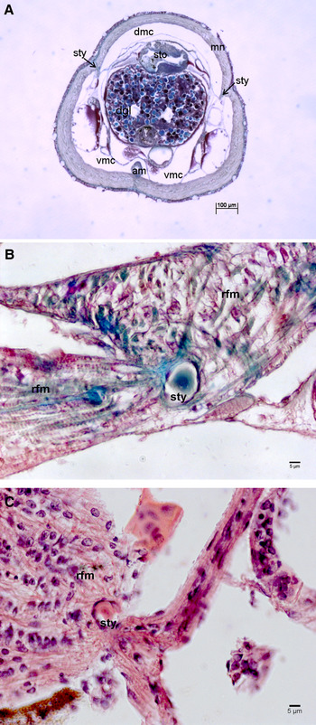
As in adults, the stylets of O. vulgaris paralarvae were located in the insertion of the funnel retractor muscles, in the posterior region of the mantle. Compared with adults, these structures were situated more dorsally and mid region of the mantle (Figure 1A). In the paralarvae, the stylet bend (where the primordium of the structure is located) was found to lie between 100 and 200 μm from the tip of the mantle in paralarvae measuring between 0.71 and 1.88 mm of ventral mantle length.
Transversal section (A) of an Octopus vulgaris paralarva (magnification: 40×). The stylets are well inserted in the antero-dorsal region of the mantle. Detail of a cross-section of an Octopus vulgaris stylet (B and C, magnification: 400×) obtained through a transversal section of the paralarva. am, adductor muscle; dgl, digestive gland; dmc, dorsal mantle cavity; mn, mantle; rfm, funnel retractor muscle; sto, stomach; sty, stylet; vmc, ventral mantle cavity (after Bizikov, Reference Bizikov2004).

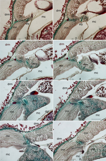
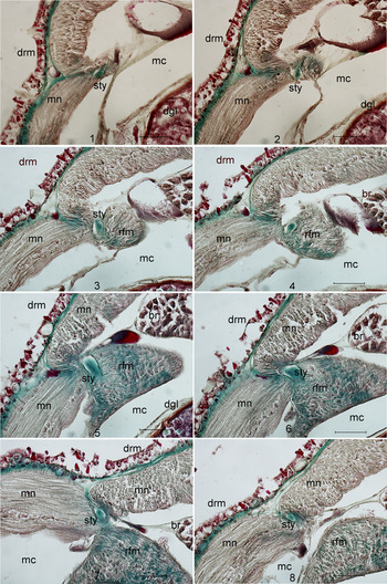
The use of Masson trichrome as a stain clearly improved the ability to locate the stylet inside the insertion between the mantle and the funnel retractor muscle (mantle–funnel retractor muscle insertion). Using this stain the stylet appeared in most paralarvae sections as green/blue contrasting with the surrounding tissue (Figure 1B). The transversal sectioning plane gave best results to obtain good cross-sections of the stylet near the bend where it was possible to locate the stylet primordium. This transversal plane allowed firstly to identify the stylet at the bend level in the mantle–funnel retractor muscle insertion and then to identify the best cross-section where it was possible to detect the hatch check in the stylet and to measure the diameter, perimeter, area and major radius of the stylet (Figure 2). The stylet is anterior-posteriorly oriented in the mantle with the anterior branch (or rostrum) inserted deep inside the mantle muscle, the bend was located inside the mantle–funnel retractor muscle insertion, and the post-rostral branch positioned more superficially along the interior wall of the mantle (Figure 2).
Sequence of transversal sections (magnification: 400×) of a 1-day-old Octopus vulgaris paralarva indicating the sequential position of the stylet in the insertion between the mantle and retractor funnel muscle. Scale bar indicates 20 μm. drm, dermis; dgl, digestive gland; br, branchia; mc, mantle cavity; mn, mantle; rfm, retractor funnel muscle; sty, stylets (after Bizikov, Reference Bizikov2004).
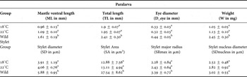
Bearing in mind that some degree of paralarvae body shrinkage can occur due to the preservation method (up to 20% with 70% ethanol according to Goto, Reference Goto2005), in the 3-day-old paralarvae, the mean diameter of the stylet (measured between the most distant points) was 3.99 ± 0.46 μm and the mean area measured was 13.00 ± 6.13 μm2. In these stylets, the nucleus was identifiable in the cross-sections near the bend. It was identified as a distinctively darker area circumscribed by one highly contrasted micro-increment (with a deeply darker zone), and within which first-order growth rings are not observed. The mean diameter of the nucleus was 2.71 ± 0.42 μm.
The nuclear area previously defined in the captive paralarvae was easily identified in the nine stylets of the wild group by its micro-structure. In this group, the diameter of the stylet measured 5.88 ± 0.95 μm and the area measured 27.54 ± 8.62 μm2. The diameter of the stylet nucleus measured 3.02 ± 0.55 μm. It was only possible to identify the deposition of one growth increment in the post-nuclear area of the stylets of two wild paralarvae (Figure 1C).
Table 1 shows the mean values obtained for each of the paralarvae and stylet dimensions studied by group. The results show that there is no statistical difference between the 18°C group and the 22°C group when comparing both stylet and paralarvae dimensions, although paralarvae from the 22°C group presented larger sizes and also bigger stylets. On the other hand, the wild group paralarvae are larger and weigh more than the 18°C group with the stylet being also bigger in the former, with the exception of the stylet nucleus diameter that did not show a significant difference between the 18°C, 22°C and wild group (Table 1).
Octopus vulgaris paralarvae and stylets mean (±SD) dimensions by group. Different superscripts indicate statistically significant differences between 18°C group and 22°C group and between 18°C and Wild group tested by Kruskal–Wallis test with significance level of P < 0.05.

The stylet area (SA) and SD (colinear with SA) correlates positively with the SRmax (SA vs. SRmax: r s = 0.63, P < 0.001). SDnucleus does not correlates with either of the other stylet dimensions (SDnucleus vs. SA, r s = 0.13, P > 0.05; SDnucleus vs. SRmax, r s = 0.22, P > 0.05). The Spearman index determined for the correlation between the stylet size and the paralarvae dimensions show that SA (colinear with SD) and SRmax correlate positively with the D_eye and with W (SA vs. D_eye: rs = 0.60, P = 0.001; SA vs. W: rs = 0.55, P = 0.002; SRmax vs. D_eye: rs = 0.60, P = 0.001; SRmax vs. W: rs = 0.58, P = 0.001), while the Srnucleus did not show any significant correlation with any of the paralarvae dimensions.
The mean SD determined in the paralarvae is statistically identical to the diameter of the nucleus identified and measured in the juveniles' stylet cross sections (k = 235, P > 0.05).
DISCUSSION
To our knowledge, this is the first time that the stylet has been identified in pelagic paralarvae of a merobenthic octopus, proving its formation in an earlier embryonic stage. In the adults of O. vulgaris, the stylet is a recognizable structure in the dorso-anterior region of the mantle, easily extracted by dissection. However, in newly hatched individuals, the body size and the fragile structure of non-mineralized chitin of the stylet make it particularly difficult to collect the stylets by dissection. Several methods to isolate and collect the stylet from the body of the larvae were tried, including staining the paralarva body with an acetic Alcian blue solution, in an adaptation of the method used by Vecchione (Reference Vecchione1991) to identify stomach contents in squids. According to that author, the Alcian blue efficiently stains eye crystalline lenses and funnel/mantle-locking cartilages in squid paralarvae. We observed that, although the Alcian blue successfully stained the eye lenses of O. vulgaris paralarvae, the staining achieved for the stylets was not effective and resulted in unclear structures.
To overcome this difficulty and considering the fragile nature of newly hatched paralarvae with the beaks and radula still under-developed, we chose to adopt a histological approach to obtain and observe cross-sections of the stylets. Nevertheless, other challenges arise with this approach. The stylets of paralarvae are, as in adults, needle-shaped rods with an irregular shape, presenting a bend in the middle region with concave and convex arms in the insertion area of the mantle–funnel retractor muscles. Both sagittal and transversal cutting planes result in good cross-sections of the stylet, but only the transversal plane allowed a greater number of sections in the vicinity of the primordium. Additionally, this sectioning plane allowed the definition of a methodology to identify the bend and the closest cross-section in which it is possible to identify the nucleus and to measure the structure in a replicable manner.
The nucleus (primordium) is visible in the nearest cross-section to the stylet bend, with a mean diameter of 2.71 μm independently of the developmental temperature, indicating that the stylet primordium size is independent of both biological and environmental factors, suggesting that the nuclear region (corresponding to stylet size at hatching) can be used as a reference point to determine age and growth and related measurements.
Under a magnification of 1000×, the stylet does not have visible growth rings in the majority of the sections. Here the size limitation factor must be considered and in only two stylets of the wild paralarvae group were post-nuclear growth increments visible. Although stylets with smooth core regions seem to be particularly common in holobenthic octopus as O. pallidus (Doubleday et al., Reference Doubleday, Semmens, Pecl and Jackson2006) and other merobenthic octopus as Macroctopus maorum (Doubleday et al., Reference Doubleday, White, Pecl and Semmens2011) one could hypothesize that the absence of visible growth increments near the nucleus may reflect an inadequate resolution power of light microscopy to resolve distances of less than 1 μm (Campana, Reference Campana, Stevenson and Campana1992; Doubleday et al., Reference Doubleday, White, Pecl and Semmens2011) rather than an actual feature of the structure. The use of scanning electron microscopy associated with cryo-sectioning of the paralarvae could be useful tools to improve the analysis of the stylet. In O. vulgaris, a merobenthic species, both stylet diameter and nuclear region of paralarvae are considerably smaller than in O. pallidus, a holobenthic species and particularly similar to stylet sizes and characteristics described by Doubleday et al. (Reference Doubleday, White, Pecl and Semmens2011) for Macroctopus maorum, a merobenthic octopus living in the temperate and the sub-Antarctic waters in Australian coastal waters. In comparison with O. pallidus, the O. vulgaris paralarvae are small and pelagic until settlement 30–60 days after hatching (Villanueva, Reference Villanueva1995; Villanueva & Norman, Reference Villanueva and Norman2008), while O. pallidus paralarvae are larger in relation to the adult size and already benthic at hatching. This results in two orders of magnitude difference in weight (2 mg weight for O. vulgaris hatchlings and 0.10–0.54 g for O. pallidus; Semmens et al., Reference Semmens, Doubleday, Hoyle and Pecl2011) at hatching and fully accounts for size differences between stylet diameter and nuclear area. Such differences illustrate the importance of investigating and validating growth structures and check marks in the stylets of each species.
We were not able to determine the age of the nine paralarvae captured in the Cies Islands. Considering the temperature conditions recorded, we can hypothesize that they developed under a temperature close to the 18°C group. Comparing these two groups, the wild paralarvae were in all cases larger in size, weight and eye diameter than the ones hatched in captivity, indicating that they may be over 3 days old (Villanueva, Reference Villanueva1995), and even though the nucleus has the same diameter for both groups, the larger stylet area in the wild paralarvae indicates that some material have been deposited in the stylet while they grow.
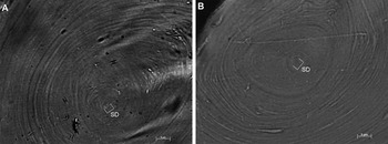
The mean diameter of the stylet in the paralarvae is close to 5 μm (4.59 ± 1.30 μm). Comparing our observations between O. vulgaris paralarva and juvenile stylet cross-sections it is possible to observe correspondences of the nuclear area among the two life stages (Figure 3). In fact, the absence of statistical differences between the SD of the paralarvae stylets with the diameter of the nucleus (mean nuclear diameter 5.80 ± 2.21 μm; see Lourenço, Reference Lourenço2014) identified in the juvenile cross-sections, give us security to use the stylet diameter in post-hatch paralarvae nuclear area to validate the limit of the nucleus in the juvenile stylet cross-sections as the first post-hatch increment. Nevertheless, more studies on the stylet structure are needed to understand how the structure grows in both width and length at this pre-settlement stage.
Octopus vulgaris juvenile stylet cross-sections showing the central area corresponding in size to the stylet diameter in the paralarvae (magnification 630×). SD – diameter of the stylet at hatching; (A) stylet cross-section of a juvenile weighing 384 g (SD = 3.5 μm); (B) stylet cross-section of a juvenile weighing 700 g (SD = 3.39 μm).

ACKNOWLEDGEMENTS
The first author is grateful to José António Durán for teaching and support with histological methodologies. The authors want to express their acknowledgements to the two anonymous referees whose comments greatly improved this manuscript.
FINANCIAL SUPPORT
This study was supported by the projects CAIBEX [CTM2007-66108-C02-01] and LARECO [CTM2011-25929]. The MultiNet® sampler used to collect wild paralarvae was funded by FEDER funds. The Portuguese Foundation for Science and Technology (FCT) supported this study through a PhD grant to Sílvia Lourenço [grant number SFRH/BD/44182/2008] and the project grant PTDC/BIA-BEC/103266/2008 to Rui Rosa.