Introduction
Notocotylus Diesing, Reference Diesing1839 is a genus of so-called monostome digeneans (without ventral sucker), which as sexual adults parasitise herbivorous and molluscivorous birds and mammals. In most traditional versions of monostome systems, based on morphological characters, this taxon was differentiated by the absence of any lateral projections at the anterior extremity and the presence of three longitudinal rows of ventral papillae (e.g., Barton and Blair Reference Barton, Blair, Jones, Bray and Gibson2005; Dubois Reference Dubois1951; Filimonova Reference Filimonova1985; Herber Reference Herber1965; Yamaguti Reference Yamaguti1971). Different species traditionally considered to be members of the genus Notocotylus use either only pulmonate or only prosobranch snails as their first intermediate host. However, the results of molecular studies do not support a direct phylogenetic relationship between the group of species of this taxon associated with pulmonate snails and that associated with prosobranchs (e.g., Capasso et al. Reference Capasso, Servián, Tkach and Diaz2020; Flores et al. Reference Flores, Hernández-Orts and Viozzi2023; Gonchar et al. Reference Gonchar, Jouet, Skírnisson, Krupenko and Galactionov2019; Gonchar and Galaktionov Reference Gonchar and Galaktionov2021; Izrailskaia et al. Reference Izrailskaia, Besprozvannykh, Tatonova, Nguyen and Ngo2019, Reference Izrailskaia, Tatonova and Belousova2024; Sasaki et al. Reference Sasaki, Kobayashi, Yoshino, Asakawa and Nakao2021).
Izrailskaia et al. (Reference Izrailskaia, Tatonova and Belousova2024) proposed a new concept for the genus Notocotylus, suggesting that this taxon includes only the species with life cycles associated with pulmonate snails, namely N. triserialis (N. attenuatus in the cited paper) as the type species, and Notocotylus ephemera Nitzsch, 1817, Notocotylus ikutai Sasaki, Kobayashi, Yoshino, Asakawa and Nakao, Reference Sasaki, Kobayashi, Yoshino, Asakawa and Nakao2021 , Notocotylus malhamensis Boyce, Hide, Craig, Harris, Reynolds, Pickles and Rogan, 2012, Notocotylus multipapillus Izrailskaia and Tatonova, Reference Izrailskaia, Tatonova and Belousova2024, Notocotylus pacifera (Noble, Reference Noble1933), Notocotylus noyeri Joyeux, 1922, Notocotylus ralli Baylis, 1936, Notocotylus regis Harwood, 1936, Notocotylus stagnicolae Herber, 1942, Notocotylus thienemanni L. Szidat and U. Szidat, 1933, and Notocotylus urbanensis (Cort, 1914).
The type species of Notocotylus has a complex taxonomic background with many years of debate about the conspecificity between Notocotylus triserialis Diesing, Reference Diesing1839 (the type by original designation) and Monostoma attenuatum Rudolphi, 1809 (e.g., Barton and Blair Reference Barton, Blair, Jones, Bray and Gibson2005; Cribb Reference Cribb1991; Dubois Reference Dubois1951; Filimonova Reference Filimonova1985; Herber Reference Herber1965; Kossack Reference Kossack1911; Lal Reference Lal1935; L. Szidat and U. Szidat Reference Szidat and Szidat1933; Pike Reference Pike1969; Yamaguti Reference Yamaguti1971). However, due to the lack of type material, the argumentation in favour of one or another point of view is subjective. We follow the authors of the last major review on the taxonomy of Notocotylidae (Barton and Blair Reference Barton, Blair, Jones, Bray and Gibson2005) and accept N. triserialis (=N. attenuatus auct.) as a valid name of the Notocotylus type species.
Recent research points to a probable error in the identification of the isolate of the Notocotylus type species for which data are available in GenBank. According to Izrailskaia et al. (Reference Izrailskaia, Tatonova and Belousova2024), this isolate actually belongs to the genus Pseudonotocotylus Izrailskaia, Tatonova and Belousova, Reference Izrailskaia, Tatonova and Belousova2024, whose reliably identified members, unlike true Notocotylus spp., use prosobranch snails as their first intermediate host.
During parasitological studies of the Greater White-fronted Goose Anser albifrons (Scopoli, 1769) from Russian Karelia and the Eurasian Coot Fulica atra Linnaeus, 1758, from different localities in Eurasia (Eastern Europe and the Far East), we have collected material on N. triserialis, N. pacifera, and Notocotylus sp. The aim was to document the morphological and molecular characteristics of these three notocotylid species and test the monophyly of the genus Notocotylus sensu Izrailskaia et al. (Reference Izrailskaia, Tatonova and Belousova2024).
Materials and methods
Sampling and morphological study
Eight sexual adult specimens of N. triserialis were collected from the caeca of one Greater White-fronted Goose shot down by licensed hunters on the south-eastern coast of Lake Ladoga, near Eroyla Village, Karelia, Russia (Table 1). A total of over a hundred sexual adult specimens of N. pacifera were collected from the caeca of Eurasian Coots taken down in four localities of Eurasia: Volga River delta near Poldnevoe Village, Astrakhan Region; Lake Naroch near Naroch Village, Minsk Region, Belarus; River Mokraya Chuburka near Leninsky Leskhoz Village, Rostov Region, Russia; and Lake Maloe Krugloe near Khasan Village, Primorsky Krai, Russia (Table 1). In total, 52 Eurasian Coot individuals were dissected: 15 birds from each of Belarus, Rostov Region, and Primorsky Krai of Russia and 7 birds from the Astrakhan Region. A single sexual adult specimen of Notocotylus sp. was collected from the caeca of one Eurasian Coot taken down in the above-mentioned locality at River Mokraya Chuburka (Table 1). All Eurasian Coot individuals were examined immediately after hunting. The Greater White-fronted Geese were frozen after hunting and dissected a while after thawing.
Samples of Notocotylus triserialis, Notocotylus pacifera, and Notocotylus sp. analysed in the study and corresponding accession numbers for sequence data submitted to GenBank

All the collected N. triserialis specimens were rinsed in fresh water and fixed in 96% ethanol without flattening. Most of N. pacifera specimens and the single specimen of Notocotylus sp. were relaxed in fresh water, initially fixed in 70% ethanol under a cover glass without additional pressure, and then transferred to 96% ethanol. Three N. pacifera specimens were rinsed in fresh water and then immediately fixed in hot 10% formalin. Morphological examination was done using optical microscopy (all collected specimens of the genus Notocotylus) and scanning electron microscopy (only N. pacifera). For optical microscopy, ethanol-fixed specimens were stained with acetocarmine, dehydrated in a graded ethanol series, cleared in dimethyl phthalate, and mounted in Canada balsam. The slides were then examined through an Olympus BX-53 light microscope (Nagano, Japan) with differential interference contrast (DIC) at the Core Facility of the Karelian Research Centre of the Russian Academy of Sciences, Petrozavodsk, Russia. Before staining, small body fragments were cut off from one specimen of N. triserialis, three specimens of N. pacifera, and the single specimen of Notocotylus sp. to be subsequently used for DNA extraction. These specimens are the hologenophores. For the scanning electron microscopy (SEM), three formalin-fixed specimens of N. pacifera were dehydrated through a graded ethanol series and acetone, and critical-point dried. After coating with gold, they were examined under a Tescan Vega TS5130MM microscope (TESCAN Brno, s.r.o.). Vouchers were deposited in the Museum of Helminthological Collections of the Centre of Parasitology of the A.N. Severtsov Institute of Ecology and Evolution RAS (IEE RAS; Moscow, Russia).
DNA extraction, amplification, and sequencing
DNA was extracted individually from digenean specimens (whole paragenophore or fragments of hologenophore bodies) according to Holterman et al. (Reference Holterman, van der Wurff, van den Elsen, van Megen, Bongers, Holovachov, Bakker and Helder2006). The following DNA loci were amplified for all the newly collected Notocotylus species by BIORAD T100 Thermal Cycler: fragments of the 28S rRNA and cox1 genes and the complete ITS2 region. However, repeated attempts to obtain cox1 gene amplicons for Notocotylus sp. from the Rostov Region of Russia have failed. In addition, a complete 18S rRNA gene was amplified only for N. pacifera. Polymerase chain reactions (PCRs) were run in a total volume of 25 μl using Encyclo Plus PCR kit (Evrogene, Moscow) according to the manufacturer’s instructions.
The partial 28S rRNA gene sequences were amplified with the digl2 (5′-AAGCATATCACTAAGCGG-3′) (Tkach et al. Reference Tkach, Pawlowski and Mariaux2000) and 1500R (5′-GCTATCCTGAGGGAAACTTCG-3′) (Tkach et al. Reference Tkach, Littlewood, Olson, Kinsella and Swiderski2003) primers. The protocol was as follows: initial denaturation at 95°C (5 min); 35 cycles of 30 s at 95°C; 30 s at 50°C; 80 s at 72°C; and 7 min at 72°C for the final extension.
The complete 18S rRNA gene sequence was amplified using the WormA (5′-GCGAATGGCTCATTAAATCAG-3′) and WormB (5′-CTTGTTACGACTTTTACTTCC-3′) primer set as described elsewhere (Littlewood and Olson Reference Littlewood, Olson, Littlewood and Bray2001) with the following protocol: initial denaturation at 94°C for 2 min, followed by 40 cycles of 30 s at 94°C, 30 s at 54°C, 2 min at 72°C, and 7 min extension at 72°C.
A nuclear DNA fragment including the ITS2 region (complete 5.8S-ITS2 locus and partial 28S rRNA gene) was amplified with the primers 3S (5′-GGTACCGGTTCACGTGGCTAGTG-3′) and ITS2.2 (5′-CCTGGTTAGTTTCTTTTCCTCCGC-3′) (Bowles et al. Reference Bowles, Blair and McManus1992) using the following protocol: cycle 1 was 95°C for 3 min, 45°C for 2 min, and 72°C for 150 s; this was followed by 4 shorter cycles with 95 °C for 45 s, 50°C for 45 s, and 72 °C for 90 s, and then 30 cycles of 95 °C for 20 s, 52 °C for 20 s, and 72 °C for 90 s, and 5 min at 72°C for the final extension.
Fragments of the cox1 mtDNA gene were amplified using the forward primer JB3 (5′-TTTTTTGGGCATCCTGAGGTTTAT-3′) and the reverse primer JB4.5 (5′-TAAAGAAAGAACATAATGAAAATG-3′) under the following settings: 5 min denaturation at 95°C, 35 cycles of 20 s at 95°C, 30 s at 55°C, 30 s at 72°C, and a 5 min extension at 72°C (Bowles et al. Reference Bowles, Blair and McManus1992).
The amplicons were purified using Cleanup S-Cap (Evrogene) and sequenced using both PCR primers. DNA sequencing was performed using ABI PRISM® BigDye™ Terminator v. 3.1 with subsequent analysis of the reaction products on an Applied Biosystems 3730 DNA Analyzer automated sequencer at the Genome Core Facility (Engelhardt Institute of Molecular Biology, Moscow, Russia). Next, the chromatograms of the newly obtained sequences were studied using Chromas Lite v. 2.6.6 software (Technelysium Pty Ltd. 2018), where, for the analysed species, we did not detect any double peaks or discrepancies in the nucleotide sequence reads obtained from either the forward or reverse primers.
Alignments and phylogenetic analysis
To assess the phylogenetic position of N. triserialis, N. pacifera, and Notocotylus sp., Bayesian inference analysis was performed against alignments of sequences of the 28S rRNA gene and the concatenated sequences of the cox1 gene and ITS2 region. These alignments included the newly obtained data and relevant sequences of notocotylids and pronocephalids from GenBank NCBI (Tables S1 and S2). The alignment of 28S rRNA gene sequences and concatenated cox1+ITS2 sequences was generated with “MAFFT” v. 7.520 (Katoh and Standley Reference Katoh and Standley2013) in the Conda environment (Anaconda Software Distribution 2020) and then checked manually in SeaView v. 4 (Gouy et al. Reference Gouy, Guindon and Gascuel2010), after which the alignment was adjusted manually. The final length of the alignment was 1210 bp and 734 bp for the partial 28S rRNA gene sequences and the cox1+ITS2 dataset, respectively. The Bayesian algorithm was run in MrBayes 3.2.7a (Ronquist et al. Reference Ronquist, Teslenko, van der Mark, Ayres, Darling, Höhna, Larget, Liu, Suchard and Huelsenbeck2012). In the case of the concatenated cox1+ITS2 sequences dataset, a partitioned model was implemented to estimate the evolution parameters separately for each locus: TIM1 + I for cox1 gene and TPM3 + I for ITS2 region. Also, the GTR + I + G model was used for aligning the 28S rRNA gene sequences. The evolutionary models were estimated as suggested by jModeltest 2.1.7 (Darriba et al. Reference Darriba, Taboada, Doallo and Posada2012). In the analysis, 15,000,000 generations of the Markov chain Monte Carlo posterior third were simulated; the selection frequency was once in every 100 generations. Members of the Pronocephalidae family were used as an outgroup for both trees based on 28S rRNA gene and cox1+ITS2 locus data (Izrailskaia et al. Reference Izrailskaia, Besprozvannykh, Tatonova, Nguyen and Ngo2019). P-distances were estimated with MEGA v. 10 (Kumar et al. Reference Kumar, Stecher, Li, Knyaz and Tamura2018). A heatmap based on pairwise distances was made using the ComplexHeatmap library (Gu Reference Gu2022) in the R programming language.
Results
Taxonomy
Order Plagiorchiida La Rue, 1957
Superfamily Pronocephaloidea Looss, 1899
Family Notocotylidae Lühe, 1909
Genus Notocotylus Diesing, Reference Diesing1839
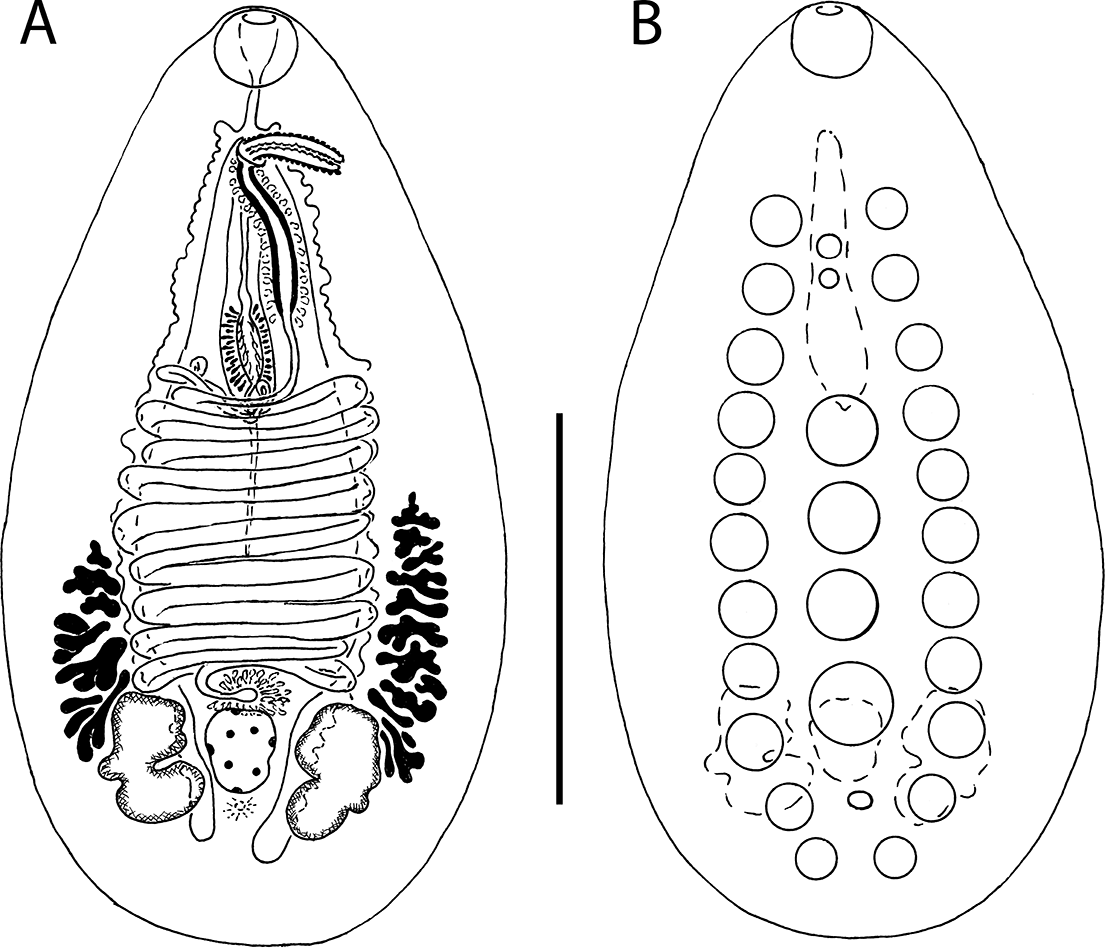
Notocotylus triserialis Diesing, Reference Diesing1839 (Figure 1)
Paragenophore of Notocotylus triserialis ex Anser albifrons, Russian Karelia; ventral view. (А) general morphology and internal anatomy; (B) ventral papillae. Scale bars: A, B = 1000 μm.

Description (based on one hologenophore and seven paragenophores). Body elongate-oval, tapering anteriorly, broadly rounded posteriorly, concave ventrally; length 4045–4466 (4264), maximal width 1125–1476 (1294) in posterior third of body. Tegumental spines not observed (as probable consequence of freezing). Ventral side with 13–16 (15) papillae in right-hand longitudinal row, 13–15 (14) papillae in median longitudinal row, and 14–17 (15) papillae in left-hand longitudinal row. Anterior right-hand papillae 788–898 (847) from anterior body extremity; this distance representing 18.7–22.2% (19.9%) of body length. Posterior right-hand papillae 142–291 (207) from posterior body extremity; this distance representing 3.3–4.8% (4.3%) of body length. Anterior median papilla 0.5–1.5 intervals anterior to anterior lateral papilla, 698–752 (731) from anterior body extremity; this distance representing 16.5–17.7% (17.2%) of body length. Posterior median papillae 173–365 (285) from posterior body extremity; this distance representing 4.1–8.2% (6.5%) of body length. Anterior left-hand papilla 787–898 (851) from anterior body extremity; this distance representing 18.6–22.2% (20.0%) of body length. Posterior left-hand papilla 134–243 (197) from posterior body extremity; this distance representing 3.7–5.4% (4.8%) of body length. Oral sucker subrounded, 199–217 (210) × 196–247 (222), mouth slightly or distinctly ventro-subterminal. Oesophagus length 142–194 (167). Caeca slightly diverticulated, terminate medianly near posterior body extremity. Testes two, lobed, opposite, extracaecal, in posterior quarter of body; right 621–893 (757) × 338–448 (402), left 694–871 (791) × 345–487 (423). Vas deferens forms convoluted external vesicle. Cirrus sac elongate, 1128–1429 (1269) × 198–355 (244), representing 27.5–30.7% (28.8%) of body length, containing curved seminal vesicle, pars prostatica surrounded by field of prostatic cells, ejaculatory duct, and long unarmed cirrus. Distance from anterior body extremity to proximal edge of cirrus sac as proportion of body length 37.2–41.3% (39.1%). Common genital atrium shallow. Genital pore median, immediately postbifurcal, 419–475 (448) from anterior extremity. Ovary entire to indented, 309–385 (339) × 142–310 (261), intercaecal, intertesticular. Mehlis’ gland median, just anterior to ovary. Uterus transversely coiled, preovarian. Metraterm length 302–460 (359), representing 21.7–39.2% (28.2%) of cirrus sac length. Eggs oval, operculate, with two polar filaments; length without filaments 16–20 (18) × 10–13 (11), length of filaments 248–505 (387). Vitellarium follicular; follicle irregular in shape, in two lateral, extracaecal, symmetrical or slightly asymmetrical fields, extending from about midlevel of body to posterior quarter of body, distinctly posterior to distal uterine coils, slightly intruding into testes area or not. Excretory vesicle indistinguishable; pore dorsal, in midlevel of postovarian space.
Taxonomic summary
Host in this study: Greater White-fronted Goose.
Locality in this study: south-eastern coast of Lake Ladoga, Karelia, Russia.
Site of infection: caeca.
Prevalence and intensity of infection: in a single examined host specimen; 8 worms per host specimen.
Accession sequences in GenBank: PV658391 (28S rRNA gene, 1325 bp); PV658397 (ITS2 region, 476 bp); PV659469 (cox1 gene, 391 bp).
Specimens deposited: IEE RAS 14379.
Remarks
Notocotylus triserialis was originally described on specimens from European anatid birds (Diesing Reference Diesing1839). To date, this species has been found (as N. triserialis or N. attenuatus auct.) in an extensive range of birds of various orders as well as in rodents, and on different continents (e.g., Aguilar-Morales et al. Reference Aguilar-Morales, Sánchez-Nava, Aguilar-Ortigoza and Rodríguez-Romero2022; Amundson et al. Reference Amundson, Traub, Smith-Herron and Flint2016; Dubois Reference Dubois1951; Filimonova Reference Filimonova1982, Reference Filimonova1985; Hanson and Gilford Reference Hanson and Gilford1961; Lumsden and Zischke Reference Lumsden and Zischke1963; Nicoll Reference Nicoll1914; Oyarzún-Ruiz et al. Reference Oyarzún-Ruiz, Muñoz, Paredes, Valenzuela and Ruiz2019; Papazahariadou et al. Reference Papazahariadou, Diakou, Papadopoulos, Georgopoulou, Komnenou and Antoniadou-Sotiriadou2008; L. Szidat and U. Szidat Reference Szidat and Szidat1933). Notocotylus triserialis has a multitude of geographical and individual variations, many of which have a complex taxonomic history (e.g., Filimonova Reference Filimonova1985).
The number of ventral papillae is one of the key features in the species diagnosis of Notocotylus spp. and Pseudonotocotylus spp. (e.g., Harwood Reference Harwood1939; Filimonova Reference Filimonova1982, Reference Filimonova1985). Notocotylus triserialis is characterised by 13–18 ventral papillae in lateral rows and 13–17 papillae in the median row (Filimonova Reference Filimonova1985). However, two other notocotylid species parasitising European anatid birds also have a similar number of papillae and are therefore superficially similar to N. triserialis, viz. P. atlanticus (Stunkard, 1966) and P. imbricatus (Looss, 1893).
Notocotylus triserialis differs from P. atlanticus in the length of egg filaments (Gonchar et al. Reference Gonchar, Jouet, Skírnisson, Krupenko and Galactionov2019). According to most authors, the length of egg filaments in N. triserialis exceeds 175 μm (Table 1). Eom et al. (Reference Eom, Rim and Jang1984) indicated 50–60 μm as the length of egg filaments but the reliability of these figures is questionable. The length of egg filaments in P. atlanticus is ≤151 μm (Gonchar et al. Reference Gonchar, Jouet, Skírnisson, Krupenko and Galactionov2019). Gonchar et al. (Reference Gonchar, Jouet, Skírnisson, Krupenko and Galactionov2019) also suggest the metraterm to cirrus sac ratio as a feature for differentiating between these species. In reality, however, the ranges for this feature in N. triserialis and P. atlanticus overlap significantly: metraterm length is 29–80% of cirrus sac length in the former and 33–51% in the latter species (Filimonova Reference Filimonova1985; Gonchar et al. Reference Gonchar, Jouet, Skírnisson, Krupenko and Galactionov2019). In addition, N. triserialis differs from P. atlanticus in the position of the anterior median papilla. In N. triserialis, this papilla is located 0.5–2.5 intervals anterior to the anterior lateral papilla (Filimonova Reference Filimonova1985). In P. atlanticus, the anterior median papilla is usually located one-half interval posterior to the anterior lateral papilla, but sometimes one-half interval anterior to it (Gonchar et al. Reference Gonchar, Jouet, Skírnisson, Krupenko and Galactionov2019).
Notocotylus triserialis differs from P. imbricatus in the position of the anterior median papilla. In last species, this papilla is normally located 0.5 interval posterior to the anterior lateral papilla and very rarely 0.5 interval anterior to it (Filimonova Reference Filimonova1985).
The digenean specimens presented in our work are characterised by a posbifurcal position of the common genital pore, 13–17 ventral papillae in lateral rows, 13–15 ventral median papillae, the anterior median papilla located 0.5–1.5 intervals anterior to the anterior lateral papilla, and vitelline fields distinctly posterior to distal uterine coils. These specimens match the description of N. triserialis in all the listed characteristics, as well as in morphometric data (Filimonova Reference Filimonova1982, Reference Filimonova1985; Pike Reference Pike1969; L. Szidat and U. Szidat Reference Szidat and Szidat1933; Table 2). We would like to especially emphasise that the anterior median papilla in the specimen we used as the hologenophore is clearly located one interval anterior to the anterior lateral papilla, which adds to the reliability of its identification to species.
Comparison of Notocotylus triserialis isolates based on some metric traits (in μm)

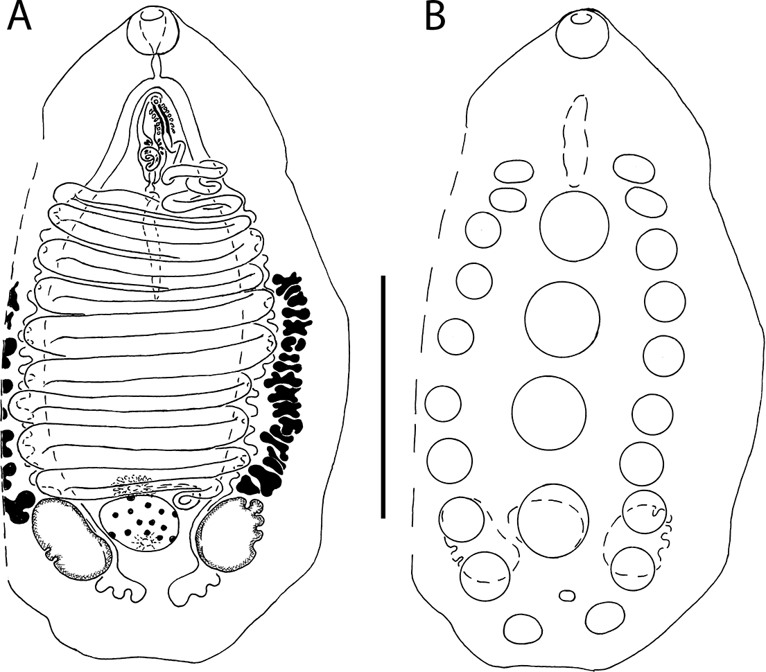
Notocotylus pacifera (Noble, Reference Noble1933) Harwood, Reference Harwood1939 (Figures 2 and 3)
Paragenophore of Notocotylus pacifera ex Fulica atra, Rostov Region, Russia; ventral view. (А) general morphology and internal anatomy; (B) ventral papillae. Scale bars: A, B = 1000 μm.

SEM photograph of paragenophores of Notocotylus pacifera ex Fulica atra, Rostov Region, Russia. (A) ventral view, showing ventral papillae (arrows); (B) everted cirrus with numerous knobs; (C) egg; (D) tegumental spines and detail of egg capsules and polar proximal part of polar filaments. Scale bars: A = 500 μm; B, C = 20 μm; D = 10 μm.

Description (based on three hologenophores and 16 paragenophores). Body ovoid or oval, tapering anteriorly, broadly rounded posteriorly, concave ventrally or not; length 1913–2760 (2290), maximal width 768–1401 (1126) in posterior third of body. Tegument body with small spines only visible under SEM. Ventral side with 10–11 (11) papillae in right-hand longitudinal row, usually 4–6 (5) papillae, occasionally 6 or 7, in median longitudinal row, and 10–11 (11) papillae in left-hand longitudinal row. Anterior right-hand papilla 370–677 (475) from anterior body extremity; this distance representing 18.6–24.5% (20.9%) of body length. Posterior right-hand papilla 95–245 (141) from posterior body extremity; this distance representing 4.0–10.7% (6.4%) of body length. Anterior median papilla 0.5–2.5 intervals posterior to anterior lateral papilla, 504–875 (719) from anterior body extremity; this distance representing 21.5–38.8% (31.3%) of body length. Posterior median papilla 215–532 (328) from posterior body extremity; this distance representing 7.8–21.8% (14.7%) of body length. Anterior left-hand papilla 331–556 (454) from anterior body extremity; this distance representing 15.2–21.9% (19.6%) of body length. Posterior left-hand papilla 76–223 (141) from posterior body extremity; this distance representing 3.5–9.7% (6.4%) of body length. Oral sucker subrounded, 152–216 (178) × 164–235 (188), mouth terminal. Oesophagus length 42–147 (103). Caeca conspicuously diverticulated, terminate medianly near posterior body extremity. Testes two, indented to lobed, opposite, extracaecal, in posterior quarter of body; right 252–440 (338) × 149–294 (231), left 278–469 (352) × 179–343 (237). Vas deferens forms convoluted external vesicle. Cirrus sac elongate, 529–807 (614) × 104–166 (132), representing 21.7–30.7% (27.3%) of body length, containing curved seminal vesicle, pars prostatica surrounded by field of prostatic cells, ejaculatory duct, and long cirrus. Cirrus armed with numerous knobs. Distance from anterior body extremity to proximal edge of cirrus sac as proportion of body length 36.9–45.8% (42.2%). Common genital atrium shallow. Genital pore median, immediately postbifurcal, 238–389 (345) from anterior extremity. Ovary entire to indented, 160–284 (214) × 148–246 (205), intercaecal, intertesticular. Mehlis’ gland median, just anterior to ovary. Uterus transversely coiled, preovarian. Metraterm length 307–517 (416), representing 53.2–80.1% (67.8%) of cirrus sac length. Eggs oval, operculate, with two polar filaments; length without filaments 16–22 (19) × 10–13 (11), length of filaments. 185–365 (267). Vitellarium follicular; follicles irregular in shape, in two lateral, extracaecal, symmetrical or slightly asymmetrical fields extending from level approximately 2/5 of body length or about midlevel of body to posterior quarter of body, distinctly posterior to distal uterine coils, slightly or deeply intruding into testes area. Excretory vesicle indistinguishable; pore dorsal, in anterior half of postovarian space.
Taxonomic summary
Host in this study: Eurasian Coot.
Locality in this study: Volga River delta near Poldnevoe Village, Astrakhan Region, Russia; River Mokraya Chuburka near Leninsky Leskhoz Village, Rostov Region, Russia; Lake Maloe Krugloe near Khasan Village, Primorsky Krai, Russia; Lake Naroch near Naroch Village, Minsk Region, Belarus.
Site of infection: caeca.
Prevalence and intensity of infection: Astrakhan Region – 3 of 7 host individuals infected; 2–7 worms per host individual; Rostov Region – 73.3% (n = 15); 1–15 worms per host individual; Primorsky Krai – 13.3% (n = 15); 1–2 worms per host individual; Belarus – 73.3% (n = 15); 2–25 worms per host individual.
Accessions sequences in GenBank: PV658390 (28S rRNA gene, 985 bp); PV657184 (18S rRNA gene, 1741 bp); PV658392–PV658395 (ITS2 region, 462–480 bp); PV659464–PV659467 (cox1 gene, 386–438 bp).
Specimens deposited: IEE RAS 14380, 14381.
Remarks
Notocotylus pacifera was originally described on specimens from the American Coot Fulica americana Gmelin, 1789, taken in the USA (Noble Reference Noble1933). Noble (Reference Noble1933) placed this species in the genus Catatropis Odhner, 1905. Szidat (Reference Szidat1936) moved it to the genus Kossackia U. Szidat, Reference Szidat1936 with the type species Monostomum gibbum Mehlis in Creplin, 1846 (under the synonym Notocotylus gibbus), and renamed C. pacifera as Kossackia pacifera. However, Hardwood (1939) did not recognise this genus and moved K. pacifera to the genus Notocotylus. For many years, Palaearctic N. pacifera isolates have often been confused with another species parasitising rallid and anatid birds, N. gibbus (e.g., Dubois Reference Dubois1951; L. Szidat and U. Szidat 1933, Reference Szidat and Szidat1961). Odening (Reference Odening1961) and Filimonova (Reference Filimonova1982, Reference Filimonova1985) clarified the morphological differences between these two digenean species. To date, N. pacifera has been found in a variety of rallid birds, including the Eurasian Coot, and at various locations in the Holarctic and even in the Neotropics (e.g., Aguilar-Morales et al. Reference Aguilar-Morales, Sánchez-Nava, Aguilar-Ortigoza and Rodríguez-Romero2022; Canaris and Waldmann Reference Canaris and Waldmann2017; Filimonova Reference Filimonova1982; Guillén and Morales 2023; Hüğül and Güçlü Reference Hüğül and Güçlü2020; Kulišić et al. Reference Kulišić, Lepojev, Aleksić-Bakrač, Jakić, Pavlović, Milutinović and Mišić2004; Larios Reference Larios1943; Lumsden and Zischke Reference Lumsden and Zischke1963; Macko Reference Macko1956; Odening Reference Odening1961; Yamaguti Reference Yamaguti1939).
Notocotylus pacifera is characterized by the immediately postbifurcal position of the common genital pore, 10–12 ventral papillae in lateral rows and normally 4–5 papillae in the median row, the position of the anterior papilla in the median row distinctly posterior to anterior papillae of both lateral rows, the position of vitelline fields distinctly posterior to distal uterine coils, slightly or deeply intruding into testes area. However, the same set of morphological characters is characteristic of two more nominal species of the genus Notocotylus, namely Notocotylus porzanae Harwood, Reference Harwood1939, and Notocotylus ratti Ye, Qiu, Wen, Li and Li, 1956 (Filimonova Reference Filimonova1982, Reference Filimonova1985; Harwood Reference Harwood1939; Ye et al. Reference Ye, Qiu, Wen, S-H and Li1956). Notocotylus porzanae was originally described on specimens found in the Carolina Crake Porzana carolina (Linnaeus, 1758) from the USA (Harwood Reference Harwood1939). Filimonova (Reference Filimonova1985) called N. porzanae a junior synonym of N. pacifera. Notocotylus ratti was originally described on specimens from the black rat, Rattus rattus alexandrines (Geoffroy, 1803), collected in China (Ye et al. Reference Ye, Qiu, Wen, S-H and Li1956). The only real differences between N. pacifera and N. ratti are the host groups (birds vs. mammals) and the ovary shape (entire or indented vs. conspicuously lobate). The position of the common genital pore, the number of ventral papillae in the lateral and median rows, the position of vitelline fields, the ovary shape, and morphometric data prompt us to affiliate our specimens with N. pacifera (cf. Filimonova Reference Filimonova1982, Reference Filimonova1985; Noble Reference Noble1933; Odening Reference Odening1961; Table 3).
Comparison of Notocotylus pacifera isolates based on some metric traits (in μm)

Notocotylus sp. (Figure 4)
Hologenophore of Notocotylus sp. ex Fulica atra, Rostov Region, Russia; ventral view. (А) general morphology and internal anatomy; (B) ventral papillae. Scale bars: A, B = 1000 μm.

Description (based on one hologenophore). Body broadly oval, conical anteriorly, and almost trapezoid posteriorly, flattened dorso-ventrally; length 2857, maximal width 1488 (excluding cut-off body part). Tegumental spines not observed. Ventral side with 10 papillae in right-hand longitudinal row, 5 papillae in median longitudinal row, and 10 papillae in left-hand longitudinal row. Anterior right-hand papilla 618 from anterior body extremity; this distance representing 21.6% of body length. Posterior right-hand papilla 112 from posterior body extremity; this distance representing 3.9% of body length. Anterior median papilla located 1.5 interval posterior to anterior lateral papilla, 759 from anterior body extremity; this distance representing 26.6% of body length. Posterior median papillae 299 from posterior body extremity; this distance representing 10.5% of body length. Anterior left-hand papilla 589 from anterior body extremity; this distance representing 20.6% of body length. Posterior left-hand papilla 161 from posterior body extremity; this distance representing 5.6% of body length. Oral sucker subrounded, 199 × 224, mouth terminal. Oesophagus length 94. Caeca conspicuously diverticulated, terminate medianly near posterior body extremity. Testes two, slightly indented, opposite, extracaecal, in posterior quarter of body; right 315 × 265, left 351 × 323. Vas deferens forms convoluted external vesicle. Cirrus sac elongate, 304 × 102, representing 10.6% of body length, containing curved seminal vesicle, pars prostatica surrounded by field of prostatic cells, ejaculatory duct, and long cirrus. Cirrus probably unarmed, spines or knobs not observed. Distance from anterior body extremity to proximal edge of cirrus sac as proportion of body length 23.7%. Common genital atrium shallow. Genital pore median, immediately postbifurcal, 372 from anterior extremity. Ovary entire, 233 × 324, intercaecal, intertesticular. Mehlis’ gland median, just anterior to ovary. Uterus transversely coiled, preovarian. Metraterm length 195, representing 64.1% of cirrus sac length. Eggs oval, operculate, with two polar filaments; length without filaments 23 × 16, full length of filaments not visible. Vitellarium follicular; follicles irregular in shape, in two lateral, extracaecal, symmetrical fields, extending from level approximately 2/5 of body length to posterior quarter of body, distinctly posterior to distal uterine coils, slightly intruding into testes area on right side and not on left side. Excretory vesicle indistinguishable; pore dorsal, at level of ovary.
Taxonomic summary
Host in this study: Eurasian Coot.
Locality in this study: River Mokraya Chuburka near Leninsky Leskhoz Village, Rostov Region, Russia.
Site of infection: caeca.
Prevalence and intensity of infection: 6.7% (n = 15); 1 worm per host individual.
Accession sequences in GenBank: PV658396 (ITS2 region, 496 bp); PV659468 (cox1 gene, 356 bp).
Specimens deposited: IEE RAS 14382
Remarks
The Notocotylus sp. presented here is very similar to N. pacifera (=N. porzanae) and Notocotylus ratti Ye, Qiu, Wen, Li and Li, 1956 in the number of ventral papillae and the position of the median papilla posterior to the anterior lateral papillae. However, Notocotylus sp. differs from these digeneans in cirrus sac length (10.6% vs. >20% of body length) and distance from anterior body extremity to proximal edge of the cirrus sac (23.7% vs. 25–46% of body length) (cf. Bychovskaya-Pavlovskaya Reference Bychovskaya-Pavlovskaya1974; Filimonova Reference Filimonova1982, Reference Filimonova1985; Harwood Reference Harwood1939; Noble Reference Noble1933; Odening Reference Odening1961; Ye et al. Reference Ye, Qiu, Wen, S-H and Li1956; data reported here). Furthermore, the newly collected Notocotylus sp. differs from N. ratti in the ovary shape: entire vs. conspicuously lobate (cf. Ye et al. Reference Ye, Qiu, Wen, S-H and Li1956).
Phylogeny
We obtained partial sequences of the cox1 gene and complete sequences of the ITS2 region from one specimen of N. triserialis (hologenophore), four specimens of N. pacifera (one hologenophore from each of Primorsky Krai, Rostov Region, and Belarus, and a paragenophore [studied only molecularly] from the Astrakhan Region), and one specimen of Notocotylus sp. (hologenophore). The p-distances between the cox1 gene sequences in different N. pacifera specimens were 0–1.14%, and the range of p-distances between ITS2 region sequences was 0–0.22% (Figure 5, 6). The sequences of both loci in Notocotylus sp. differed from those of N. pacifera (p-distances of 7.12–7.69% and 1.04–1.30%, respectively), despite the morphological similarity of the two species (Figures 5 and 6). We also obtained partial sequences of the 28S rRNA gene from one specimen of N. triserialis and one specimen of N. pacifera (Rostov Region). All attempts to amplify this gene in Notocotylus sp. were unsuccessful. In addition, a complete sequence of the 18S rRNA gene has been obtained from one specimen of N. pacifera collected in the Astrakhan Region.
Heatmap based on p-distances between newly obtained cox1 gene sequences of Notocotylus pacifera and Notocotylus sp. Values are shown in corresponding cells: lower diagonal – p-distance values, %; upper diagonal – standard error, %. Pairwise distances and standard error are encoded into colour variations. Abbreviations: A, Astrakhan Region of Russia; B, Belarus; P, Primorsky Krai of Russia; R, Rostov Region of Russia.

Heatmap based on p-distances between newly obtained ITS2 region sequences of Notocotylus pacifera and Notocotylus sp. Values are shown in corresponding cells: lower diagonal – p-distance values, %; upper diagonal – standard error, %. Pairwise distances and standard error are encoded into colour variations. Abbreviations: A, Astrakhan Region of Russia; B, Belarus; P, Primorsky Krai of Russia; R, Rostov Region of Russia.

In the phylogenetic reconstruction based on the partial sequences of the 28S rRNA gene, N. triserialis formed the poorly supported clade together with N. malhamensis, N. ikutai, N. multipapillus, and several unidentified Notocotylus spp. (Figure 7). This clade appeared as a poorly supported sister to the Notocotylus pacifera + Pseudocatatropis dvoryadkini Izrailskaia, Besprozvannykh, Tatonova, Nguyen and Ngo, Reference Izrailskaia, Besprozvannykh, Tatonova, Nguyen and Ngo2019, clade. The N. pacifera + P. dvoryadkini clade also had poorly support. In turn, the clade containing all the above-mentioned specimens was a strongly supported sister to the equally strongly supported clade formed by Quinqueserialis spp., N. ephemera, Pseudocatatropis sp., and some representatives of the genus Notocotylus not identified to the species level. The digenean specimen available in GenBank under the name N. attenuatus (AF184259) did not cluster together with the newly collected N. triserialis but was nested among members of the genus Pseudonotocotylus. The Pseudonotocotylus spp. clade had no direct phylogenetic relationship with the large clade of Notocotylus, Pseudocatatropis, and Quinqueserialis members described above.
Phylogenetic relationships of Notocotylus triserialis and Notocotylus pacifera based on Bayesian inference analysis of partial 28S rRNA gene sequences. Posterior probability values below 0.9 are not shown. The newly obtained sequences are highlighted in bold. Abbreviations: K, Russian Karelia; R, Rostov Region of Russia.

In the tree based on the concatenated sequences of the cox1 gene and ITS2 region, N. triserialis formed a poorly supported clade together with N. malhamensis and N. pacifera (Figure 8). This clade was a poorly supported sister to a poorly supported clade consisting of two groups. The first of these groups had moderate support and comprises N. ephemera, N. ikutai, and N. multipapillus. The second group has poor support and was represented by P. dvoryadkini and Pseudocatatropis sp. The clade containing all the mentioned species had a poorly supported sister relationship with the newly collected Notocotylus sp.
Phylogenetic relationships of Notocotylus triserialis, Notocotylus pacifera, and Notocotylus sp. based on Bayesian inference analysis of concatenated cox1 + ITS2 gene sequences. Posterior probability values below 0.9 are not shown. The newly obtained sequences are highlighted in bold. Abbreviations: A, Astrakhan Region of Russia; B, Belarus; K, Russian Karelia; P, Primorsky Krai of Russia; R, Rostov Region of Russia.

Since data on the 18S rRNA gene of notocotylids available in GenBank are scarce, our analysis of the newly obtained sequence of this marker was limited to calculating the pairwise distance with the only one available, deposited under the name N. pacifera (AY245765). The compared sequences have a fairly high degree of difference for this marker (p-distance 1.1%).
Discussion
In the current study we identified three members of the genus Notocotylus: N. triserialis, N. pacifera, and Notocotylus sp. Our data support the findings of Izrailskaia et al. (Reference Izrailskaia, Tatonova and Belousova2024) on the misidentification of the digenean deposited in GenBank as N. triserialis (under the synonym N. attenuatus auct.; AF184259). In recent reconstructions, this digenean has been shown to be nested among the notocotylids which develop in prosobranch snails during parthenogenetic stages (Izrailskaia et al. Reference Izrailskaia, Tatonova and Belousova2024; data reported here). At the same time, it is well known that the first intermediate host for the true N. triserialis is pulmonate snail (e.g., L. Szidat and U. Szidat Reference Szidat and Szidat1933; Skála et al. Reference Skála, Bulantová, Walker and Horák2014). The newly collected Notocotylus sp. is probably a new species. This conjecture is supported by an interspecific level of the p-distance (cf. Vilas et al. Reference Vilas, Criscione and Blouin2005; Chan et al. Reference Chan, Chaisiri, Saralamba, Morand and Thaenkham2021, Reference Chan, Thaenkham, Wichaita and Saralamba2025) from N. pacifera, to which Notocotylus sp. is similar in both morphology and the host. In addition, despite the general morphological similarity of these two parasites, they still differ from each other in the cirrus sac length.
Our phylogenetic analysis based on 28S rRNA gene sequences does not support the monophyly of the genus Notocotylus within the taxonomic boundaries recognised by Izrailskaia et al. (Reference Izrailskaia, Tatonova and Belousova2024). We show that N. pacifera is more closely related to P. dvoryadkini than to the genus Notocotylus group formed by N. triserialis, N. ikutai, N. malhamensis, N multipapillus, and several other representatives of this genus not identified to species. However, the clade of N. pacifera and P. dvoryadkini does not have reliable support. Notocotylus ephemera is more closely related to Quinqueserialis spp. than all the above-mentioned members of the genus Notocotylus. However, the identification of the digenean appearing on GenBank under the name N. ephemera is not reliable because it is based on the larval stage (cercariae). The tree generated using concatenated sequences of mitochondrial and nuclear DNA markers did not resolve the phylogenetic position of N. pacifera, N. triserialis, and the newly collected Notocotylus sp. with reliable support. Thus, the genus affiliation of N. pacifera and the newly collected Notocotylus sp. needs further clarification.
Wide geographic ranges are characteristic of many notocotylids (e.g., Filimonova Reference Filimonova1985). Molecular data presented by Gonchar et al. (Reference Gonchar, Jouet, Skírnisson, Krupenko and Galactionov2019) confirm the broad range for one such species, P. atlantucus. However, for most other widely distributed notocotylids, including N. triserialis and N. pacifera, it remains unclear whether they represent a single widespread species or an array of morphologically poorly distinguishable endemic species. Our molecular and morphological data prove the conspecificity of different N. pacifera isolates from Eastern Europe (Belarus and Astrakhan and Rostov Regions of Russia) and the Russian Far East and thus confirm the wide distribution of this species in Eurasia. An 18S rRNA gene sequence obtained from a specimen presumably collected in the USA and originally identified as N. pacifera is available in GenBank (AY245765), but without specifying the host and the precise locality. The P-distances between this sequence and those of our N. pacifera isolates are quite high, which casts considerable doubt on the possible conspecificity of the digeneans from which they were obtained. These findings indicate an urgent need for a molecular comparison of Eurasian isolates of N. pacifera with reliably identified American isolates of this species.
Supplementary material
The supplementary material for this article can be found at http://doi.org/10.1017/S0022149X25100849.
Acknowledgements
The study was conducted using equipment of the Core Facility of the Karelian Research Centre RAS (Petrozavodsk, Russia) and the Instrumental Methods in Ecology Core Facility at IEE RAS (Moscow, Russia). We are also grateful to Olga Kislova for language editing.
Financial support
The study was funded by the Russian Academy of Sciences, project no. FMEN-2022-0005 (Yakovleva GA), and FFER-2024-0027 (Sokolov SG, Vlasenkov SA).
Competing interests
The authors declare that they have no known competing financial interests or personal relationships that could have influenced the work reported in this paper.
Ethical standard
The authors assert that all procedures contributing to this work comply with the ethical standards of the relevant national and institutional guidelines on the care and use of laboratory animals.