


Highlights
• This is the first report of TDP-43 pathology in ARSACS, a neurodegenerative disease not classically associated with TDP-43 proteinopathy.
• This finding raises the hypothesis that shared molecular mechanisms may exist between ARSACS and other TDP-43-driven diseases.
• Linking ARSACS to the TDP-43 proteinopathy spectrum may open new research and therapeutic avenues.
Introduction
Autosomal recessive spastic ataxia of Charlevoix–Saguenay (ARSACS) is a rare, early-onset neurodegenerative disorder characterized by progressive cerebellar ataxia, spasticity and peripheral neuropathy. Reference Salem, Blais, Zuluaga-Sánchez, Rouleau, Becker and Dupré1 First described in 1978 in Quebec, Canada, it has since been identified worldwide, including the USA, India, Turkey, Japan and across Europe. The incidence in the Quebec founder population is estimated at 1 in 1932 live births, though the global prevalence is still unknown. Reference Bagaria, Bagyinszky and An2
Clinical onset typically manifests in early childhood, with delayed walking and gait instability between 12 and 18 months in Quebec patients, while appearing later (at about 5 years old) in non-Quebec cases. Reference Salem, Blais, Zuluaga-Sánchez, Rouleau, Becker and Dupré1,Reference Takiyama3 ARSACS features significant motor system involvement, including lower-limb stiffness, weakness and hyperreflexia due to pyramidal tract degeneration. Reference Salem, Blais, Zuluaga-Sánchez, Rouleau, Becker and Dupré1 Combined with cerebellar and peripheral nerve damage, these changes create a complex motor phenotype. Widespread neurodegeneration affects the cerebellum, brainstem, spinal cord and peripheral nerves, severely impairing mobility and independence over time. Cerebellar ataxia usually appears first, followed by lower-limb spasticity and later by peripheral neuropathy. The latter contributes to muscle wasting, high-arched feet and loss of ankle reflexes. Reference Salem, Blais, Zuluaga-Sánchez, Rouleau, Becker and Dupré1 Additional signs may include upper-limb incoordination, dysarthria, nystagmus, cognitive impairment, hearing loss and bladder dysfunction.
ARSACS is caused by biallelic mutations in the SACS gene on chromosome 13q12, which encodes sacsin, a large protein involved in neurofilament (NF) organization and mitochondrial function. Reference Khaled, Mohamed Taha, Mara, Sadhna and Mohan4,Reference Perna, Salsbury and Dushti5 A well-characterized founder mutation in the SACS gene (c.8844delT) accounts for the majority of ARSACS cases in Quebec, where patients typically present with a relatively homogeneous and early-onset clinical phenotype. However, ARSACS is now recognized as a panethnic morphotype neurodegenerative disorder, with numerous mutations identified in diverse populations worldwide. These non-Quebec variants are often associated with broader clinical heterogeneity, including atypical presentations and later disease onset, reflecting the extensive allelic variability observed across different populations. Reference Takiyama3 Neural cell metabolism dysfunctions caused by loss of sacsin have been identified as a key pathological mechanism in ARSACS. Reference Perna, Salsbury and Dushti5,Reference Galatolo, Rocchiccioli and Di Giorgi6 These dysfunctions include alterations in lysosomal transport and microtubule dynamics, which are mediated by sacsin. Reference Francis, Alshafie, Kumar, Girard, Brais and McPherson7 In sacsin-deficient neurons, intermediateNF assembly and dynamics are impaired as well, leading to the formation of overabundant NF bundles near dendrites, disrupting neuronal functions and survival. Reference Gentil, Lai and Menade8,Reference Larivière, Gaudet and Gentil9 Recent studies have also suggested that autophagy pathways may be impaired, with increased expression of markers such as p62/SQSTM1, pointing to defects in neuronal protein degradation. Reference Bitto, Lerner, Nacarelli, Crowe, Torres and Sell10
Diagnosis combines clinical, imaging, electrophysiological and genetic findings. Brain MRI typically reveals cerebellar vermis atrophy, Reference Martin, Bouchard, Sylvain, St-Onge and Truchon11 pontine linear T2 hypointensities and thalamic hyperintensities. Reference Salem, Blais, Zuluaga-Sánchez, Rouleau, Becker and Dupré1 Optical coherence tomography often shows thickened retinal nerve fiber layers. A mixed axonal and demyelinating polyneuropathy is observed on nerve conduction studies. Definitive diagnosis requires the identification of pathogenic SACS variants through molecular testing. Loss of motor neurons, atrophy in the superior and anterior cerebellar vermis associated with Purkinje cell death, cortical atrophy and demyelinating sensory-motor neuropathy were reported on histological analysis as well. Reference Salem, Blais, Zuluaga-Sánchez, Rouleau, Becker and Dupré1
Case history
The patient was a male in his early forties and exhibited a typical clinical phenotype of ARSACS, with disease onset in early childhood characterized by delayed motor milestones, progressive gait ataxia and spasticity. Over time, his condition evolved to include distal muscle wasting, cerebellar ataxia, dysarthria and brisk tendon reflexes, without clinical evidence of lower motor neuron involvement. The ARSACS diagnosis was genetically confirmed by genotyping. The patient exhibited the homozygous c.8844delT Quebec founder mutation in the SACS gene. Electrophysiological data acquired during childhood revealed severe, predominantly demyelinating sensory and motor neuropathy. Follow-up evaluations conducted later in adulthood confirmed the presence of severe sensory and motor neuropathy, with marked involvement in both the ulnar and median nerves. Sensory nerve responses were absent in the ulnar, median and sural nerves, consistent with the characteristic peripheral nerve involvement observed in ARSACS. Despite stable supportive care and multidisciplinary follow-up, the patient experienced severe psychological distress related to his progressive physical decline and loss of autonomy, leading to repeated suicide attempts. These occurred despite ongoing psychiatric and psychological support. In his early forties, after a careful evaluation and in accordance with provincial laws and ethical guidelines, the patient elected to undergo medical assistance in dying (MAiD) due to intractable psychological suffering associated with the loss of perceived quality of life and dignity. There were no atypical clinical features, and his disease course remained consistent with classical ARSACS until the time of death.
To validate phosphorylated TAR DNA-binding protein 43 (pTDP-43) findings, cerebellar tissue from a second genetically confirmed ARSACS patient was analyzed. This patient was a woman in her early sixties with a clinical presentation consistent with ARSACS who also underwent MAiD, allowing access to well-preserved postmortem tissue.
Methods
Postmortem brain biopsies and ethics
This study received approval from the national ethical review boards in Canada (Ethical Research Board of the CHU de Québec, protocol number: 2012-1286 [PEJ-235], contact: gurecherche@chuq.qc.ca). Informed consent for tissue donation, including the brain and spinal cord, was obtained from the patient prior to participation in the MAiD program. In accordance with the patient’s expressed wishes, brain tissue was collected postmortem through an organ donation procedure rather than a conventional autopsy. For comparative and validation purposes, postmortem tissue from a sporadic amyotrophic lateral sclerosis (ALS) patient and cerebellar tissue from a second genetically confirmed ARSACS patient were obtained through the same MAiD-associated donation process, ensuring comparable tissue collection conditions. Upon reception, one hemisphere from the index ARSACS and the ALS cases was snap-frozen and stored at –80°C for future molecular and biochemical analyses. The contralateral hemisphere was partially sectioned, and selected brain regions were immersion-fixed in 4% paraformaldehyde (Electron Microscopy Sciences, Hatfield, PA, USA), paraffin-embedded and sectioned at 5 µm thickness for histological analysis. For the second ARSACS case, only fixed cerebellar tissue was available, precluding biochemical analyses but allowing independent histopathological validation of the pTDP-43 findings.
Immunohistochemistry/immunofluorescence
Immunohistochemistry (IHC) was conducted on 5 µm cross-sections of the prefrontal and motor cortex, the cerebellum and the spinal cord of the patient’s brain with antibodies against pTDP-43 Ser409/410 (66318-1-Ig; Proteintech, Rosemont, IL, USA), p62 (610833; BD Biosciences, Heidelburg, Germany) and neurofilament light (NF-L) (GA607, Dako). All IHC staining procedures were carried out by the pathology department staff at the Québec University Hospital.
For immunofluorescence (IF), paraffin was removed in a 100% toluene bath for 5 minutes, followed by a 100% ethanol bath for 5 minutes. These tissues underwent antigenic unmasking in a 10 mM citrate buffer (pH 6.0) at 95°C for 15 minutes and blocked with 5% goat serum (Invitrogen, Waltham, Massachusetts, USA) and 0.3% Triton X-100 (Bio-Rad Laboratories Inc., Hercules, CA, USA) in phosphate-buffered saline (PBS) for 1 hour at room temperature. Specific primary antibodies against native and phosphorylated TDP-43 (1:500; 60019-2; Proteintech, Rosemont, IL, USA) and beta-tubulin III (TUJ-1) (1:500; S3886; Cell Signaling Technology, Danvers, MA, USA) were incubated on the tissues for 16 hours at 4°C. After washing, the tissues were then incubated for 1 hour at room temperature with Alexa Fluor 594-labeled goat anti-mouse immunoglobulin G (IgG) (H+L) (1:500; A11012; Invitrogen) for TAR DNA-binding protein 43 (TDP-43) and Alexa Fluor 488-labeled goat anti-mouse IgG (1:500; A21131; Invitrogen) for TUJ-1. Finally, tissues were incubated with 0.1% Sudan Black B (MilliporeSigma, Burlington, MA, USA; S0593) at room temperature for 30 minutes and mounted with fluoromont-G with 4’,6-diamidino-2-phenylindole (Invitrogen; 00-4959-52). The immunostained tissues were visualized using an LSI 700 confocal microscope with a Zeiss Axio Imager (Carl Zeiss Microscopy, Jena, Germany).
Quantitative assessment of cytoplasmic pTDP-43-positive cells
Quantitative assessment of pTDP-43-positive neurons was performed on brain sections from both the ARSACS patient and a 52-year-old ALS patient. For each case, and in the ALS brain, quantification was performed on a single section per brain region. Immunohistochemical staining for pTDP-43 was conducted using the same protocol and imaging conditions for both cases. For each tissue (prefrontal cortex, motor cortex and cerebellum), 50 randomly selected microscopic fields were acquired at 400× magnification using brightfield microscopy. Two independent observers, blinded to the diagnosis, manually counted neurons displaying cytoplasmic pTDP-43 immunoreactivity, defined by visible redistribution of the protein from the nucleus to the cytoplasm, with or without inclusion-like aggregates. Cells were considered pTDP-43-positive when they exhibited cytoplasmic staining that was clearly more intense than the background. Examples of neurons lacking this abnormal cytoplasmic staining, illustrating typical background levels, are shown in Supplementary Figure S1. The percentage of pTDP-43-positive cells was calculated for each region and compared between the ARSACS and ALS cases. Statistical analyses were performed using one-way Analysis of Variance (ANOVA) followed by Tukey’s post hoc test to assess regional differences between the two patients. Representative images shown in the figures were selected based on staining quality and cellular morphology but were not used for quantification.
Preparation of sarkosyl-insoluble fractions from ARSAC and ALS donors
Sarkosyl-soluble and sarkosyl-insoluble protein fractions were prepared from human postmortem frozen frontal cortex using a sequential detergent extraction protocol adapted from Tamaki et al. (2023). Reference Tamaki, Ross and Alipour12 Briefly, tissue was sonicated (three 5-second pulses, with 10-second intervals between, at 20% amplitude) and incubated in high-salt buffer (10 mM Tris-HCl pH 7.4, 0.5 M NaCl, 2 mM ethylenediaminetetraacetic acid, 10% sucrose, 1 mM dithiothreitol) containing 1% Triton X-100 and protease/phosphatase inhibitors for 30 min at 37°C. Following centrifugation (21,000 g, 30 min, 4°C), the supernatant was collected as the high-salt Triton X-100 (HS-TX) soluble fraction. The pellet was subjected to sucrose flotation for myelin removal, treated with benzonase (500 U/g) to eliminate nucleic acids and subsequently incubated in high-salt buffer containing 2% sarkosyl for 30 min at 37°C. After centrifugation (21,000 g, 30 min, 22°C), the supernatant was collected as the sarkosyl-soluble fraction. The remaining pellet was washed in cold PBS, resuspended, briefly sonicated and designated as the sarkosyl-insoluble fraction.
Western blotting for TDP-43
Protein concentrations for the fractions were determined using the Bradford assay with Quick Start Bradford Dye Reagent (Bio-Rad). Different amounts of proteins (20 µg for HS-TX fractions or 40 µL for the sarkosyl- soluble and sarkosyl-insoluble fractions) were resolved by sodium dodecyl sulfate polyacrylamide gel electrophoresis on 12% polyacrylamide gels and transferred onto polyvinylidene fluoride membranes (Bio-Rad). Gels have been added with 2,2,2-trichloroethanol to allow total protein visualization with Gel DocTM EZ Imager (Bio-Rad). Membranes were blocked in Tris-buffered saline (TBS) containing 5% nonfat dry milk and 0.05% Tween-20 and then incubated overnight at 4°C with primary antibodies against TDP-43 (1:2000; 10782-2-AP; Proteintech). After washing in TBS with 0.05% Tween-20, membranes were incubated for 1 hour at room temperature with horseradish peroxidase-conjugated anti-rabbit secondary antibody (1:5000; Jackson ImmunoResearch). Protein signals were visualized using enhanced chemiluminescence (Amersham ECL, GE Healthcare) and imaged with a Fusion Fx7 system (Vilber Lourmat).
Results
IHC and IF analyses revealed the presence of cytoplasmic dot-like pTDP-43 aggregates, also referred to as punctate or dash-like aggregates of TDP-43 in neurons from various brain regions of the index ARSACS patient (Figure 1 A–C and Figure S2). Across all examined regions, affected neurons displayed these distinctive cytoplasmic inclusions accompanied by reduced or absent nuclear staining, consistent with pathological mislocalization of pTDP-43. The cerebellum, a region prominently affected in ARSACS, also exhibited the expected reduction in Purkinje cell density and abnormal dendritic arborization, together with widespread pTDP-43-positive dot-like aggregates, suggesting that pTDP-43 accumulation may contribute to cerebellar pathology in ARSACS. Although detectable, pTDP-43 immunoreactivity in the spinal cord was limited to a small number of neurons (Figure S4 A–C). No evident enlarged neurons or abnormal p62 staining was detected in the spinal cord (Figure S4 G). To further validate these observations, cerebellum tissue from a second genetically confirmed ARSACS patient, a woman in her early sixties, was stained using the same anti-pTDP-43 antibody. Similar dot-like pTDP-43-positive aggregates were observed in this independent case, supporting the reproducibility of the findings (Figure S3).
Immunohistochemistry (IHC) staining using different antibodies in different brain regions. (A, B, C) IHC staining revealed aggregates of positively labeled phosphorylated TAR DNA-binding protein 43 (pTDP-43) (dot-like inclusions; arrows) in the cytoplasm of neurons in prefrontal cortex, motor cortex and cerebellum from an autosomal recessive spastic ataxia of Charlevoix–Saguenay (ARSACS) patient (D, E, F). Increased neurofilament light (NF-L) expression was detected in the prefrontal cortex, motor cortex and cerebellum in the brain of ARSACS patient. Intense somatodendritic neurofilament accumulations were also detected, with abnormal clear vacuole-like structures within neurons (arrow heads). Some neurons were also found to be enlarged (*). (G, H, I) Positively labeled p62 in neural cells in the brain of an ARSACS patient in the prefrontal cortex, motor cortex and cerebellum. Cerebellar cells featured in (C), (F) and (I) are, respectively, Purkinje neuron, neurons from the dentate nucleus and neurons from the molecular layer. Magnification: 200× (G, H, I) and 400× (A–F). Scale bar = 20 µm.
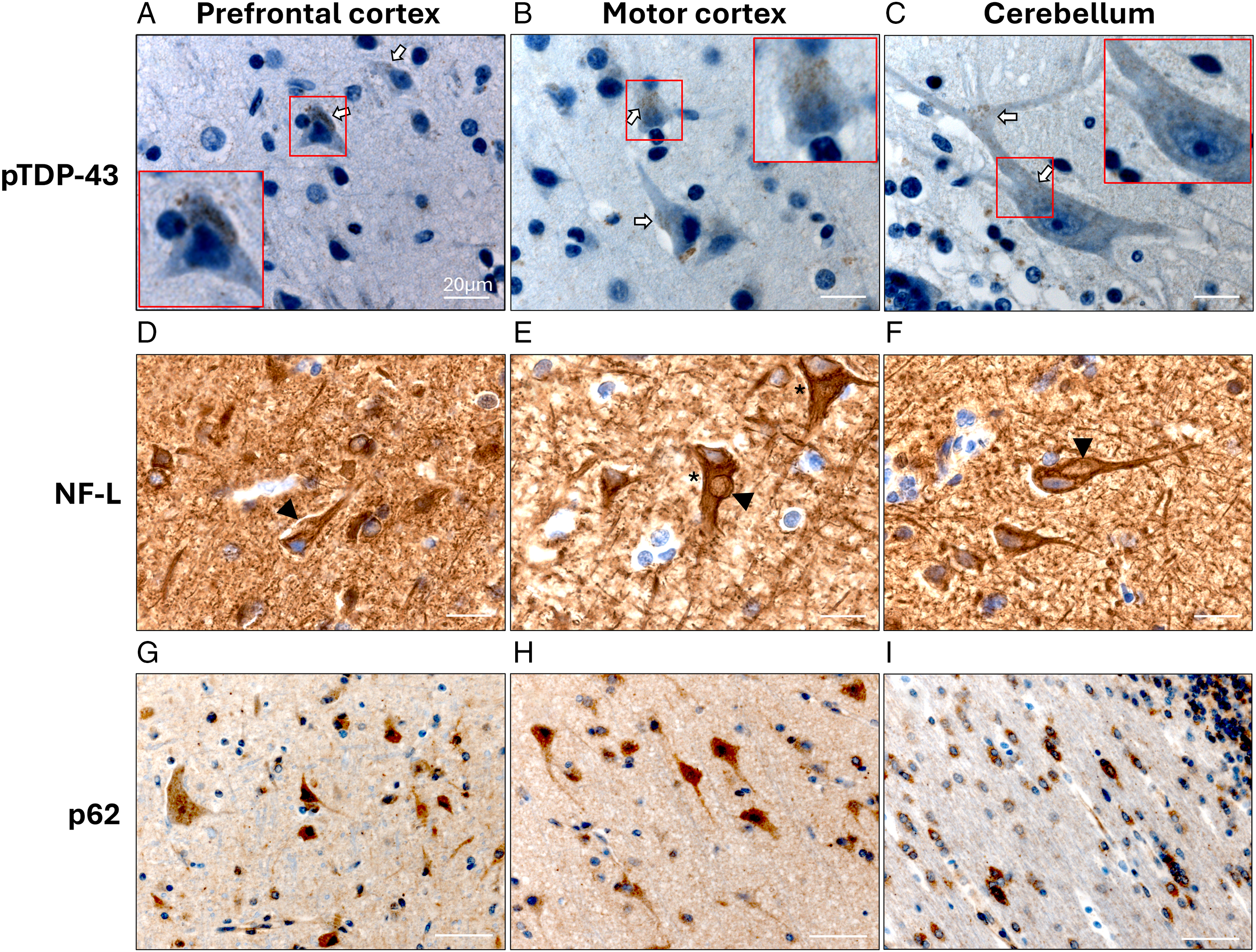
IHC also revealed, as expected, increased NF-L immunoreactivity in different brain regions of the index ARSACS patient, including prefrontal cortex (Figure 1 D), motor cortex (Figure 1 E) and cerebellum (Figure 1 F). Consistent with known ARSACS neuropathological features, Reference Larivière, Gaudet and Gentil9 remaining Purkinje cells and neurons of the external pyramidal layer 3 (Lamina III) of the cerebral cortex exhibited prominent somatodendritic NF accumulations (Figure 1 and Figure S5 A–H). Enlarged neurons containing vacuole-like areas within the soma were also observed, particularly in the prefrontal and motor cortices, in association with marked NF-L accumulation and focal NF-negative bulges in the cell body, suggestive of abnormal cytoplasmic protein accumulation (Figure 1 D–F and Figure S5 A–H). A reduced number of NF-L-positive Purkinje cells was also noted, consistent with Purkinje cell degeneration (Figure S5 G, H). Furthermore, abnormal accumulation of p62 in neurons was observed (Figure 1 G–I). p62 is a key protein marker of autophagy, and its cytoplasmic aggregation indicates impaired autophagic degradation. Such accumulation is often associated with chronic proteostatic stress, neuroinflammation and neurodegeneration. Reference Bitto, Lerner, Nacarelli, Crowe, Torres and Sell10
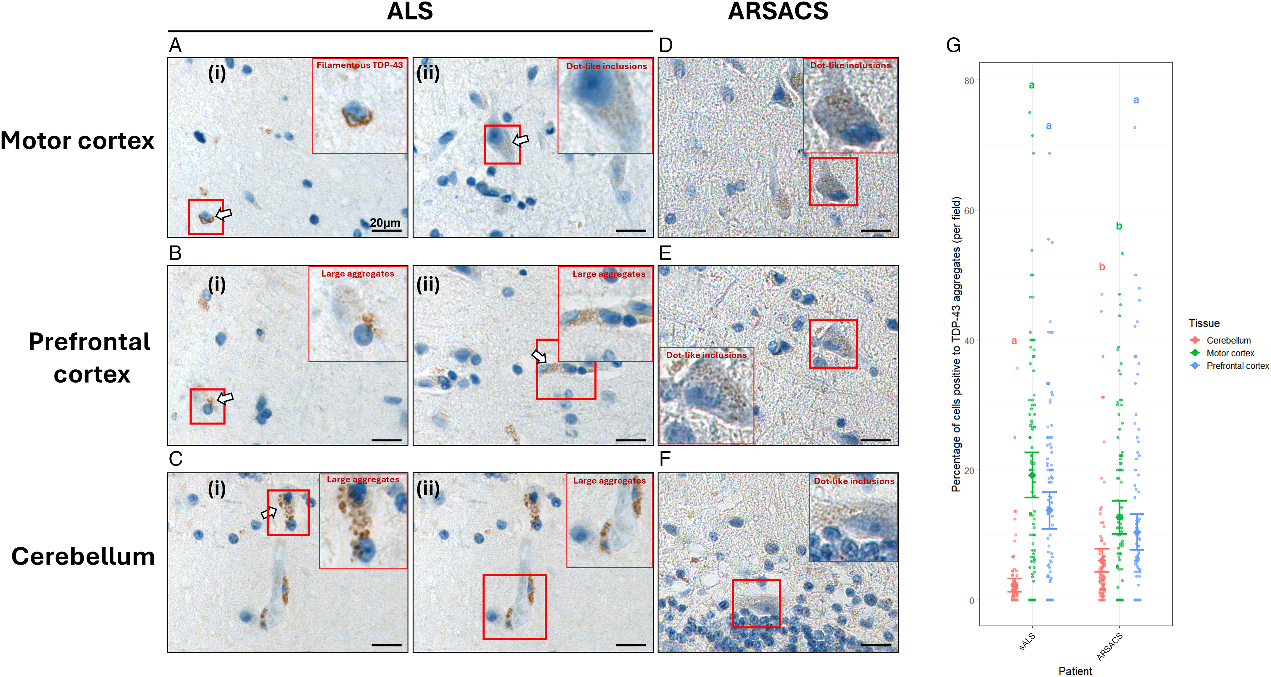
To better contextualize the pTDP-43 pathology observed in ARSACS, the same brain regions from a sporadic ALS patient were analyzed by IHC in parallel as a positive control (Figure 2 A–C). The staining revealed distinct regional patterns of pTDP-43 pathology across the motor cortex, prefrontal cortex and cerebellum. In the ALS motor cortex, abundant cytoplasmic inclusions were observed, often presenting as filamentous or large granular aggregates within neurons (Figure 2 A). These aggregates were less frequent in the prefrontal cortex and rare in the cerebellum (Figure 2 B, C). In contrast, the index ARSACS patient displayed prominent TDP-43 immunoreactivity in the cerebellum (Figure 2 F), where small, compact dot-like aggregates were observed within Purkinje cell layers. Similar punctate aggregates were also occasionally detected in the motor and prefrontal cortices (Figure 2 D, E). Unlike the ALS case, the ARSACS tissue exclusively displayed this small punctate pattern of pTDP-43 aggregation. Quantitative analysis supported these regional differences (Figure 2 G). The proportion of pTDP-43-positive cells was significantly higher in the motor cortex of the ALS patient compared to the ARSACS patient, whereas the latter exhibited a greater proportion of TDP-43-positive cells in the cerebellum. Statistical comparisons using one-way ANOVA followed by Tukey’s post hoc test confirmed these regional trends (p < 0.05).
Comparison of phosphorylated TAR DNA-binding protein 43 (pTDP-43) protein inclusions in the motor cortex, prefrontal cortex and cerebellum of an autosomal recessive spastic ataxia of Charlevoix–Saguenay (ARSACS) and an amyotrophic lateral sclerosis (ALS) patient. Representative immunohistochemical staining shows distinct morphologies of pTDP-43-positive inclusions in the motor cortex (A, D), prefrontal cortex (B, E) and cerebellum (C, F) from an ALS patient (A–C) and an ARSACS patient (D–F). Insets highlight examples of filamentous or large aggregated forms of pTDP-43 (i–ii). Scale bar = 20 µm. Magnification: 400×. (G) Quantification of the percentage of cells containing pTDP-43-positive inclusions across 50 microscopic fields per region with 2 separate observations made by distinct observers. Data are expressed as mean ± 95% confidence interval. Each dot represents an observation. Letters (a, b) indicate statistically significant differences between groups as determined by one-way Analysis of Variance (ANOVA) followed by a post hoc Tukey test. sALS = sporadic amyotrophic lateral sclerosis.
Sequential detergent extraction followed by Western blotting was performed to assess the presence of insoluble TDP-43 in human postmortem frontal cortex tissue from the index ARSACS patient and the sporadic ALS patient. As expected, the ALS sample exhibited a strong TDP-43 signal in the sarkosyl-insoluble fraction. The ARSACS sample also displayed detectable insoluble TDP-43, although at lower levels than the ALS case (Figure S6). These findings support the immunohistochemical evidence of TDP-43 mislocalization and aggregation observed in ARSACS tissue.
Discussion
TDP-43 is a DNA/RNA-binding protein that normally shuttles between the nucleus and cytoplasm, where it regulates multiple aspects of RNA metabolism. Reference Zeng, Luo and Jiang13 However, mutations or aberrant conformation in TDP-43 disrupt this dynamic, impairing nuclear import and promoting the formation of pathological, irreversible cytoplasmic inclusions in neurons, Reference Jo, Lee, Jeon, Kim, Kwon and Kim14,Reference Barmada, Skibinski, Korb, Rao, Wu and Finkbeiner15 a hallmark of several neurodegenerative disorders. Pathological TDP-43 species display considerable conformational and morphological heterogeneity, ranging from diffuse cytoplasmic staining and large amorphous inclusions to filamentous, punctate or dot-like aggregates. Reference Kawakami, Arai and Hasegawa16–Reference Balendra, Sreedharan and Hallegger18 Such diversity likely reflects differences in disease mechanisms, affected brain regions and selectively vulnerable neuronal populations. Reference Kawakami, Arai and Hasegawa16,Reference Balendra, Sreedharan and Hallegger18,Reference Meneses, Koga, OLeary, Dickson, Bu and Zhao19
In the present study, we identified cytoplasmic pTDP-43 aggregates in multiple central nervous system (CNS) regions of a genetically confirmed ARSACS patient. These aggregates exhibited a predominantly punctate or dot-like morphology and were associated with reduced nuclear staining, consistent with pathological TDP-43 mislocalization. Similar pTDP-43 aggregates were also observed in a second independent ARSACS case, supporting the reproducibility of this finding. Although TDP-43 pathology is best characterized in ALS and frontotemporal lobar degeneration (FTLD), it has also been reported in a growing number of neurodegenerative disorders. Reference Nelson, Dickson and Trojanowski20–Reference Liao, Ma and Dou25 The present observations therefore suggest that TDP-43 dysregulation may occur in ARSACS, a disease not currently recognized as a TDP-43 proteinopathy. The presence of total TDP-43 in the sarkosyl-insoluble fraction has been assessed, as done in anterior studies, Reference Feneberg, Charles and Finelli26–Reference Geser, Winton and Kwong28 which further supports this finding. Comparison with a sporadic ALS case highlighted clear differences in both the regional distribution and morphology of pTDP-43 pathology. Although the pathological significance of these differences remains uncertain, these findings suggest that TDP-43 abnormalities in ARSACS differ from the classical inclusions observed in ALS and may represent an atypical or early-stage form of TDP-43 pathology.
The co-occurrence of pTDP-43 with p62 accumulation, NF abnormalities and Purkinje cell degeneration is particularly noteworthy. These features are consistent with previous studies implicating impaired proteostasis, autophagy and cytoskeletal disorganization in ARSACS. Reference Francis, Alshafie, Kumar, Girard, Brais and McPherson7,Reference Morani, Doccini and Sirica29 Consequently, the disorganization of the observed TDP-43 abnormalities may represent a downstream consequence of sacsin deficiency and altered protein quality-control mechanisms rather than a primary pathogenic event. At present, the data do not allow discrimination between these possibilities. Nevertheless, the findings raise the possibility that disturbances in protein clearance pathways contribute to TDP-43 mislocalization in ARSACS.
Several observations argue against the presence of concomitant or subclinical ALS in the index ARSACS case, including the absence of lower motor neuron signs, the lack of classical skein-like ALS-associated TDP-43 inclusions and the distinct regional distribution and morphological characteristics of the observed TDP-43 pathology. Rather, our findings suggest that TDP-43 abnormalities can occur in ARSACS and may represent either a rare manifestation of ARSACS or an underrecognized pathological feature, as TDP-43 staining is not routinely assessed in this disease. This study raises, however, the possibility that shared molecular mechanisms may exist between ARSACS and other TDP-43 proteinopathies, including ALS and FTLD. This parallel is also of interest given that cognitive impairment has been reported in advanced stages of ARSACS and may share some features with the behavioral and executive dysfunction observed in FTLD. Both disorders also exhibit motor neuron involvement, with shared clinical features such as spasticity, distal muscle wasting and brisk reflexes. While ALS typically manifests in adulthood with rapidly progressive weakness, ARSACS presents in childhood. Despite differences in age of onset and clinical presentation, these overlapping neurological features point to the possibility of shared disease mechanisms, a hypothesis further supported by the present findings. While further validation is required, these observations suggest that mechanisms related to TDP-43 mislocalization and clearance should be explored in future ARSACS studies.
While the inclusion of a second independent ARSACS case strengthens the present findings, detailed biochemical and regional analyses were restricted to a single index case due to tissue availability. Additional studies incorporating larger numbers of ARSACS patients, broader CNS sampling and relevant cellular and animal models will be required to determine the prevalence, distribution and mechanistic significance of TDP-43 abnormalities in ARSACS.
Supplementary material
The supplementary material for this article can be found at https://doi.org/10.1017/cjn.2026.10695.
Data availability statement
Postmortem CNS tissue from the ARSACS patient is not available for distribution.
Acknowledgments
We would like to thank the ARSACS Foundation for their helpful discussion and support. Link: https://arsacs.com/.
Author contributions
Conceptualization, A.P. S.S. and F.G.-L.; methodology, A.P., L.T.-D. and V.R.; formal analysis, A.P., L.T.-D., S.S. and F.G.-L.; writing – original draft preparation, A.P., S.S. and F.G.-L.; writing – review and editing, A.P., L.T.-D., V.R., J.-D.B., J.M., N.D., S.S. and F.G.-L.; supervision, N.D. and F.G.-L.; funding acquisition, F.G.-L. All authors have read and agreed to the published version of the manuscript.
Funding statement
F.G.-L. is the recipient of a tier-1 Canada Research Chair. F.G.-L. has received funding from the ARSACS Foundation. A.P. is the recipient of scholarship support from CHUQ Research Center Foundation/Desjardins. We therefore gratefully acknowledge the ARSACS Foundation and also the support of the Canada Foundation for Innovation, which provided essential funding for the research infrastructure that enabled this work.
Competing interests
The authors declare no conflict of interest.
Language editing statement
The authors used ChatGPT (OpenAI, San Francisco, CA, USA) for basic assistance with English language checking. All scientific content, data interpretation and conclusions were conceived, written and verified solely by the authors.
Institutional review board statement
All procedures were conducted in accordance with ethical guidelines and approved by the institutional ethics committee of the CHU de Québec – Université Laval Research Center (protocol number 2012-1286 [PEJ-235]).
Informed consent statement
Informed consent for publication of clinical data, postmortem analyses of brain and spinal chod and related findings was obtained from the patient prior to undergoing MAiD.


 Open access

