Introduction
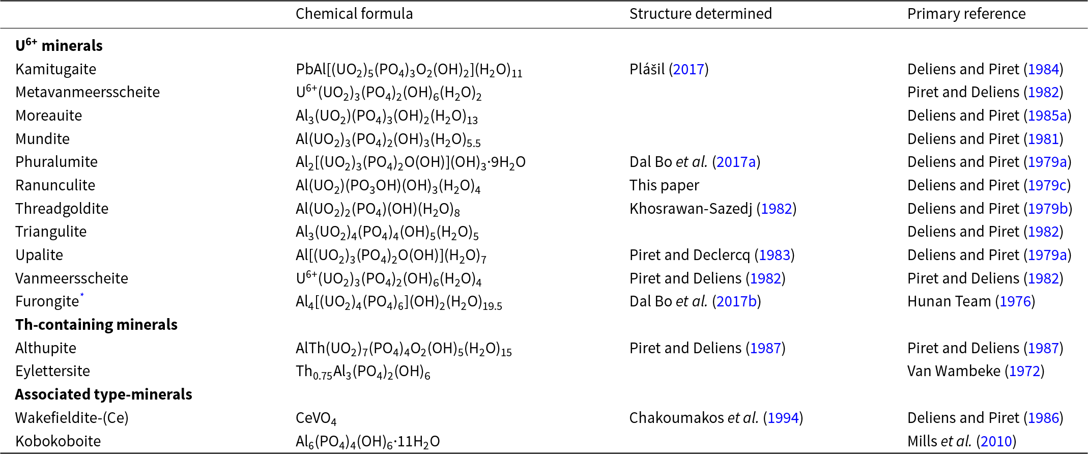
The Kobokobo pegmatite in the Sud-Kivu province, Democratic Republic of Congo, Africa, was one of the largest beryllium deposits in the World during the 1950s, producing a tremendous amount of beryl, columbite and cassiterite (Safiannikoff and Van Wambeke, Reference Safiannikoff and Van Wambeke1967; Van Wambeke, Reference Van Wambeke1987). It is one of the most interesting mineralogical localities known, mainly due to occurrences of rare uranium and thorium phosphates within this U-bearing quartz–albite–muscovite pegmatite. These minerals formed during the last evolutionary stages when the phosphate-rich solutions altered the primary Th- and Pb-rich uraninite and zircon. This pegmatite is a type locality for 12 actinoid-bearing phosphates, most of which contain Al3+ (Table 1). Despite some recent crystallographic studies conducted on the Kobokobo material deposited in museums, the structures of many of the Kobokobo minerals remain unknown. Here, we present a crystal structure determination on the rare uranyl phosphate ranunculite, originally described by Michel Deliens and Paul Piret in 1979 (Delins and Piret, Reference Deliens and Piret1979c). Since their study, no additional analytical data have been reported for this phase (!). Ranunculite has a peculiar structure that contains a structural sheet of a novel topology. Along with the first structure determination from the electron diffraction, we also provide reliable Raman data and experimental powder diffraction data.
Actinoide-bearing minerals and associated type minerals discovered from the Kokokobo pegmatite, Kivu Province, Democratic Republic of Congo

* Originally described from Hunan province, China; from Kobokobo reported by Deliens and Piret (Reference Deliens and Piret1985b).
Occurrence
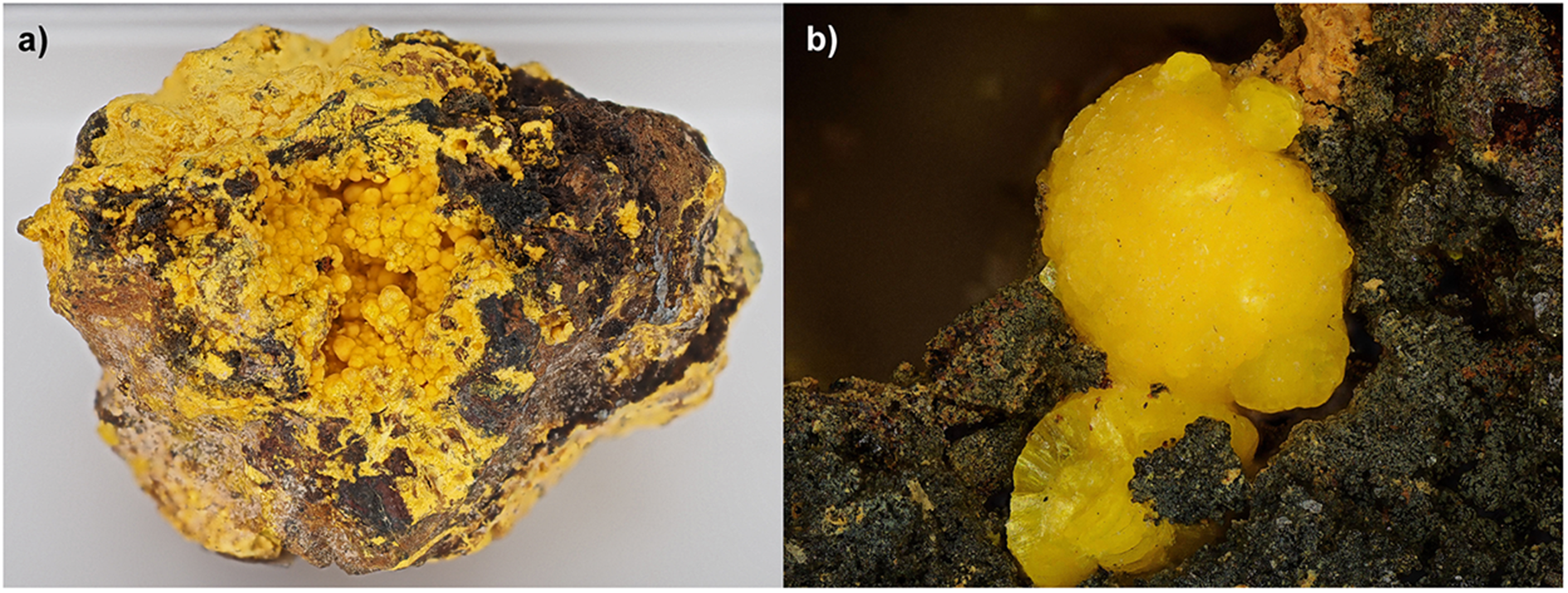
The specimen we used for the investigations comes from the collection of Luc Vandenberghe (Belgium) and was previously in the collection of Leopold van Wembeke, a Belgian collector who received the specimen around 1960. The specimen measures 4.4 × 3.2 × 2.8 centimetres in overall size. At the centre of the specimen, there is a cavity of around 2 × 2 centimetres, which is completely filled with yellow spherical ranunculite aggregates (Fig. 1a). Isolated crystalline aggregates are also found in high relief on the matrix (Fig. 1b). These crystal aggregates form radial spheres that show a layered, finely tabular structure. The isolated crystal aggregates that reach a size of up to 0.5 millimetres (Fig. 1b) are particularly suitable for further crystal structural investigations and perfectly fit the description by Deliens and Piret (Reference Deliens and Piret1979c). Notably, in this case, phuralumite, often closely associated with ranunculite, is absent on the specimen.
The ranunculite specimen from Kobokobo investigated in this study. (a) The entire specimen (∼4.4 cm across) with a large cavity filled with ranunculite. (b) Detail (FOV 1.5 mm) of the ranunculite aggregate studied by 3D ED and other methods (photo by A. Matthies).

The ranunculite powder in the capillary used in this study has been registered as a structure type (paratype) in the Museum of Natural History in Luxembourg under the catalogue number PV038.
Ranunculite is only known with certainty from one locality, the type locality of the Kobokobo pegmatite in the Democratic Republic of Congo. We emphasize that we tried to get access to the type specimen deposited in the collections of the Royal Museum of Central Africa, Tervuren, Belgium (RGM 6201), but without success.
Raman spectroscopy
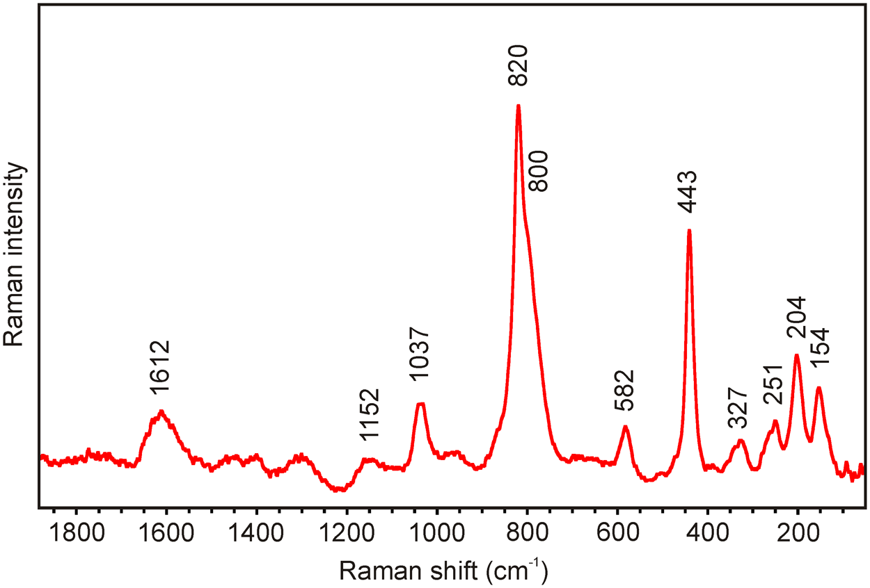
The Raman spectrum of ranunculite (Fig. 2) was collected in the range 1800–40 cm–1 (high-resolution settings, grid grating: 830 lines/mm; ∼2 cm–1) using a DXR dispersive Raman Spectrometer (Thermo Scientific) mounted on a confocal Olympus microscope. The Raman signal was excited by an unpolarised near-infrared 780 nm solid-state diode-pumped laser and detected by a CCD detector. The experimental parameters were: 50× objective, 2 s exposure time, 64 exposures, 50 μm slit, aperture and 8 mW laser power level. The spectra were repeatedly acquired from different grains to obtain a representative spectrum with the best signal-to-noise ratio. No thermal damage was observed by visual inspection of the excited surface after measurement, observation of possible decay of spectral features at the start of excitation, and checking for thermal downshift of Raman lines. The instrument was set up using a software-controlled calibration procedure using multiple neon emission lines (wavelength calibration), multiple polystyrene Raman bands (laser-frequency calibration), and standardized white-light sources (intensity calibration). Spectral manipulations were performed using the Omnic 9 software (Thermo Scientific). The Raman spectrum of ranunculite is deposited and available as supplementary material (*.csv file).
The Raman spectrum of ranunculite measured with a 780 nm laser.

The main bands observed in the Raman spectrum of ranunculite (Fig. 2) in the region between 50 and 1800 cm−1 are: 154, 204, 251, 327, 443, 582, 820, 1037, 1152 and 1612 cm−1. To our knowledge, no Raman spectrum of natural and synthetic ranunculite has been published. The following interpretation of the ranunculite spectrum is based on the papers by Čejka (Reference Čejka1999), Nakamoto (Reference Nakamoto2009), Sejkora et al. (Reference Sejkora, Kristek, Škácha and Dolníček2024), and references therein. A weak band at 1612 cm−1 is attributed to the ν2 bending vibrations of water molecules. A very weak band at 1152 cm−1 and a weak band at 1037 cm−1 correspond to the ν3 (PO4)3– asymmetric stretching and ν1 (PO4)3– symmetric stretching vibrations, respectively. The prominent Raman band at 820 cm−1 with a shoulder at 800 cm−1 are attributed to the ν1 (UO2)2+ symmetric stretching vibrations. Using the empirical relationship of Bartlett and Cooney (Reference Bartlett and Cooney1989) we have calculated corresponding approximate U–O bond lengths as 1.791 Å (820 cm−1) and 1.810 Å (800 cm−1), which are in line with values from the structure refinement. A weak band at 582 cm–1 is connected with the triply degenerate ν4 (δ) (PO4)3– bending vibrations, and a strong one at 443 cm−1 to the doubly degenerate ν2 (PO4)3− bending vibrations. A weak band at 251 cm−1 is related to the ν2 (UO2)2+ doubly degenerate bending vibrations. Other observed bands, 204 and 154 cm–1 may be attributed to Oeq–U–Oeq bending vibrations (Ohwada, Reference Ohwada1976), UO22+ translations and rotations, and external lattice vibration modes (Plášil et al., Reference Plášil, Buixaderas, Čejka, Sejkora, Jehlička and Novák2010).
Powder X-ray
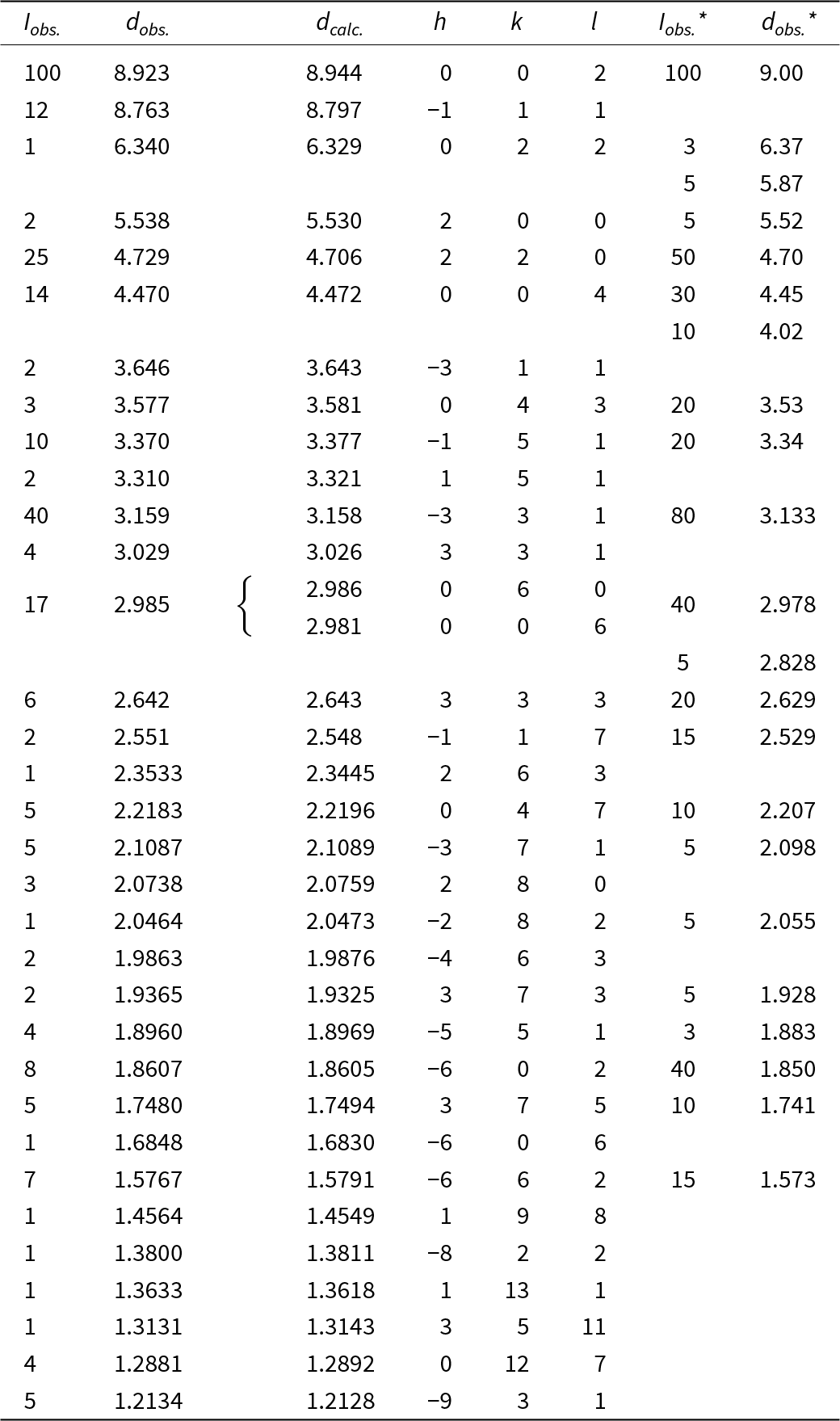
Powder X-ray diffraction data were collected at room temperature by a Rigaku SmartLab 9 kW rotation anode diffractometer (CuKα1,2 by curved Göbel mirror providing focused beam for Debye-Scherrer geometry) equipped with D/tex Ultra 250 detector. Ranunculite powder was loaded into a 3 mm borosilicate-glass capillary and data were collected in 20 repeating runs from 3–80°2θ (with an integrated step size of 0.01°2θ and a variable counting time. Positions and intensities of diffractions were found and refined using the Pearson VII profile-shape function of the ZDS program package (Ondruš, Reference Ondruš1993). The unit-cell parameters of barronite were refined by the least-squares program of Burnham (Reference Burnham1962). The powder X-ray diffraction pattern of ranunculite is reported in Table 2 compared to data from the original description of ranunculite (Deliens and Piret, Reference Deliens and Piret1979c). The refined unit-cell parameters (for the space group C2/c) are a = 11.183(6) Å, b = 17.918(8) Å, c = 18.086(13) Å, β = 98.50(8)°, with V = 3584(3) Å3 (Z = 4). The PowderCell program was used to calculate and visualize the theoretical powder pattern of ranunculite (Kraus and Nolze, Reference Kraus and Nolze1996).
Powder X-ray diffraction data for ranunculite from Kobokobo

Iobs.*, dobs.* - ranunculite from Kobokobo, 114.6 mm camera, CuKα radiation, Ni filter (Deliens and Piret, Reference Deliens and Piret1979c).
Electron crystallography
Data collection
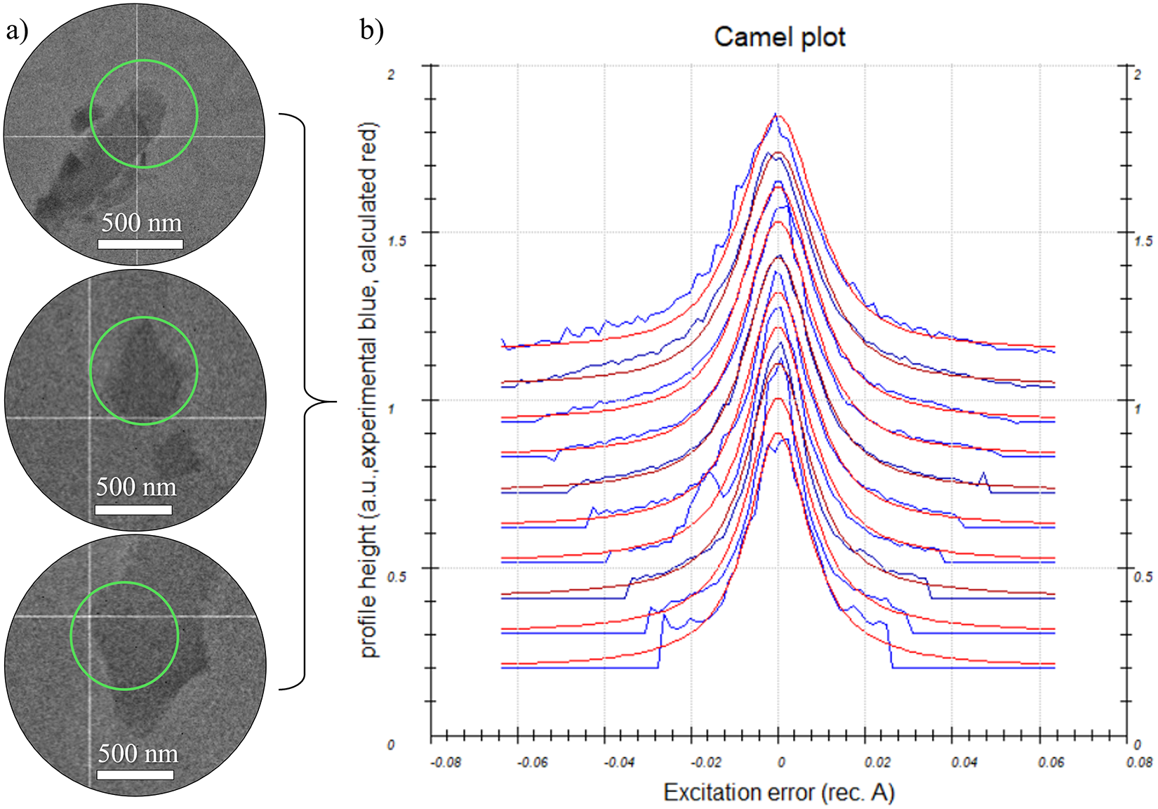
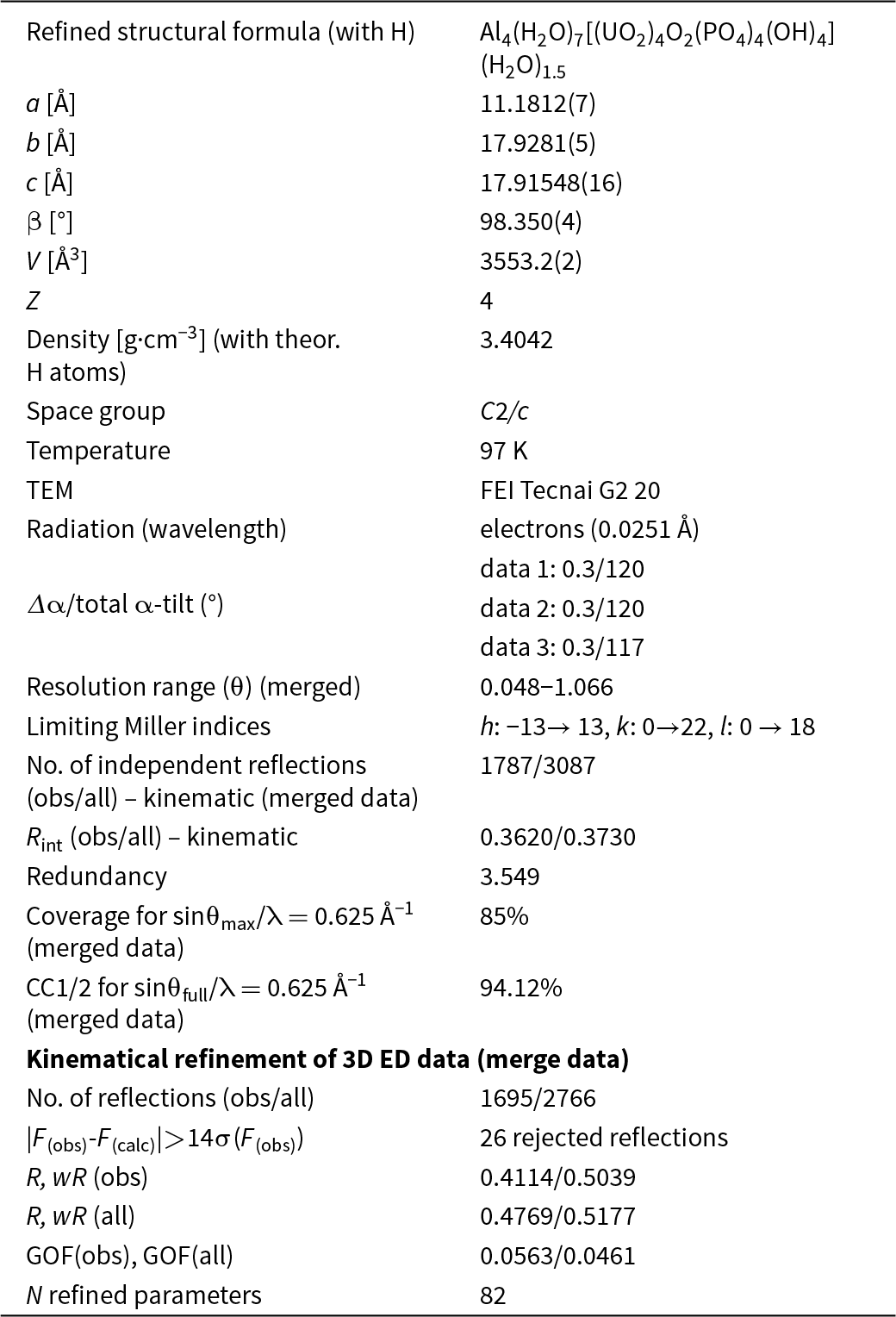
To reveal the structure of ranunculite, we employed three-dimensional electron diffraction (3D ED) techniques using a transmission electron microscope (TEM) (Gemmi and Lanza, Reference Gemmi and Lanza2019; Gemmi et al., Reference Gemmi, Mugnaioli, Gorelik, Kolb, Palatinus, Boullay, Hovmöller and Abrahams2019). A spherical aggregate of ranunculite crystals was gently crushed in a mortar in propanol and deposited on a Cu-grid coated with a thin film of holey amorphous carbon. To prevent dehydration caused by the TEM’s high vacuum, the grid was plunged into liquid nitrogen and then transferred to the TEM using a Gatan cryo-plunging transfer holder (Mugnaioli et al., Reference Mugnaioli, Lanza, Bortolozzi, Righi, Merlini, Cappello, Marini, Athanassiou and Gemmi2020; Steciuk et al., Reference Steciuk, Sejkora, Čejka, Plášil and Hloušek2021, Reference Steciuk, Majzlan, Rohlíček, Škoda, Sejkora and Plášil2024). The 3D ED data were collected at 97 K with a continuous rotation mode in an FEI Tecnai G2 TEM (acceleration voltage of 200 kV, LaB6) equipped with a side-mounted hybrid single-electron detector ASI Cheetah M3, 512 × 512 pixels with high sensitivity and fast readout. For each selected crystal (Fig. 3a), a series of non-oriented patterns was continuously collected by steps of 0.3°, on all the accessible tilt ranges of the goniometer. The data collections were automated by the in-house software, including the tracking of the crystal (Plana-Ruiz et al., Reference Plana-Ruiz, Portillo, Estradé, Peiró, Nicolopoulos and Kolb2018). In addition to the low-temperature experiment, low-illumination settings were used to further limit the beam-induced crystal damage. Continuous-rotation 3D ED data (cRED) reduction was performed using the computer program PETS2 (Palatinus et al., Reference Palatinus, Brázda, Jelínek, Hrdá, Steciuk and Klementová2019; Brázda et al., Reference Brázda, Klementová, Krysiak and Palatinus2022; Klar et al., Reference Klar, Krysiak, Xu, Steciuk, Cho, Zou and Palatinus2023). It includes the correction of geometrical and optical distortions to get accurate lattice parameters and better integration of the reflections (Brázda et al., Reference Brázda, Klementová, Krysiak and Palatinus2022). Three data sets were merged in PETS2 to improve the data coverage and get better statistics on lattice parameters. The quality of the merged data set is represented on the plots of the rocking curves (Camel plot) by the rocking-curve width as well as the estimation of the crystal mosaicity (Fig. 3b). The specific data processing for cRED data used in the structure solution and the refinement (with and without the dynamical approaches) was given by Klar et al. (Reference Klar, Krysiak, Xu, Steciuk, Cho, Zou and Palatinus2023). Overlapping virtual frames (OVFs) were produced from the tilt series of frames to model experimental intensities from continuous rotation data by summing consecutive experimental diffraction patterns into a set of virtual frames. Each OVF was characterized by its angular range Δαv covered by the virtual frame and the angular step between two virtual frames (Table 3). The solution and the refinement using the kinematical approximation were based on three merged datasets (Rint (obs/all) = 0.3620/0.3730). The structure was solved using Superflip (Palatinus and Chapuis, Reference Palatinus and Chapuis2007) implemented in Jana2020 (Petříček et al., Reference Petříček, Dušek, Plášil and Palatinus2023) and refined using DYNGO and Jana2020. Some polyhedral measures were calculated using the Vesta program (Momma and Izumi, Reference Momma and Izumi2011). The Topos program (Blatov et al., Reference Blatov, Shevchenko and Proserpio2014) was used for complexity calculations.
(a) Three crystals that were used for the structure characterization by 3D ED. (b) Rocking curve plot of the merged data. Plots of the rocking-curve profiles (Camel plot) of the experimental 3D ED merged data. The lowest blue curve is the average observed rocking curve in the range of 0.2 to 0.3 Å−1, and the next ones are obtained by steps of 0.1 Å−1. The red dotted curves are calculated from the rocking curve width = 0.0118 Å−1, the apparent mosaicity = 0.197°, and the tilt semi-angle = 0.15°. I > 10*σ(I) reflections are involved in the Camel plot.

3D ED data collection and structure refinement details for ranunculite

Structure solution and refinement
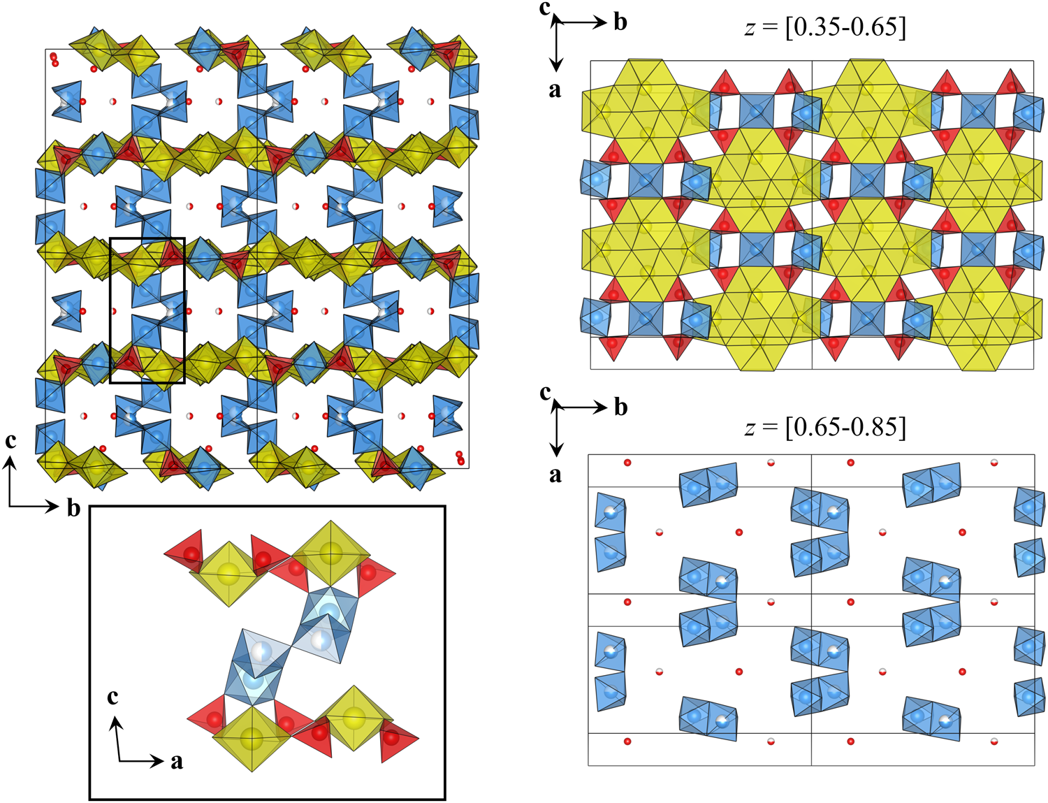
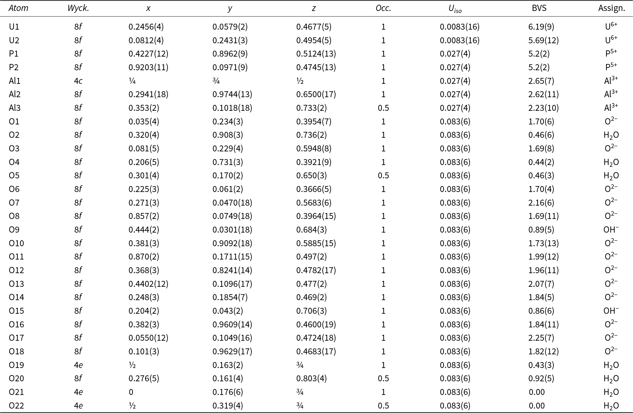
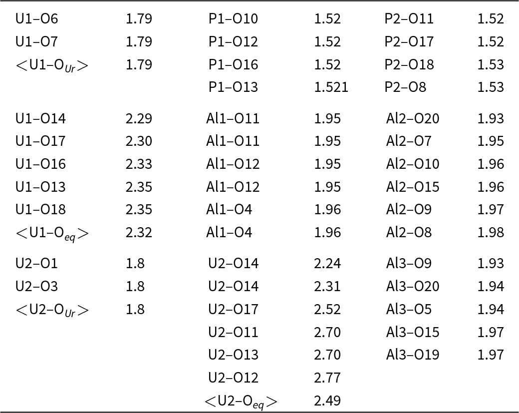
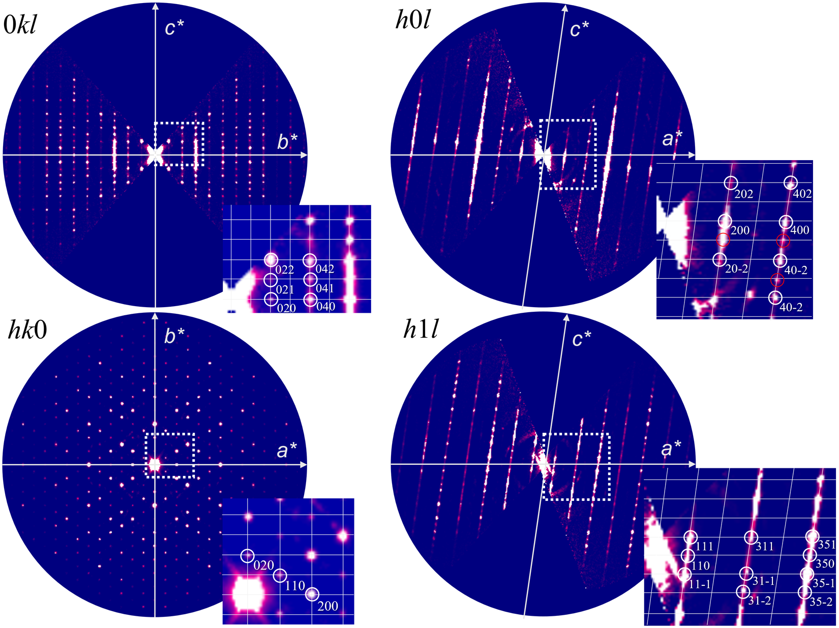
According to 3D ED data, ranunculite is monoclinic with a C-centred cell: a = 11.1812(7) Å, b = 17.9281(5) Å, c = 17.91548(16) Å, β = 98.350(4)° and V = 3553.2(2) Å3. Despite the preferential (001) orientation on the grid, merging data sets allowed an increase in the data completeness to reach 85% data coverage for sinθmax/λ = 0.625 Å–1 (Laue class: 2/m). Merging data also allowed for more accurate lattice parameters and better statistics. The diffuse rows of reflections along c on the h0l section make the symmetry determination ambiguous. The sections of the reciprocal space suggest the possible presence of a c-glide perpendicular to b (l = 2n on h0l). Possible space groups were C2/c, C2/m, C2, Cm, and P  $\overline{1}$ (in the primitive cell). The different symmetries and solutions were tested in the Superflip program. A complete solution describing the two dense layers was exclusively obtained in the space group C2/c. This is in line with the Superflip space-group determination (from the symmetry of the flipped electron density). The initial electrostatic potential map was re-interpreted manually based on the visible cation coordination and the result of the WDS analysis. Aluminium was placed at the centre of an octahedra, a uranium atom in pentagonal or hexagonal bipyramids, and phosphorus in tetrahedral coordination. Both kinematical and dynamical approaches were tested to refine the model against 3D ED data of the first data set. Possibly, due to imperfect crystallization resulting in stacking faults along c (visible on the (a,c) plane), none of those approaches lead to satisfying R-values. However, the refined model and the topology make sense when applying cation–oxygen distance restrictions. In this study, only the kinematical refinement is presented because the dynamical theory did not yield structure improvements on the relatively poor data set and was computationally demanding. The details of the kinematical refinement are given in Table 3. Restrictions were applied to ensure reasonable cation–oxygen distances and O···O distances in some polyhedra (P–O = 1.49 Å, U–Oyl = 1.8 Å, Al–O = 1.9 Å). Note that lower symmetries were also tested during the refinement. However, none improved the model, indicating that the refinement limitations were due to poor data rather than incorrect space group attribution. The final model is presented in Fig. 4, positional parameters and atomic displacement parameters in Table 4, and selected interatomic distances in Table 5. The limited data quality did not allow the determination of the hydrogen positions from the difference-Fourier maps. The nature of the oxygen atoms (distribution of O2−, hydroxyl, or water molecules in the structure) was based on the bond-valence analysis (Table 4; using bond-valence parameters given by Gagné and Hawthorne, Reference Gagné and Hawthorne2015). The resulting formula is electro-neutral, Al4(UO2)4O2(PO4)4(OH)4(H2O)7(H2O)1.5 for Z = 4. The cation ratio corresponds to the original description of ranunculite as a mineral by Deliens and Piret (Reference Deliens and Piret1979c).
Crystal structure of ranunculite (represented approximately as 2a × 2b × 2c along the main directions. The uranyl–phosphate sheet is shown between 0.35 < z < 0.65, and the Al dimers are found in the interlayers for 0.65 < z < 0.85. Colour scheme: U – yellow; PO4 – red; Al-polyhedra blue; and O atoms of the H2O molecules – red spheres.

Atom coordinates, displacement (in Å2), occupational parameters, and bond-valence sums (in valence units) for the ranunculite structure obtained from 3D electron diffraction

Selected interatomic (bond) distances (in Å) for ranunculite

Notice: the restrictions on bond lengths were applied during the structure refinement.
Description of the structure
Based on the 3D ED data, the structure of ranunculite (Fig. 4) contains two independent U sites, two P sites, three Al sites and 22 O sites in the asymmetric unit (Table 4). The U1 site is coordinated by seven O atoms, two with short bonds forming a uranyl ion UO22+, and five located at the horizontal vertices of the pentagonal uranyl bipyramid. The U2 site is linked to eight O atoms, two forming a uranyl ion UO22+, and six located at the horizontal vertices of the hexagonal uranyl bipyramid. The U–O bond lengths were bound by soft restrictions (see above); the refined values (see Table 5) are consistent with most typical lengths observed for uranyl coordination polyhedra in minerals and inorganic compounds (Lussier et al., Reference Lussier, Lopez and Burns2016). The P1 and P2 sites are coordinated as regular tetrahedra by four O atoms (none protonated as PO3OH). The Al1 site is coordinated by six O atoms (all of them O2–, based on BVS; Table 4) forming an octahedron (EcoN = 5.9998, bond-angle variance = 7.296°^2, average Al–O = 1.95 Å, expected Al–O = 1.89 Å, BVS = 2.65 vu). The Al2 site is coordinated by six O atoms (from them, O9 and O15 are OH–; Table 4), also forming an octahedron (EcoN = 5.9885, bond-angle variance = 7.7037°^2, average Al–O = 1.96 Å, expected Al–O = 1.89 Å, BVS = 2.62 vu). The Al3 site is coordinated by five ligands (of them, O9 and O15 are OH–; Table 4 as a tetragonal pyramid (EcoN = 4.988, average Al–O = 1.95 Å, expected Al–O = 1.82 Å, BVS = 2.23 vu). Based on the bond-valences analysis, the following O atoms, including O2, O4, O5, O19, O20, O21 and O22, belong to molecular H2O (in sum, 8.5 H2O, of which 1.5 H2O is hosted in the voids of the structure = O21, O22; see further).
Structure connectivity and sheet topology
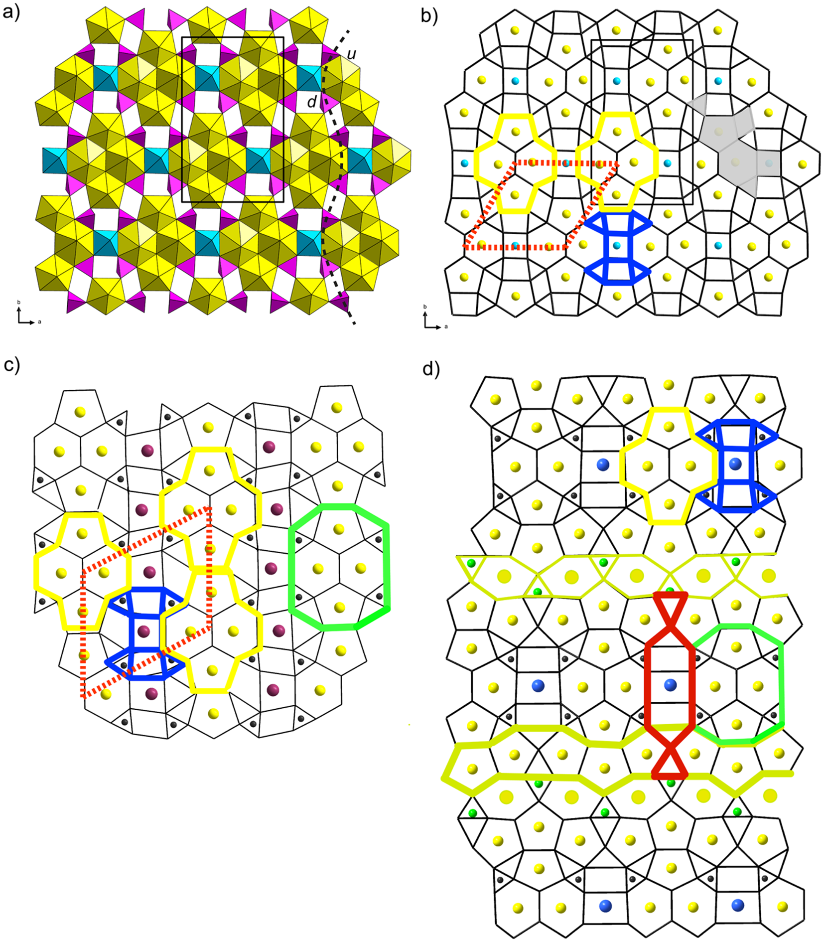
Two oppositely oriented pentagonal bipyramids (UPB, U1) form dimers of edge-sharing uranyl hexagonal bipyramids (UHB; U2) (Fig. 5a), resulting in a cluster of uranyl coordination polyhedra (in yellow colour, Fig. 5b). Those clusters share two edges of the UHB with the octahedra of Al1 (along [100]). The Al1-octahedron shares four equatorial vertices with PO4 tetrahedra, which in turn share one edge with UHB and one vertex with neighbouring UPB. The remaining vertex of each PO4 is not linked within the resulting sheet and is staggered into the interlayer, providing a linkage with Al2 octahedra. The Al2 octahedron is linked to the uranyl phosphate sheets by sharing three O atoms (apical O7yl, equatorial O8PO4, equatorial O10PO4). The other apex (vertex) of the Al2-octahedron is the O2 atom, which points to the interlayer space of the ranunculite structure and is an H2O group based on the BVS analysis. The remaining ligands of the Al2 octahedra are O9 and O15 atoms, belonging to the OH groups (Table 4) and linking Al2 octahedra to Al3 pyramids. The three remaining ligands of the Al3-pyramids are H2O molecules (O5, O19 and O20).
The uranyl-phosphate sheet present in the ranunculite structure. (a) Detailed view of the polyhedral representation of the sheet. The colour scheme for polyhedra: U – yellow, P – purple, Al – azure. The dashed line outlines the PO4 tetrahedra and the orientation of their vertices concerning possible orientational stereoisomerism (here ud stereoisomer). (b) the graph representation of its topology. Colour scheme: HxPt cluster – yellow; SqTr cluster – blue; red dotted line connects the idealized centres of HxPt clusters. (c) Bijvoetite topology. The colour scheme is similar to the previous graph; the green border outlines the HxPtTr cluster, which is common with (d) lepersonnite topology, where additional infinite chains of polyhedra are present (two types of yellow borders outlining HxTr chain and PtTr chain).

The infinite sheets of polyhedra (structural units) are perpendicular to c, and adjacent sheets are linked in two ways, as some of the O atoms within the Al-polyhedra are partially occupied. Regions (unit cells) in which adjacent sheets are linked by hydrogen bonds only alternate with those where sheets are linked via tetramers of Al-polyhedra (through shared O19). H-bonds emanate from the H2O groups that are of the transformer type (for terminology, see Hawthorne and Schindler, Reference Hawthorne and Schindler2009; [3]-coordinated O atoms): O2, O5, O19, O20 (in bold are those that secure interactions with adjacent sheets). The H atoms of the OH groups (donors are O9 and O15) interact either with the O atoms (acceptors) of the H2O groups in the interlayer (O21 and O22) or with apical Ouranyl atoms of adjacent layers. Both O21 and O22 are non-transformer H2O groups with [4]-coordinated O atoms (bonded to two H-atoms as H2O and accepting two weak H-bonds, 2 × 0.8 vu + 2 × 0.2 vu = 2.00 vu). Hydrogen bonds emanating from the O2 atom are accepted by O21/O22 or apical Ouranyl atoms of adjacent layers. The alternation of regions with stronger and weaker interlayer bonding leads most probably to the statistically high distribution of stacking faults, which contributes significantly to the diffraction profiles (Fig. 6).
0kl, h0l, h1l, and hk0 sections of the reciprocal space reconstructed from the merged data set. Red reflections are forbidden due to the c-glide mirror.

The uranyl–phosphate sheet in ranunculite (Fig. 5a) is a novel topological type, consisting of hexagons, pentagons, squares and triangles (TrSqPtHx sheet). The ring symbol (derived after Krivovichev, Reference Krivovichev2004) is 61514232. In the case of ranunculite, hexagons of the topology are occupied by U6+ and pentagons as well; Al3+ populates part of the squares, and part is a void. Triangles of the topology here are occupied by P5+. Despite being novel, the topology found in ranunculite shares some similarities with other known, complex topologies. The bijvoetite topology (Fig. 5c) found in the structures of bijvoetite-(Y) (Li et al., Reference Li, Burns and Gault2000) and kamotoite-(Y) (Plášil and Petříček, Reference Plášil and Petříček2017) is very similar to the ranunculite topology. The main difference is that clusters of hexagons and pentagons (HxPt, yellow modules in Fig. 5c) are linked into infinite chains and shifted along the direction of the elongation of chains. Thus, the adjacent HxPt clusters are shifted to each other in bijvoetite (this corresponds to ~30° rotation of the red rectangles, Figs 5b, c). In ranunculite, HxPt clusters are separated by the cluster of squares and triangles (blue cluster in Figs 5b, c). In ranunculite, the central square of that cluster is occupied by Al3+, while in bijvoetite, it is occupied by Y3+. Triangles (blue cluster) are occupied by C4+ in bijvoetite. A similar HxPt cluster exists in the lepersonnite topology, present in the structure of lepersonnite-(Gd) (Plášil et al., Reference Plášil, Steciuk, Sejkora, Kampf, Uher, Ondrejka, Škoda, Dolníček, Philippo, Guennou, Meisser, Rohlíček and Mees2025). Nevertheless, the topology is even more complex in that case, hosting two additional infinite chains: one of pentagons and triangles and the second of hexagons and triangles (Si4+ populates those associated with hexagons). Bijvoetite and lepersonnite topologies contain the same arrangement of HXPT with triangles (green cluster in Figs 5c, d).
Stereoisomerism within ranunculite sheets
As the fourth corner of the tetrahedra in the ranunculite-type sheets is not involved in the intra-sheet polymerization of coordination polyhedra, it may thus be oriented either up or down relative to the plane of the sheet. This results in different orientational geometrical isomers (Krivovichev, Reference Krivovichev2010). Here, in the studied structure (Fig. 7a), it is the ud isomer, where the orientation of the vertices alternates alongside the respective chain.
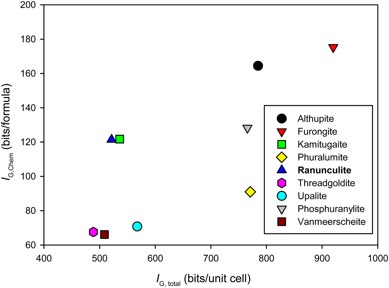
Complexity measures (structural vs. chemical) plot for actinoid-bearing phases from Kobokobo pegmatite.

Kobokobo pegmatite – home of unusual uranyl minerals – a few concluding remarks
The Kobokobo pegmatite (Safiannikoff and Van Wambeke, Reference Safiannikoff and Van Wambeke1967; Van Wambeke, Reference Van Wambeke1987) is among the most peculiar mineralogical localities in the World. The incorporation of Th within the otherwise U6+-dominated minerals is particularly unusual. It indicates rather special conditions during the crystallization, as Th4+ is mobile under different conditions from U6+. Recently, intriguing discoveries of rare Th4+-containing minerals, two molybdates and halide minerals from the small Mo–Bi occurrence at Su Seinargiu in Sardinia, Italy (Orlandi et al., Reference Orlandi, Biagioni, Bindi and Nestola2014, Reference Orlandi, Biagioni, Bindi and Merlino2015, Reference Orlandi, Biagioni and Zaccarini2017) have been made. However, our knowledge of the Th4+ behaviour during alteration processes is still not well understood.
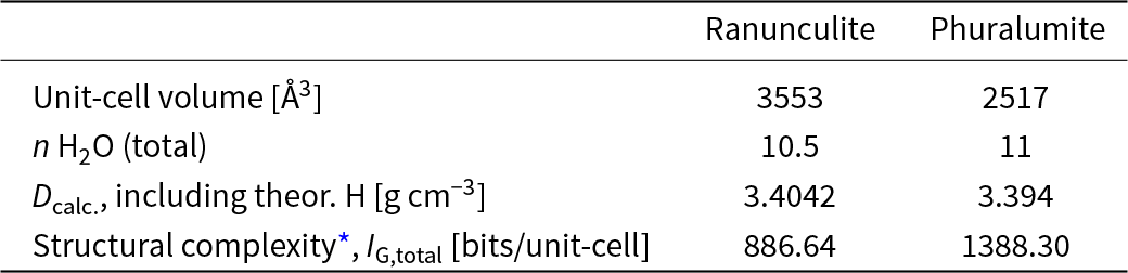
In the study of Dal Bo et al. (Reference Dal Bo, Hatért and Philippo2017b), it has been documented that phuralumite, Al2[(UO2)3(PO4)2O(OH)](OH)3(H2O)9 (Z = 4), is often closely associated with ranunculite. Usually, phuralumite crystals grow on the underlying globular aggregates of ranunculite. Interestingly, the ranunculite formula derived from our study is Al4(UO2)4O2(PO4)4(OH)4(H2O)7(H2O)1.5 (Z = 4). These two minerals have very similar densities (Table 6) and molecular H2O content. This resembles the case of stable and metastable polymorphs. Krivovichev (Reference Krivovichev2017) demonstrated for polymorphic minerals, gilmarite and clinoclase, that the metastable phase (gilmarite), which is structurally simpler than the stable one (clinoclase), crystallized prior to the stable phase under some kinetically favoured conditions. He also demonstrated that according to the Ostwald–Volmer rule (Holleman et al., Reference Holleman, Wiberg and Wiberg2001), for metastable crystallization, the less dense metastable phase forms first because, under supersaturated conditions, its nuclei reach their critical sizes faster than those of the stable phase (Fischer and Jansen, Reference Fischer and Jansen2002; Bach et al., Reference Bach, Fischer and Jansen2013). Although ranunculite and phurcalite are not polymorphs (as phuralumite is based upon phosphuranylite topology), their proximity in chemistry, density, and other properties is worth investigating and discussing further. Ranunculite is a much simpler phase from the point of view of the structural complexity concept (Krivovichev, Reference Krivovichev2012, Reference Krivovichev2013, Reference Krivovichev2016) (Table 6). From all the observed features of the structure and properties of ranunculite, we can conclude that the structure of ranunculite can be considered as less ordered compared to that of phuralumite; ranunculite contains many stacking faults (manifested in the powder diffraction data by the presence of anisotropic broadening); ranunculite crystallizes, in contrast to phuralumite, in minute crystals in radial aggregates. This noticeable difference in appearance suggests that ranunculite is most probably the result of quick crystallization from saturated/oversaturated solutions. After that, crystallization of a more ordered, more structurally complex phase – phuralumite proceeds.
Selected data for ranunculite and phuralumite from Kobokobo

* including theoretical H atoms contribution.
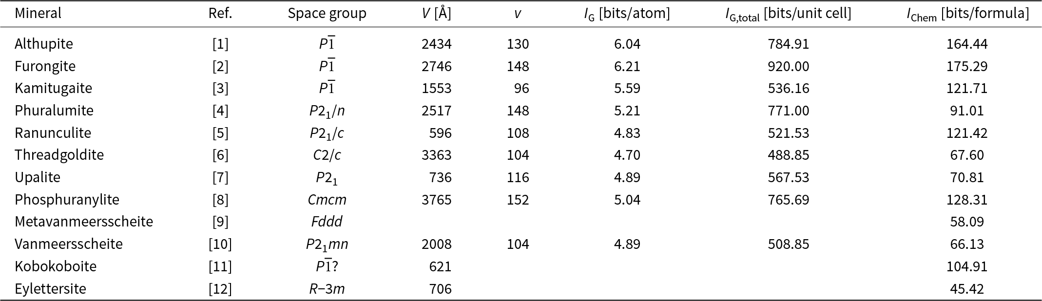
It is worth considering the structural complexities of some other actinoid-bearing mineral species from Kobokobo. The calculated structural and chemical complexity measures are reported in Table 7 and are displayed for comparison in Fig. 7. The chemical complexity (after Siidra et al., Reference Siidra, Zenko and Krivovichev2014) is estimated by considering the chemical formula as a message, where symbols correspond to different chemical elements. Interestingly, ranunculite has very similar figures (both structurally and chemically) as kamitugaite, another very peculiar phase from Kobokobo (the only phase in that system containing Pb2+ and As5+). Phuralumite, which is often associated with ranunculite (never associated with kamitugaite and vice versa), is more complex structurally and less so chemically. Furongite, althupite and phosphuranylite are structurally and chemically complex minerals, reaching the highest values within the given mineral system. In general, they are also very stable alteration phases formed from primary U minerals (see Krivovichev and Plášil, Reference Krivovichev, Plášil, Burns and Sigmon2013; Plášil, Reference Plášil2014). This is probably in line with the fact that they are the most complex in the given mineral system (the most stable ones = with the highest complexity).
Actinide-bearing and associated minerals from Kobokobo pegmatite and their structural and chemical complexity measures

[1] – Piret and Deliens (Reference Piret and Deliens1987), [2] – Dal Bo et al. (Reference Dal Bo, Hatért and Philippo2017a), [3] – Plášil (Reference Plášil2017), [4] – Dal Bo et al. (Reference Dal Bo, Hatért and Philippo2017b), [5] – this paper, [6] – Khosrawan-Shazedj (Reference Khosrawan-Sazedj1982), [7] – Piret and Declerq (Reference Piret and Declercq1983), [8] – K2Ca[(UO2)7(PO4)4O2(OH)2](H2O)8 – the electroneutral formula proposed for phosphuranylite, which was derived from new structure determinations (unpublished data of authors), [9] – Piret and Deliens (Reference Piret and Deliens1982), [10] – Piret and Deliens (Reference Piret and Deliens1982), [11] – Mills et al. (Reference Mills, Birch, Kampf and van Wambeke2010), [12] – Van Wambeke (Reference Van Wambeke1972).
Supplementary material
The supplementary material for this article can be found at https://doi.org/10.1180/mgm.2025.10147.
Acknowledgements
The helpful comments of two anonymous reviewers, as well as editors, are greatly appreciated. This study was supported by the CzechNanoLab Research Infrastructure supported by MEYS CR (LM2023051). Additionally, we acknowledge the support by the Ministry of Culture of the Czech Republic (long-term project DKRVO 2024-2028/1.II.b; National Museum, 00023272) for JS.
Competing interests
The authors declare none.