Clostridium difficile infection (CDI) is a major cause of healthcare-associated diarrhea.Reference Kyne, Hamel, Polavaram and Kelly 1 In a recently published multistate prevalence survey, CDI was responsible for 12.1% of healthcare-associated infections,Reference Magill, Edwards and Bamberg 2 pointing to its ongoing significance and the need for further investigation regarding prevention. In intensive care unit (ICU) patients, CDI has been associated with prolonged length of hospital and ICU stay, and increased need for skilled nursing care or rehabilitation following discharge.Reference Micek, Schramm and Morrow 3 CDI in hospitalized patients is generally attributed to the current hospital stay. Recent studies, however, challenge this concept and reveal high C. difficile colonization rates upon hospital admission.Reference McDonald 4 In patients known to be colonized with toxigenic C. difficile, a key component for prevention of progression to CDI is to avoid, when possible, agents known to incite clinical expression of C. difficile—emphasizing the importance of antibiotic stewardship. If this principle is true, it is key to identify those patients who are colonized in order to define risk and optimal methods of patient-specific prevention interventions in high-risk populations.
We sought to determine the rate of colonization with toxigenic C. difficile both at hospital admission and acquired during hospitalization, as well as the risk of subsequent CDI, among a cohort of high-risk patients in the ICU.
METHODS
Design and Setting
We performed a prospective, observational cohort study of patients at least 18 years of age admitted to ICUs within 48 hours of admission to the Johns Hopkins Hospital, a 900-bed tertiary care academic medical center in Baltimore, Maryland, from April 15 through July 8, 2013.
Patients were screened by means of rectal swabs for colonization with toxigenic C. difficile on admission to the ICU and weekly thereafter until discharge from the ICU. C. difficile screening results were not shared with clinicians. No enhanced infection control practices were undertaken for patients identified as colonized with toxigenic C. difficile on the basis of rectal swab results. Patients diagnosed with CDI within 48 hours of hospital admission were excluded from this study because the infection likely was acquired before admission.Reference Cohen, Gerding and Johnson 5 Information regarding demographic variables, clinical characteristics, and exposures to established risk factors 4 weeks prior to admission and during hospitalization (up to development of CDI) was obtained by electronic medical chart review, including records on outpatient visits in Johns Hopkins–affiliated practices. Patients were longitudinally followed up for development of CDI during their hospital stay and up to 1 month after discharge by medical chart review. Data regarding symptoms and complications attributable to CDI were recorded. Results from all stool samples submitted for C. difficile testing as part of routine clinical care throughout the Johns Hopkins Hospital and its outpatient clinics as standard of care were collected. This study was approved by the Johns Hopkins University School of Medicine institutional review board with a waiver of informed consent.
Definitions and Categorization of Patients
Patients were considered colonized on admission if rectal swabs performed within 48 hours of admission were positive for toxigenic C. difficile. A patient was classified 6 as colonized with toxigenic C. difficile during hospitalization if (1) the rectal swab upon admission to the ICU was positive for toxigenic C. difficile, (2) a subsequent rectal swab during a patient’s ICU stay was positive for toxigenic C. difficile, or (3) stool sent as part of routine clinical care was positive for toxigenic C. difficile in the absence of clinical findings suggestive of CDI.
CDI was defined using standard definitions.Reference Cohen, Gerding and Johnson 5 Development of CDI was evaluated during hospitalization and during a 1-month follow-up period after hospital discharge.
Microbiological Analyses
Rectal surveillance swab specimens were obtained on ICU admission and weekly thereafter until ICU discharge and were immediately sent to the microbiology laboratory for processing. Both rectal swabs and stool specimens submitted for C. difficile detection as standard of care underwent polymerase chain reaction (PCR) analysis using the GeneOhm C. difficile assay (BD). Toxigenic bacterial culture was performed for PCR-positive samples. All isolated strains were prospectively collected.
PCR ribotyping was performed by capillary electrophoresis as described elsewhere.Reference Stubbs, Brazier, O'Neill and Duerden 7 Capillary electrophoresis was conducted using the automated sequencer ABI-PRISM 3130 Genetic Analyzer and fragments were analyzed using GeneMapper software (both, Applied Biosystems).
Statistical Analyses
Relative risks for colonization with toxigenic C. difficile on admission and development of CDI were estimated using univariable Poisson regression with robust error variance.Reference Zou 8 Only variables found to be significant in univariable analyses were included in a multivariable model. P≤.05 was considered significant. The Pearson and deviance goodness-of-fit tests were performed to assess the fit of data to a Poisson distribution in the final regression model. A priori power calculations were performed using data from a prior studyReference Hung, Tsai and Hung 9 that showed that 182 patients are necessary to detect a significant difference in the risk of CDI on the basis of toxigenic C. difficile colonization status, assuming a 2-sided significance level of.05 and power of 0.8. All analyses were performed using Stata, version 12.0 (StataCorp).
RESULTS
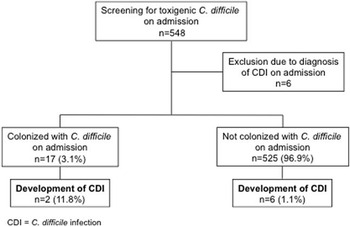
During the study period, 548 consecutive patients were screened for colonization with toxigenic C. difficile on admission. Six patients were excluded from further analyses because they were diagnosed with CDI during the first 48 hours of admission (Figure 1).
Colonization with toxigenic Clostridium difficile on admission and subsequent development of C. difficile infection.

Reasons for ICU admission included monitoring after neurosurgery (239), respiratory failure (46), monitoring after cardiac surgery (33), sepsis (29), intracranial hemorrhage (28), trauma (25), stroke (24), monitoring after vascular surgery (23), cardiac failure (23), monitoring after abdominal surgery (17), gastrointestinal bleeding (16), encephalopathy (7), liver or renal failure (6), meningoencephalitis (6), seizures/status epilepticus (5), monitoring after thoracic surgery (4), and other reasons (11).
Among the remaining 542 patients, 17 (3.1%) were colonized with toxigenic C. difficile on admission. Three of 35 patients with weekly follow-up swabs, after admission, were identified as subsequently colonized with toxigenic C. difficile. Follow-up information 1 month after discharge was available for 393 patients (72.5%). Patients with and without follow-up information on CDI status 1 month after discharge were similar regarding demographic characteristics, preexisting medical conditions, and exposures to known risk factors for the development of CDI (data not shown).
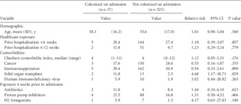
Neither age, nor prior exposure to healthcare (up to 12 weeks prior to admission), nor receipt of antibiotics or gastric acid suppressants were associated with an increased risk for colonization with toxigenic C. difficile on admission. Patients with solid organ transplant were more commonly colonized on ICU admission (relative risk, 4.68 [95% CI, 1.17–18.71]) (Table 1).
Risk Factors for Colonization With Toxigenic Clostridium difficile on Intensive Care Unit Admission

NOTE. Data are no. (%) of patients unless otherwise indicated.
CDI developed in 8 patients (1.5%) during hospitalization (Figure 1). An additional 4 patients were diagnosed with CDI within 1 month after discharge (overall, 12 [2.2%] of 542 patients). None of the patients with CDI were diagnosed with severe complicated CDI (ie, septic shock, toxic megacolon) and no patients experienced a CDI-attributable death.
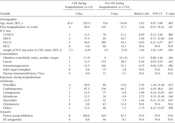
In univariable analyses, exposures to penicillins (82.1% being penicillin and beta-lactamase inhibitor combinations) or proton pump inhibitors during hospitalization were associated with an increased subsequent risk for CDI (Table 2). For each additional day in the ICU, there was a 4% increase in the risk of CDI (relative risk, 1.04 [95% CI, 1.01–1.07]).
Risk Factors for Development of Clostridium difficile Infection (CDI) During Hospitalization

NOTE. Data are no. (%) of patients unless otherwise indicated. CVSICU, cardiovascular surgical intensive care unit; ICU, intensive care unit; MICU, medical intensive care unit; NCCU, neuroscience critical care unit; SICU, surgical intensive care unit.
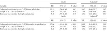
Both colonization with toxigenic C. difficile on admission and colonization during hospitalization were associated with an increased risk for development of CDI in univariable analyses (Table 3). After adjusting for exposures to penicillins and length of ICU stay before CDI, colonization with toxigenic C. difficile upon admission or hospitalization remained associated with an increased risk of development of CDI during hospitalization (Table 3) and for up to 1 month after discharge (data not shown).
Crude and Adjusted Relative Risks (RRs) for Development of Clostridium difficile Infection (CDI) During Hospitalization

NOTE. ICU, intensive care unit.
a The multivariable model includes exposure to penicillins during hospitalization, length of ICU stay prior to CDI, and colonization with toxigenic C. difficile on admission.
b The multivariable model includes exposure to penicillins during hospitalization, length of ICU stay prior to CDI, and colonization with toxigenic C. difficile during hospitalization.
The Pearson and deviance goodness-of-fit tests for the multivariable regression models revealed insignificant P values indicating adequate model-fit (χ2=419.2, P>.99, and χ2=57.7, P>.99, respectively, for the model including colonization with toxigenic C. difficile on admission; χ2=548.1, P=.372, and χ2=59.7, P>.99, respectively, for the model including colonization during hospitalization). Receipt of proton pump inhibitors during hospitalization was not included in the multivariable regression model because all patients with CDI were exposed.
Strains were available for ribotyping from 14 of 17 patients found to be colonized on admission. PCR ribotypes 001 and 027 were most commonly identified, accounting for 4 (29%) and 3 (21%) of the 14 strains, respectively. Ribotype 014 was determined in 2 patients and ribotypes 126, 056, 015, 003, and 002 in 1 patient each.
DISCUSSION
In ICU patients, colonization with toxigenic C. difficile on admission or during hospitalization increases the risk for development of CDI during hospitalization and up to 1 month after discharge.
The rate of colonization with toxigenic C. difficile on admission identified in our study was low—reflecting colonization rates described for the general populationReference Alasmari, Seiler, Hink, Burnham and Dubberke 10 but also similar to the 4% reported in a large multicenter trial conducted in Canada.Reference Loo, Bourgault and Poirier 11 One explanation for the low proportion of patients colonized upon admission may be the high proportion of patients without prior exposure to healthcare facilities and with none or few comorbid conditions, reflected by the low mean Charlson comorbidity index identified in our study. We cannot rule out that our low prevalence of toxigenic C. difficile upon hospital admission may be due to the use of rectal swab rather than stool specimens and the use of PCR rather than anaerobic culture for detection.Reference Curry, Schlackman and Hamilton 12 Other studies report asymptomatic carriage with toxigenic C. difficile on admission for up to 14% of patients.Reference McFarland, Mulligan, Kwok and Stamm 13 – Reference Samore, DeGirolami, Tlucko, Lichtenberg, Melvin and Karchmer 18 Even higher rates of colonization with C. difficile have been identified on admission to rehabilitation facilities, with 16% of stool samples testing positive.Reference Marciniak, Chen, Stein and Semik 19 Similarly, a C. difficile colonization rate of 15% has been described in general medical and surgical patients.Reference Alasmari, Seiler, Hink, Burnham and Dubberke 10
Although earlier studies revealed recent exposure to antibiotics and healthcare facilities as important risk factors for colonization with toxigenic C. difficile on hospital admission,Reference Loo, Bourgault and Poirier 11 , Reference McFarland, Mulligan, Kwok and Stamm 13 , Reference Hutin, Casin and Lesprit 15 , Reference Leekha, Aronhalt, Sloan, Patel and Orenstein 17 , Reference Samore, DeGirolami, Tlucko, Lichtenberg, Melvin and Karchmer 18 both our study and another recent investigationReference Alasmari, Seiler, Hink, Burnham and Dubberke 10 were not able to replicate these findings. Apart from solid organ transplantation, we were not able to identify any risk factors for colonization with toxigenic C. difficile on admission; however, we cannot rule out that our sample size is too small to detect such associations. Nevertheless, a growing body of knowledge points to C. difficile being acquired in the community setting without any clear antibiotic or healthcare exposuresReference Khanna, Pardi and Aronson 20 —affecting patients previously considered as being at low risk as otherwise healthy people without prior exposure to antibiotics 21 , 22 as well as peripartum womenReference Garey, Jiang, Yadav, Mullins, Wong and Dupont 23 , Reference Rouphael, O'Donnell and Bhatnagar 24 and children.Reference Benson, Song, Campos and Singh 25 Possible sources may arise from the food chain; there is growing concern that C. difficile may be acquired from ingestion of spores in contaminated foods.Reference Hoover and Rodriguez-Palacios 26
Importantly in this study, colonization with toxigenic C. difficile on admission was identified as a strong and independent predictor for development of subsequent CDI. This finding suggests that traditional infection control measures may not suffice to avoid further increase in disease-burden. For patients colonized with toxigenic C. difficile, the challenge for disease prevention is not to prevent exposure but to reduce the risk of C. difficile toxin expression by restricting use of antibiotics,Reference Loo, Bourgault and Poirier 11 acid-suppressive agents,Reference Deshpande, Pant and Pasupuleti 27 and narcotics.Reference Dubberke, Reske and Olsen 28
Furthermore, it needs to be acknowledged that, in contrast to these recent findings, an earlier study, based on data collected in the 1980s and early 1990s, reported that primary symptomless C. difficile colonization was associated with a decreased risk of CDI. Risk reduction was found with colonization with either nontoxigenic or toxigenic strains.Reference Shim, Johnson, Samore, Bliss and Gerding 29 Novel diagnostic approaches—including real-time PCR—and changing epidemiology may explain these conflicting results. One possible underlying mechanism for the finding of C. difficile colonization as a protective factor for CDI-development may be that lack of IgG against C. difficile toxins was described as an important risk factor for CDI for hospitalized patients with C. difficile colonization.Reference Kyne, Warny, Qamar and Kelly 16 , Reference Kyne, Warny, Qamar and Kelly 30 Subsequent studies also showed monoclonal antibodies to C. difficile toxins A and B to be protective against CDI relapse, indicating the defensive role of serologic response and thus suggesting a mechanism for carriage plus risk without CDI.Reference Lowy, Molrine and Leav 31
Our study has some important limitations. These include its conduction at a single center and its reliance on clinical chart review to identify both exposures to known risk factors for development of CDI and diagnosis of CDI. We used the definitions outlined in the Society for Healthcare Epidemiology of America/ Infectious Diseases Society of America guidelinesReference Cohen, Gerding and Johnson 5 to differentiate CDI likely acquired before hospital admission rather than during hospitalization, suggesting a cut-off of 48 hours from admission. Our results and conclusions, however, did not change when applying the National Healthcare Safety Network reporting criteria for healthcare facility onset of CDI of greater than 3 days from admission. The low number of patients colonized with toxigenic C. difficile in our population may result in the study being underpowered for detection of risk factors for colonization and limits the precision of our point estimates, reflected by broad confidence intervals. We were not able to assess the interaction between carriage at admission and subsequent exposure to antibiotics because all 17 patients colonized on admission received antibiotics during their hospital stay. Due to the short mean duration of ICU stay, weekly follow-up swabs could be performed in only a very small proportion of patients. Toxigenic C. difficile strains identified at diagnosis of CDI in the 2 patients found to be colonized at admission were not available for ribotyping, therefore identity of strains could not be assessed. Further, follow-up information was missing at 1 month after discharge for 149 (27.5%) of the 542 patients included in this study and we cannot rule out that CDI was missed in some patients with symptom onset after discharge. We were, however, not able to identify any differences in baseline characteristics, exposures, or colonization with toxigenic C. difficile between patients with and without follow-up information.
In conclusion, in ICU patients, colonization with toxigenic C. difficile is an independent risk factor for development of subsequent CDI. The low rate of colonization found in a general ICU population questions the utility of this screening strategy for identification of patients at risk for C. difficile in this setting. Further studies are needed to identify populations with higher toxigenic C. difficile colonization rates possibly benefiting from screening or avoidance of agents known to promote CDI.
ACKNOWLEDGMENTS
Financial support. The Sherrilyn and Ken Fisher Center for Environmental Infectious Diseases, Division of Infectious Diseases of the Johns Hopkins University School of Medicine.
Potential conflicts of interest. S.T.-S. reports that she is funded by the Swiss National Science Foundation; the Medical Division of the Lichtenstein Foundation of the University of Basel, Switzerland; and the Scientific Society Basel, Switzerland.
All other authors report no conflicts of interest relevant to this article.
Disclaimer: This publication was made possible by support from the Sherrilyn and Ken Fisher Center for Environmental Infectious Diseases, Division of Infectious Diseases of the Johns Hopkins University School of Medicine. Its contents are solely the responsibility of the authors and do not necessarily represent the official view of the Fisher Center or Johns Hopkins University School of Medicine.