Introduction
Alternative approaches to chemotherapy for the control of nematodes and trematodes in ruminants are urgently needed in India and elsewhere, because of rapidly developing anthelmintic resistance in parasitic helminths of ruminants and increasing concern about chemical residues in livestock products and the environment (Sanyal, Reference Sanyal2001). Biological control methods using nematophagous fungi could be one step in this direction (Sanyal, Reference Sanyal2005). Biological control is aimed at exploiting natural enemies to reduce the number of target organisms to a level less than that occurring in the absence of the predators. Egg-parasitic fungi are one of the potent candidates for biological control. Egg-parasitic fungi are characterized by their ability to colonize parasite cysts and eggs. They generally attack by penetrating the eggs using specialized perforating organs or lateral branches from the mycelium (Stirling, Reference Stirling and Stirling1991). Recently, two egg-parasitic fungi, Paecilomyces lilacinus and Verticillium chlamydosporium, were isolated from a manure heap of the Chhattisgarh region in India (Sanyal et al., Reference Sanyal, De, Sarkar, Patel, Mandal and Pal2008).
As all the species of fungi commonly found in nature may not necessarily be efficient biocontrol agents against animal parasites, they should be screened for their growth, predatory activity, ability to produce chlamydospores which are easy to isolate and germinate, and ability to survive intestinal transit in animals (Peloille, Reference Peloille1991; Sanyal, Reference Sanyal2005). Different physical and chemical factors influence the growth and spore production of nematophagous fungi (Gronvold et al., Reference Gronvold, Korsholm, Wolstrup, Nansen and Henriksen1985, Reference Gronvold, Wolstrup, Nansen, Larsen, Heriksen, Bjorn, Kichheiner, Lassen, Rawat and Kristiansen1999). Among many parameters suggested by Peloille (Reference Peloille1991) for the use of fungi as biocontrol agents against zooparasites, two important criteria are that they should grow well in a temperature range of 15–35°C and significantly reduce the number of prey in the environment.
The present paper deals with studies on the influence of different media types, temperature and pH on in vitro growth profiles of P. lilacinus and V. chlamydosporium. Predatory activity of the fungi was evaluated in agar plates baited with eggs of Fasciola gigantica (liver fluke) and Gigantocotyle explanatum (hepatic amphistome).
Materials and methods
Fungi, culture media, temperature, pH
Wild isolates of V. chlamydosporium and P. lilacinus recently isolated from organic samples of Durg, Chhattisgarh (Sanyal et al., Reference Sanyal, De, Sarkar, Patel, Mandal and Pal2008) were maintained as stock cultures in 2.5% corn-meal agar (CMA) medium at 26°C.
Five different types of in vitro culture media – CMA, potato-dextrose agar (PDA), nutrient agar (NA), Sabouraud glucose agar (SGA) and water agar (WA) (Hi-Media, Mumbai, India) – were used in this study; 2.5% solutions of the media were prepared in conical flasks and autoclaved. Antibiotic solutions (0.02% tetracycline and neomycin (w/v)) were added to the media to suppress bacterial growth and media were poured into radiation-sterilized Petri plates (90 mm diameter) in a laminar air flow chamber and allowed to solidify.
The preferred medium observed from the experiments on media type, was taken as the standard medium. In separate experiments, the effect of six different temperatures (4°C, 10°C, 18°C, 26°C, 34°C or 40°C) and pHs of 4, 5, 6, 7 or 8, set up by using 1 m HCl and 1 m NaOH, were monitored by observing the growth profiles of the fungi.
Growth assay
An agar disc of about 6 mm diameter, scooped from the periphery of 15-day-old stock cultures of P. lilacinus and V. chlamydosporium, was placed at the centre of media plates (90 mm) in all the experiments (five replicates). The cultures were incubated and colony diameters of the fungi were measured at regular intervals. Two measurements of colony diameter of mycelia spread on agar surface, each perpendicular to the other, were taken from each plate.
Increase in the colony diameter of the fungi with respect to time was taken as a criterion for assessment of growth on different media as well as in studies on the effect of different treatments on the growth. Generally, no, or less, increase in the diameter in the early period of incubation after inoculation constituted a lag phase. Subsequently, at least a 5 mm increase in the diameter of the fungal colony within an interval of 8–10 h was judged to be a phase of rapid growth, and the overall period during which identical and sustained increase in the colony diameter occurred was taken as a log phase. Finally, after the end of the log phase, a slow increase in the colony diameter was taken as a stationary phase.
Predatory activity
Full-grown agar plates of P. lilacinus and V. chlamydosporium were baited with viable eggs of F. gigantica and G. explanatum collected from the gall bladder of buffaloes from a local abattoir and plates were examined under the microscope twice daily for signs of egg destruction by each fungal species.
Statistical analysis
Means of data associated with all parameters were tested by two-way analysis of variance using GraphPad Prism software (GraphPad Software Inc., San Diego, California USA, 1990–1994) and considered significant at P < 0.05.
Results
Influence of media type
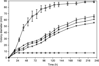
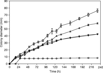
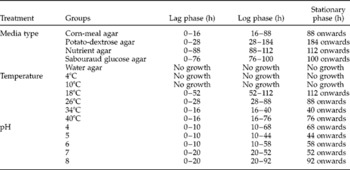
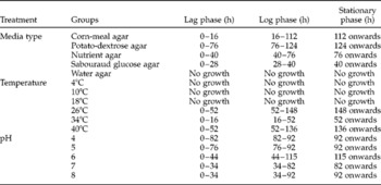
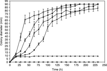
Observations on the influence of media type on in vitro growth profiles of P. lilacinus are presented in fig. 1. Maximum growth was obtained in CMA compared to any other media types (P < 0.001). PDA, SGA and NA provided similar growth rates, and after an initial growth of 7.7 mm at 16 h of incubation, no further growth of P. lilacinus was observed in WA. The preferred medium of V. chlamydosporium was CMA, followed by PDA (fig. 2). NA and SGA provided much lower (P < 0.001) but uniform growth compared to CMA and PDA. Similar to P. lilacinus, an initial growth of 7.7 mm at 16 h of incubation was observed in WA for V. chlamydosporium and no further growth was observed. Colony diameters of P. lilacinus indicated that after an initial lag phase of up to 16 h, log phase began and continued for up to 88 h, followed by a stationary phase that lasted until the end of the observation period (table 1). Similarly for V. chlamydosporium, the log phase began after an initial lag phase of 16 h and continued up to 112 h, followed by a stationary phase until the end of the observation period (table 2). The observations about the growth of both fungi on other media were not satisfactory. Considering the sustenance and uniformity in the pattern of growth, CMA was the preferred medium in all the subsequent experiments.
In vitro growth profiles (mean ± SEM) of Paecilomyces lilacinus in corn-meal agar (□), potato-dextrose agar (△), nutrient agar (▿), Sabouraud glucose agar (⋄) and water agar (○).

In vitro growth profiles (mean ± SEM) of Verticillium chlamydosporium in corn-meal agar (□), potato-dextrose agar (△), nutrient agar (▿), Sabouraud glucose agar (⋄) and water agar (○).

Growth phases of Paecilomyces lilacinus in different media types, temperature and pH.

Growth phases of Verticillium chlamydosporium in different media types, temperature and pH.

Influence of incubation temperature and pH
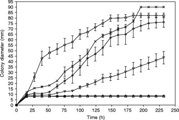
Observations on the influence of incubation temperature on in vitro growth profiles of P. lilacinus and V. chlamydosporium are presented in figs 3 and 4, respectively. While no and very limited growth of both the fungi were observed at 4°C and 10°C, growth profiles were optimal at 26–40°C for P. lilacinus. As the temperature was increased from 26°C, growth became much faster initially at 34°C (P < 0.001), but growth was accelerated in the 40°C group compared the 34°C group by 124 h and both attained the maximum of 90 mm by 184 h of incubation. Growth of V. chlamydosporium was very sluggish initially at 18°C (P < 0.001) but increased after 100 h of incubation. Growth profiles were initially fastest at 34°C, but the 40°C group increased growth and matched that of the 34°C groups by 184 h and attained the maximum of 90 mm by 196 h of incubation. The optimal growth of both the fungi was observed at 26°C with the maintenance of a lag phase, a log phase and a stationary phase (tables 1 and 2). At 34°C, the stationary phase began at 40 h onwards, while at 40°C, the stationary phase started at 76 h. At temperatures above 26°C, there was a marked reduction in the time taken to attain maximum growth, which is indicative of the ability of the fungi to adapt even at higher temperatures.
In vitro growth profiles (mean ± SEM) of Paecilomyces lilacinus in corn-meal agar plates incubated at 4°C (□), 10°C (△), 18°C (▿), 26°C (⋄), 34°C (○) or 40°C ( × ).

In vitro growth profiles (mean ± SEM) of Verticillium chlamydosporium in corn-meal agar plates incubated at 4°C (□), 10°C (△), 18°C (▿), 26°C (⋄), 34°C (○) or 40°C ( × ).

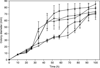
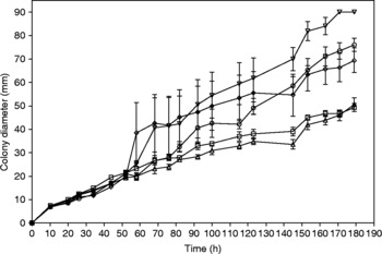
Observations on the influence of pH on in vitro growth profiles of P. lilacinus and V. chlamydosporium are presented in figs 5 and 6, respectively. While a range of pH (pH 4–8) supported growth of P. lilacinus, significantly increased colony diameter was observed at pH 6 following 58 h of incubation and both at pH 6 and 7 following 52 h of incubation. While no difference in growth profiles of V. chlamydosporium was observed at all the pH tested up to 100 h of incubation, significantly increased growth (P < 0.05) was observed at pH 6 and 7 following 115 and 123 h of incubation, respectively. Later on growth at pH 6 increased steadily (P < 0.001) and achieved a maximum diameter of 90 mm growth by 171 h of incubation. Lag, log and stationary phases in media of different pH are presented in tables 1 and 2 for P. lilacinus and V. chlamydosporium, respectively. There was not much variation in growth phases for P. lilacinus at pH 6 and pH 7 except that at pH 6 the lag phase continued for 48 h as against 32 h at pH 7.
In vitro growth profiles (mean ± SEM) of Paecilomyces lilacinus in corn-meal agar plates at pH 4 (□), 5 (△), 6 (▿), 7 (⋄) or 8 (○), incubated at 26°C.

In vitro growth profiles (mean ± SEM) of Verticillium chlamydosporium in corn-meal agar plates at pH 4 (□), 5 (△), 6 (▿), 7 (⋄) or 8 (○), incubated at 26°C.

Predatory activity
Verticillium chlamydosporium appeared to be more vigorous in its egg-parasitic ability compared to P. lilacinus. Clumping of eggs of F. gigantica and G. explanatum was observed on day 2 of egg baiting in CMA plates of V. chlamydosporium, followed by the distortion of eggshells on day 3 onwards. On the contrary, P. lilacinus exhibited very limited egg-destroying ability, some of the baited eggs even showed development of miracidia on day 7 of baiting.
Discussion
The present study reconfirmed earlier results that CMA, followed by PDA, is the best culture medium suited for in vitro growth of the Indian isolates of the fungi imperfecti Arthrobotrys oligospora (Mukhopadhyaya et al., Reference Mukhopadhyaya, Nagee, Soni and Sanyal2001) and Duddingtonia flagrans (Sanyal, Reference Sanyal2000). Equivalent growth of P. lilacinus and V. chlamydosporium in a semi-synthetic medium such as CMA and PDA and no growth in WA hinted at marked dependence of these species on vitamins and trace element supplements. No growth in synthetic medium such as WA, indicted prototype attributes of these species, indicative of a distinct ecological disadvantage compared to A. oligospora and D. flagrans which grew very well in synthetic media such as WA and Czapex Dox (CD) medium (Mukhopadhyaya et al., Reference Mukhopadhyaya, Nagee, Soni and Sanyal2001). Based on the results, all the subsequent experiments were conducted in CMA medium.
The Indian subcontinent experiences a wide range of temperature throughout the year. Certain regions experience diurnal temperature of 40°C and above. The optimal growth of P. lilacinus and V. chlamydosporium was obtained at 26°C, a finding also observed earlier with the Indian isolates of nematode-trapping deuteromycetes (Sanyal, Reference Sanyal2000; Mukhopadhyaya et al., Reference Mukhopadhyaya, Nagee, Soni and Sanyal2001). However, significant growth of P. lilacinus and V. chlamydosporium at 34°C and 40°C is encouraging from the point of view of their ecological adaptation in a tropical climate. This is indicative of the possibility of survival of a basal level of a fungal population that would otherwise have been extinct due to temperature extremes. However, no visible growth at 4°C and 10°C is indicative of their sensitivity to low temperatures. pH has an indirect effect on the growth of the fungi. Variation in pH often leads to alteration of media components to yield moieties of greater or lesser toxicity. Significant growth of P. lilacinus and V. chlamydosporium in acidic and basic environments is encouraging from the point of view of their future application in soil, irrespective of its pH status, where the possibility of their survival ensures efficient biological control.
Few attempts have been carried out to test the egg-destroying ability of the egg-parasitic fungi against long-lasting egg stages of animal parasites. Verticillium chlamydosporium and other Verticillium spp. were able to degrade the eggshells of Ascaris lumbricoides of pigs (anticipated to be A. suum) enzymatically and infect the eggs (Lysek & Krajci, Reference Lysek and Krajci1987; Lysek & Sterba, Reference Lysek and Sterba1991; Kunert, Reference Kunert1992). Short exposure to high temperature and/or UV-irradiation rendered the eggs more susceptible to fungal attack (Lysek & Bacovsky, Reference Lysek and Bacovsky1979). Araujo et al. (Reference Araujo, Santos and Ferraz1995) tested three fungal species (the net-forming predacious fungi Arthrobotrys conoides, A. robusta and the egg-parasitic fungus P. lilacinus) for activity against the eggs of Toxocara canis, and observed very little impact of nematode-trapping fungi, while P. lilacinus showed some predatory activity (16% eggs infected after 7 days). Quoting from the work done in the USA, Larsen (Reference Larsen2000) reported that V. chlamydosporium attacked and destroyed the eggs of Ascaridia galli and Parascaris equorum but only rarely invaded Trichuris suis. In Denmark, a range of different fungal species and isolates are under investigation as potential biocontrol agents against A. suum, T. suis, Nematodirus spp. and Fasciola hepatica. Preliminary results indicated fairly good egg-destroying activity of V. chlamydosporium against relatively thin-shelled eggs of N. filicollis and N. battus, and to a certain extent also against A. suum, while T. suis eggs appeared to be much more resistant to fungal attack (Larsen, Reference Larsen2000).
The present study revealed that the Indian isolates of egg-parasitic fungi grew well in CMA at a temperature range of 26–40°C and at a pH range of 6–7. Verticillium chlamydosporium appeared to be more vigorous in its egg-parasitic ability compared to P. lilacinus.
Acknowledgements
The research was the result of the All India Network Project on Gastrointestinal Parasitism of the Indian Council of Agricultural Research, India. Facilities provided by the Dean, College of Veterinary Science and Animal Husbandry, Anjora, Durg, Chhattisgarh, India are thankfully acknowledged.