1. Introduction
Aglaspidids are relatively rarely encountered Cambrian–Ordovician arthropods that have been variously placed systematically within the crown-group Euarthropoda (see Ortega-Hernández, Reference Ortega-Hernández2016 for use of terminology). A comprehensive recent assessment (Ortega-Hernández, Legg & Braddy, Reference Ortega-Hernández, Legg and Braddy2013) has recovered them within Artiopoda, a clade comprising the sister groups Trilobitomorpha and Vicissicaudata. The latter includes xenopodans and cheloniellids, together with aglaspidids sensu stricto and associated genera.
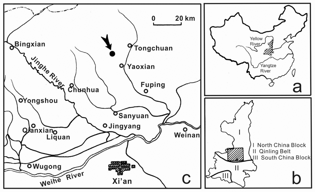
Aglaspidids were recognized from China only very recently, with the description of a new species of Aglaspella (Lerosey-Aubril, Ortega-Hernández & Zhu, Reference Lerosey-Aubril, Ortega-Hernández and Zhu2013), together with Glypharthrus? sp. and an aglaspidid-like euarthropod (Zhu et al. Reference Zhu, Peng, Zamora, Lefebre and Chen2016), from the upper Cambrian, Furongian, Sandu Formation of Guangxi Province. The material discussed in the present account was collected in 1977 during an investigation of the Ordovician stratigraphy of the western margin of the North China Platform by members of the Nanjing Institute of Geology and Palaeontology, Chinese Academy of Sciences (NIGPAS) and the Changqing Research Institute of Petroleum Exploration and Development. The specimens were obtained from the type section of the Upper Ordovician Yaoxian Formation at Taoqupo, 11 km NW of Yaoxian, Shaanxi Province (Fig. 1).
Occurrence maps. (a) China; (b) Shaanxi Province (cross-hatched area in (a)). (c) Xi'an and the area north of it (cross-hatched in (b)), containing the position of the measured geological section through the Yaoxian Formation (Fig. 2), which also contains the locality (arrowed) that has yielded Gogglops ensifer.

Aglaspidids apparently always occur in marine sediments. Almost all previous discoveries of such euarthropods have been preserved in fine- to medium-grained clastics (e.g. Hesselbo, Reference Hesselbo1992; Fortey & Rushton, Reference Fortey and Rushton2003; Rak et al. Reference Rak, Bergström, Fatka and Budil2009; Lerosey-Aubril et al. Reference Lerosey-Aubril, Ortega-Hernández, Kier and Bonino2013; Ortega-Hernández, Van Roy & Lerosey-Aubril, Reference Ortega-Hernández, Van Roy and Lerosey-Aubril2016). An exception is the material of Chlupacaris dubia Van Roy, Reference Van Roy2006 from the Upper Ordovician First Bani Group, likely from the Katian Upper Tiouririne Formation within this unit, of Morocco, which is preserved in sandstone. Aglaspidids are also frequently flattened post-mortem, while their cuticle material may have been dissolved (e.g. Fortey & Rushton, Reference Fortey and Rushton2003; Van Roy, Reference Van Roy2006) or replaced by subsequent mineralization. The fossil material described here is in a fine-grained limestone and, although it is disarticulated, it is the morphologically best-preserved, undistorted aglaspidid known. It preserves a cuticle composed of calcite, and demonstrates what we believe is an uncommon phosphatization of the visual surface. It also provides further evidence that at least some aglaspidids have considerable original convexity (e.g. Van Roy, Reference Van Roy2006; Rak et al. Reference Rak, Bergström, Fatka and Budil2009). This in turn affects the way shale-preserved specimens might be interpreted in future. Here we describe the new species and place it in the context of aglaspidid phylogeny, and re-evaluate the chemical composition of the cuticle which has been controversial.
2. Stratigraphy and age
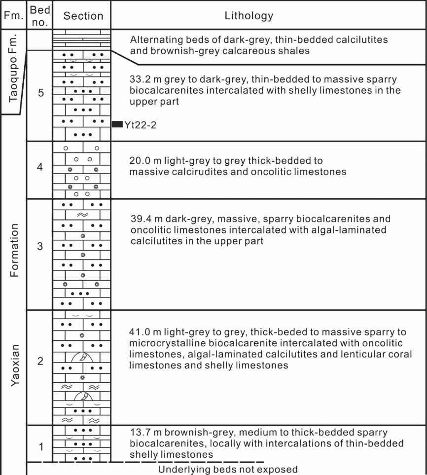
The Yaoxian Formation at its type locality is overlain by the Taoqupo Formation, a suite of medium- to thin-bedded calcilutites intercalated with shales and mudstones, although the beds underlying it are covered by recent fluvial deposits and are unexposed. It is composed mainly of thick-bedded to massive biocalcarenites, calcirudites and algal limestones. In addition to the aglaspidids, it yields abundant nautiloids, conodonts and brachiopods, together with trilobites and corals (Chen et al. Reference Chen, Zhou, Lin, Yang, Zou, Wang, Luo, Yao and Shen1984).
Aglaspidids occur only at Horizon Yt 22-2 of Bed 5, in the upper part of the formation (Fig. 2). The associated conodonts, for example Tasmanognathus carayi Burrett, Belodina compressa (Branson & Mehl), Pseudobelodina dispansa (Glenister), Dapsilodus mutatus (Branson & Mehl) and Periodon gracilis (Ethington), are quite representative of the Yaoxian Formation, and these are also accompanied by some endemic forms (see Wang & Luo, Reference Wang and Luo1984; An, Zhang & Zhu, Reference An, Zhang and Xu1985; An & Zheng, Reference An and Zheng1990).
Geological section through the Ordovician Yaoxian Formation, showing the bed numbers referred to in the text and the occurrence (black bar) of the Gogglops ensifer aglaspidid material.

The Yaoxian Formation was considered to be of Blackriverian (Wang & Luo, Reference Wang and Luo1984), or late Blackriverian to early Trenton age (An, Zhang & Zhu, Reference An, Zhang and Xu1985; An & Zheng, Reference An and Zheng1990). However, according to the conodont species listed from 47 successive horizons of the formation by An & Zheng (Reference An and Zheng1990), Tasmanognathus carayi Burrett and Belodina compressa (Bransson & Mehl) – the index forms for the Gordon Subgroup (Gisbornian) of central Tasmania, Australia and the Plattin Formation (Blackriverian-middle Shermanian) of Missouri, USA, respectively – make their first appearance in the uppermost part of Bed 1. The evidence is therefore further indicative of the B. compressa Zone, latest Blackriverian, in terms of the zonal scheme of the North American Midcontinent Province. This conclusion is consistent with the occurrence of Phragmodus undatus Branson and Mehl in the basal part of the overlying Taoqupo Formation (see An & Zheng, Reference An and Zheng1990). Wang, Bergström & Lane (Reference Wang, Bergström and Lane1996) believed that the B. compressa Zone might correspond approximately to the Prioniodus alobatus Subzone of the Amorphognathus tvaerensis Zone in terms of the zonal scheme of the North Atlantic Province, a subzone that is of latest Sandbian to earliest Katian age (Zhen et al. Reference Zhen, Wang, Zhang, Bergström, Percival and Cheng2011).
Correlation of the Yaoxian Formation with the graptolite sequence is still problematical. However, Cooper (Reference Cooper1999) suggested that the compressa conodont zone may fall within the multidens (= foliaceus) graptolite zone, the latter, in turn, belonging largely to the Burrellian Stage (late early Caradoc) of the British chronostratigraphical standard (Fortey et al. Reference Fortey, Harper, Ingham, Owen and Rushton1995, Reference Fortey, Harper, Ingham, Owen, Parkes, Rushton and Woodcock2000). This evidence indicates, in terms of the global standard stages, an early Late Ordovician, Sandbian Stage age (Cocks, Fortey & Rushton, Reference Cocks, Fortey and Rushton2010) for Horizon Yt 22-2.
3. Biofacies and environmental setting
Trilobites of the Yaoxian Formation include species of Illaenus, Sphaerexochus, Pliomerina and Glaphurina, with Illaenus specimens making up to 70% of all trilobite specimens collected (Zhou, Zhou & Zhang, Reference Zhou, Zhou and Zhang1989). This generic association is suggestive of the Illaenid-cheirurid community, which is characteristic of carbonate build-ups deposited at or near wave base in shallow-water environments along platform margins (Fortey, Reference Fortey1975, Reference Fortey1980). The brachiopod fauna accompanying the trilobites is of low diversity, and comprises Didymelasma, Orthambonites and Sowerbyella (see Chen et al. Reference Chen, Zhou, Lin, Yang, Zou, Wang, Luo, Yao and Shen1984), Didymelasma specimens outnumbering those of the others. This brachiopod fauna may have lived in shallow water, probably at depths of 30–40 m, and formed part of Benthic Assemblage 2 or the upper part of Benthic Assemblage 3 (pers. comm. Rong Jiayu, Nanjing Institute of Geology and Palaeontology, 1990). Nautiloids have been assigned to the Liulinoceras-Kotoceras Assemblage, which includes mostly endemic forms (Chen & Zou, Reference Chen and Zou1984). Based on a few specimens from the Yaoxian Formation, the strength index of nautiloids indicates that they have a depth limitation of about 70 m (Zhang, Reference Zhang1998) although the depth that they occupied in life would be expected to be much shallower (Westermann, Reference Westermann1973).
All the trilobites and aglaspidids are disarticulated and some are locally concentrated, and some aglaspidid specimens apparently represent moults (see below, species description). Most nautiloid specimens are preserved sub-parallel with each other on bedding surfaces. Bioclastic beds of 0.3–1.5 m thick appear at several levels; these are formed of abundant shelly debris that consists mainly of brachiopods, algae and corals. The taphonomic evidence suggests a high-energy environment.
The Yaoxian Formation has been interpreted as bank facies (Feng, Chen & Zhang, Reference Feng, Chen and Zhang1991). This view is supported by the lack of clayey lime in most beds of the formation, such as in the sparry calcarenites, calcirudites and oncolitic limestones. However, some intercalated lenses of algal-laminated calcilutites and coral-constructed limestones, which occur at different horizons, indicate that small, sparse mud mounds and reefs may have periodically developed on the banks. Late early Caradoc age carbonate build-ups were intermittently distributed along the western margin of the North China Platform (Zhou, Zhou & Zhang, Reference Zhou, Zhou and Zhang1989, figs 7, 8; Zhou et al. Reference Zhou, Ni, Lin, Zhou, Yu, Zhou and Dean1996, fig. 5-2), but were well developed only on its southwestern edge north of the Weihe River, at Tongchuan, Yaoxian, Chunhua, Yongshou, Liquan and Qianxian (Ye et al. Reference Ye, Yang, Xu, Zheng, Zuo, Song, Li and Fan1996).
4. Cuticle of aglaspidids reconsidered
The new aglaspidid described in this paper is preserved in full relief in limestone and is among the best material yet discovered for understanding the cuticle composition of these interesting euarthropods. This subject has been a matter of longstanding controversy from at least the time of Raasch (Reference Raasch1939), who conducted the most significant pioneer research on the group and suggested that the cuticle of aglaspidids was of primary phosphatic composition. The most common preservation of aglaspidid fossils is in shale and other fine to medium clastics, and original cuticle material is often lacking. The question of its original cuticle composition is further complicated by claims that Ordovician phosphatic fragments described as early vertebrate bone (Anatolepis, see Bockelie, Bruton & Fortey, Reference Bockelie, Bruton and Fortey1976; Bockelie & Fortey, Reference Bockelie and Fortey1976) were in fact pieces of aglaspidid euarthropod cuticle (Peel & Higgins, Reference Peel and Higgins1977; Peel, Reference Peel1979). The most recent studies (e.g. Smith, Sansom & Repetski, Reference Smith, Sansom and Repetski1996) nevertheless support a vertebrate interpretation. These unrelated taxa may indeed have similar-looking external tubercles. This claim led Briggs & Fortey (Reference Briggs and Fortey1982) to re-examine apparently well-preserved phosphatic cuticles of the typical aglaspidid Aglaspis (the species A. spinifer Raasch, Reference Raasch1939 and A. barrandei Hall, Reference Hall1862) from the late Cambrian St Lawrence Formation of Wisconsin, USA. That these cuticles were made of calcium phosphate was confirmed by X-ray diffraction. Thin-sections showed what was regarded as typical laminated euarthropod endocuticle, which was differentiated from the structures shown by Bockelie & Fortey (Reference Bockelie and Fortey1976) in Anatolepis. However, Briggs & Fortey (Reference Briggs and Fortey1982, p. 25) were careful to state that ‘the possibility of replacement of the Aglaspis cuticle [by calcium phosphate] cannot be eliminated on the basis of the very perfect preservation of the fabric alone’, although on field evidence they opted for an originally phosphatic composition. They also entertained the possibility that their descriptions might have been based on ecdysial material rather than that from complete carapaces, and that there was a possibility that they were sampling the outer part only of the cuticle (their material was only 10–35 µm thick, and its inner surface was not well-defined and showed truncated laminae). Since then, many discoveries have pointed to the perfection of calcium phosphate replication. In the Upper Cambrian Orsten fauna of Scandinavia replication of the finest details to the micron level is well known, and this can apply to both calcified and uncalcified tissues (e.g. in Agnostus; Muller & Walossek, Reference Muller and Walossek1987). Experimental demonstrations of the phosphatization process have also been carried out in the laboratory (e.g. Briggs & Kear, Reference Briggs and Kear1993; Briggs et al. Reference Briggs, Kear, Martill and Wilby1993).
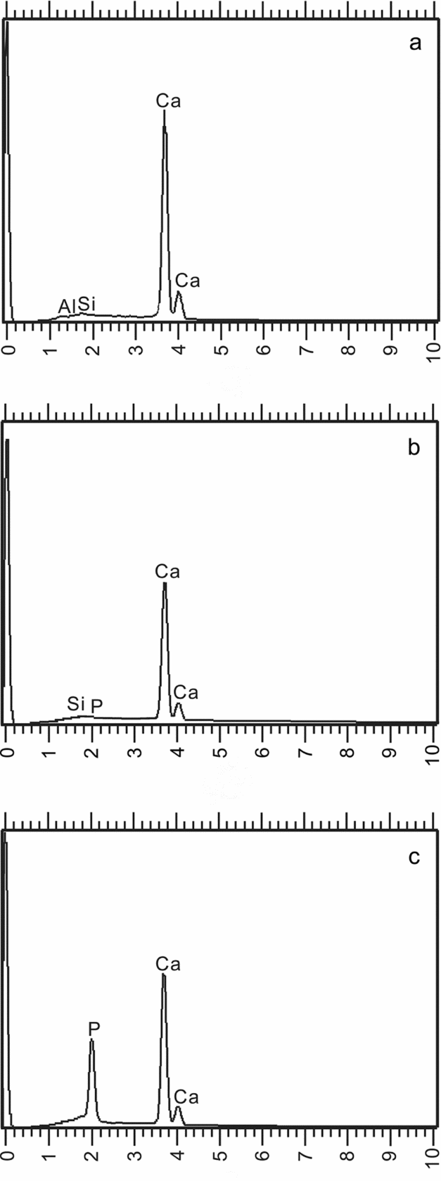
The Chinese aglaspidid cuticle is rather narrow; as measured in thin-section, its width ranges over c. 15–35 µm. This is similar in width to the material studied by Briggs & Fortey (Reference Briggs and Fortey1982), and we also appear to have some exuviae in our samples (see Sections 3 and 6). However, in terms of composition it is incontrovertible that the bulk of the dorsal exoskeleton and the surrounding sediment is composed of calcium carbonate (calcite), as confirmed by scanning electron microscope with energy-dispersive X-ray (SEM EDX) analysis (Fig. 3a, b; OUM BY.1337) together with thin-section analysis (OUM BY.1368/p - BY.1370/p; Fig. 4a–c, g, j, k). In contrast, the visual surfaces of the eyes are preserved in calcium phosphate (Figs 3c, 4 a, b, d, l, m). This is consistent through a sample of over 30 specimens. We believe that this differential preservation reflects an original compositional difference: that while the dorsal exoskeletal cuticle was made of calcite, the eye itself was uncalcified. The eyes then apparently became a preferential site for phophatization, this process being supported by knowledge of probable bacterially mediated, early phosphatization of retinal structures in Jurassic conchyliocaridid euarthropods (Fröhlich et al. Reference Fröhlich, Mayrat, Riou and Secretan1992). The pearly, dark appearance of the eye in the Chinese aglaspidid specimens, which is typically that of a phosphatic surface, is readily recognizable. If the aglaspidids comprise a natural group, then it is appropriate to ask if calcite cuticle was characteristic of the whole clade, since the new material is unambiguously calcitic (the possession of a calcified cuticle was also deduced for Chlupacaris dubia from Upper Ordovician strata of Morocco; Van Roy, Reference Van Roy2006). If this were the case then the Aglaspis cuticles studied by Briggs & Fortey (Reference Briggs and Fortey1982) were likely to have been phosphatized at an early stage in diagenensis, sufficient to preserve the microfabric. However, it should be noted that a possible primary phosphatic cuticle has also subsequently been described in the cuticles of other aglaspidids, for example Quasimodaspis brentsae from Cambrian sediments of Nevada (Waggoner, Reference Waggoner2003) and Tremaglaspis vanroyi from Cambrian deposits of Utah (Lerosey-Aubril et al. Reference Lerosey-Aubril, Ortega-Hernández, Kier and Bonino2013). A primary phosphatic composition for aglaspidids-based palaeoenvironmental factors has also been suggested (Ortega-Hernández et al. Reference Ortega-Hernández, Braddy, Jago and Baillie2010: Australaglaspis). Most recently, during a very late stage review of our paper, a cuticle comprising a thin (of unspecified measurement), biomineralized, purportedly primary phosphatic outer layer above a clay, originally organic inner layer has been described in Glypharthrus magnocolus from the Cambrian of British Columbia (Lerosey-Aubril et al. Reference Lerosey-Aubril, Paterson, Gibb and Chatterton2017). There are very few euarthropod groups with phosphatic exoskeletons, most notably Phosphatocopina and Bradoriida. Other accepted clades with calcitic exoskeletons (Ostracoda, Decapoda) are consistent in this character, although it has been claimed that one trilobite, Ellipsocephalus, is phosphatic (Dalingwater et al. Reference Dalingwater, Hutchinson, Mutvei and Siveter1991), among an overwhelmingly calcitic group. However, Lin, Ivantsov & Briggs (Reference Lin, Ivantsov and Briggs2011) have described in detail the cuticle of an early Cambrian euarthropod, Phytophilaspis; these authors conclude this was originally phosphatic, even though it sits within a larger clade including unmineralized xandarellids and calcified trilobites. These authors suggest that a ‘switch’ to a phosphatic exoskeleton might have been relatively easily accomplished, which would potentially diminish its systematic weight.
Graphs representing the results of SEM energy-dispersive system geochemical analysis on Gogglops ensifer and surrounding sediments. (a) the surrounding sediments; (b) the cuticle; and (c) the surface of the eye.

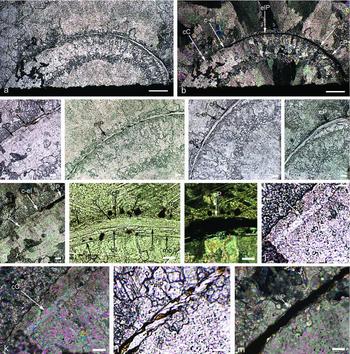
Petrographical thin-section (of OUMNH BY.1368/p) of the cuticle of Gogglops ensifer gen. et sp. nov. (a, b) Eye lens and cuticle either side; (c, e, g) part of cuticle and part of eye lens; (d, l, m) eye lens; and (f, h–k), cuticle. Note that the cuticle imaged in (h, i) has some internal dislocation that runs parallel to the lamination, labelled ‘di’. (a, c–f, h, j, l) Imaged under plain-polarized light; and (b, g, i, k, m) imaged under crossed polars. Abbreviations: cP – phosphatic cuticle; cC – calcitic cuticle; d – duct; di – dislocation of cuticle; elP – phosphatic eye lens; c–el – region where cuticle meets eye-lens. Scale bars: (a, b) 250 µm; (c–g) 50 µm; and (h–m) 25 µm.
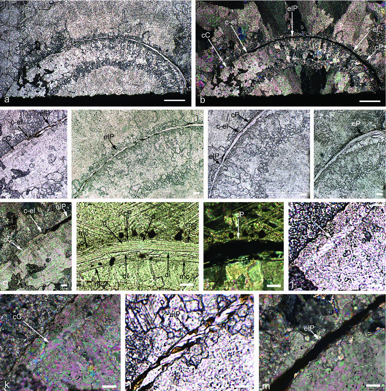
In addition to all of the above, the cuticle of Gogglops also shows another intriguing feature. In a thin-section (OUM BY 1368/p; Fig. 4), one part of the cephalon that abuts the eye is clearly calcitic (Fig. 4a–c, g, j, k), while the other side adjacent to the eye is phosphatic. This phosphatic part also shows micro-laminae (at least 20, apparently assembled, at least in the upper part of the cuticle into two or three 3–5 µm wide bundles) and vertical ducts passing through them, features typical of original euarthropod ultrastructure (Fig. 4a, b, e, f, h, i; e.g. Dalingwater et al. Reference Dalingwater, Hutchinson, Mutvei and Siveter1993; see also Briggs & Fortey, Reference Briggs and Fortey1982). We consider two possible explanations of this feature. The phosphatic cuticle preserved could be a fragmentary survivor of the original cuticle, in which case the sparry calcite forming the rest of the cuticle in this specimen and in other specimens would be a diagenetic replacement. Alternatively, if the bacterial mediation that produced phosphatization of the eye lens liberated free phosphate this would also have promoted localized phosphatization of the adjacent, originally calcitic cuticle. The second of these options appears more probable. First, among a large collection of specimens, all the eye surfaces – and only the eye surfaces – are obviously phosphatized. Second, of the major shell-building minerals, phosphate is more diagenetically stable than calcite (e.g. Bengtson & Conway Morris, Reference Bengtson, Conway Morris, Lipps and Signor1992). The preservation of the microstructure of the cuticle itself in the small phosphatized portion proves that the cuticle structure is conventionally euarthropodan. While the external detail remains perfectly preserved, the recrystallization of the rest of the calcitic cuticle is common in trilobite examples where original internal cuticle preservation is generally extraordinarily rare (e.g. Dalingwater et al. Reference Dalingwater, Hutchinson, Mutvei and Siveter1993).
Concerning certain other aglaspidid fossils, there is some evidence that they can be preserved in a similar fashion to contemporary fossils known to be calcitic. One of us (RAF) has studied an Early Ordovician aglaspidid from North Wales, Tremaglaspis Fortey & Rushton, Reference Fortey and Rushton2003, which occurs in the same strata as numerous trilobites (of which Angelina sedgwicki is most familiar) and other fossils. In this slightly metamorphosed shale the trilobites have, for the most part, decalcified. Tremaglaspis is preserved in the same way, with no evidence of original cuticle. On the other hand, lingulate brachiopods from the same localities still retain their original calcium phosphate valves, giving a field appearance of a typical pearly lustre. Were they originally phosphatic, we might have expected Tremaglaspis to present the same way. Similarly, we have also seen lingulid brachiopod examples from Lower Ordovician sediments of Morocco that show similar features. However, we note that material of the recently established aglaspidid Brachyaglaspis, admittedly known from only one specimen but also from Lower Ordovician deposits of Morocco (Ortega-Hernández, Van Roy & Lerosey-Aubril, Reference Ortega-Hernández, Van Roy and Lerosey-Aubril2016), is preserved not in phosphate but in iron oxides (relating to the original soft-tissue morphology) and clay minerals (that have replaced the biomineralized cuticular component). This provides some evidence that the circumstances preserving phosphatized Aglaspis barrandei in Wisconsin were actually unusual, possibly associated with soft-part preservation in a Konservat-Lagerstätte (Briggs, Bruton & Whittington, Reference Briggs, Bruton and Whittington1979). On the other hand, we show that our new Chinese taxon, Gogglops ensifer gen. et sp. nov., may belong to a group of closely related, stratigraphically young aglaspidids from the Sino-Siberian platform, and it is not inconceivable that this represents a clade that switched from phosphatic to calcite exoskeletons.
It is certain that the phosphatized eyes of our new species were the product of secondary mineralization. The compositional differences between the visual surface and the adjacent cuticle made the eyes the centre for bacterially mediated alteration, uniquely so for aglaspidids. Many other modes of aglaspidid eye preservation have been reported, such as where the eye is apparently compositionally the same as the cuticle (see examples in Hesselbo, Reference Hesselbo1992) or where the eyes alone are not fossilized (Lerosey-Aubril et al. Reference Lerosey-Aubril, Ortega-Hernández, Kier and Bonino2013). Further, differential preservation of euarthropod eyes is not unique to our material. Although a majority of trilobites had calcified eye lenses in vivo (Clarkson, Levi-Setti & Horvath, Reference Clarkson, Levi-Setti and Horvath2006), many others apparently did not. The visual surface of the Silurian trilobite Calymene, for example, is usually represented by an eye-shaped hole in the otherwise well-calcified dorsal exoskeleton (e.g. Siveter, Reference Siveter1996, fig. 5 m, Calymene aspera Shirley, Reference Shirley1936). No isolated lenticular surfaces have been found. The Devonian trilobite Phacops had well-developed calcitic schizochroal eyes with large lenses. However, Klug, Schultz & De Baets (Reference Klug, Schultz and De Baets2009) have illustrated examples from Morocco where the normally calcitic eye lenses – and only that part of the exoskeleton – have been replaced by a green glauconitic mineral. The eyes evidently had properties sufficiently different from the rest of the exoskeleton to encourage specific mineralization pathways to target them. We must also note that there is no possibility that the aglaspidid eye could have functioned with calcium phosphate as an optical medium in the way that calcite works for trilobites. A reasonable presumption is that the original chitinous apposition eyes in Gogglops were similar to those of living and fossil limulids, which are also set within a prosomal shield.
5. Materials and methods
The specimens were all concentrated, coquina-like, in a few limestone blocks, and were prepared under a binocular microscope using a Burgess vibrating engraving tool (‘Vibrotool’) mounted with a very fine needle. After being covered with a thin coat of photographic black opaque, they received a fine dusting of ammonium chloride sublimate before being photographed by a stand-mounted Leitz Aristophot with a Leica MD camera and a Leitz Summar 12 cm lens. Chemical analysis of the aglaspidids and surrounding matrix was enabled through use of an ISI-40 SEM with Link Systems 860 Series Si(Li) Energy Dispersive System.
Petrological thin-sections were examined through a Nikon Optiphot-Pol Petrological microscope with transmitted illumination, with images obtained using the following objectives: ×4/0.1 160/-, ×10/0.25 160/- and ×40/0.65 160/0.17. Images were obtained using a Q-imaging digital camera system attached via a trinocular and a 0.5 inch C-mount optical format with a CCD sensor specification of 1392×1040 1.4 million pixels, with a pixel size of 4.65 µm × 4.65 µm. Two software packages produced by Syncroscopy were used for image capturing. AcQuis (v. 4.0.1.8) was used for preview and capture of live single images. AutoMontage captures a succession of digital images through the z-axis of each thin-section, and automatically combines and generates a single focused montaged image by combining in-focus regions from each of the source images.
All type and figured specimens are in the Nanjing Institute of Geology and Palaeontology, Chinese Academy of Sciences (NIGPAS). Additional specimens and thin-sections are housed in the Oxford University Museum of Natural History (OUMNH).
6. Systematic palaeontology
Class Artiopoda Hou & Bergström, Reference Hou and Bergström1997
Order Aglaspidida Walcott, Reference Walcott1912
Family Aglaspididae Miller, Reference Miller1877
Genus Gogglops gen. nov.
Type species. The genus is monotypic.
Derivation of name. A combination of the vernacular ‘goggle’, staring, with ‘ops’, Greek, for eyes. Gender: feminine.
Diagnosis. Cephalon dorso-ventrally deep with very steeply sloping outer part. Genal spine moderately long. Central cephalic area divided by small- to medium-sized sub-circular to sub-ovoid raised areas. The occipital ring bears a pair of low nodes near its posterior margin. The eyes are large, situated at two-thirds anteriorly of the cephalic length. The cephalon is covered with tiny, closely spaced scales, the cheek doublure carries terrace lines, and the tergites and tail spine variously bear very small scales and pustules. Tail spine displays a median dorsal ridge.
Discussion. Gogglops is clearly very different from the Cambrian aglaspidid from China described by Lerosey-Aubril et al. (Reference Lerosey-Aubril, Ortega-Hernández, Kier and Bonino2013). The closest forms to our new Chinese genus from the North China Platform appear to lie among a clutch of variously three-dimensionally preserved genera erected as aglaspidids from the Siberian Platform, which was relatively close during Ordovician time (Torsvik & Cocks, Reference Torsvik, Cocks, Harper and Servais2013). The literature on these Siberian forms ranges from the late nineteenth to the early twenty-first century, although it is mostly in Russian, and it has been mentioned only relatively briefly in a few modern western publications (e.g. Van Roy, Reference Van Roy2006). It is therefore appropriate to further outline here the nature of this literature prior to a discussion of the Siberian genera with respect to Gogglops.
In his account of the southern and western parts of the Tungus Coal Basin, Obruchev (Reference Obruchev1932) listed various species of the euarthropods Angarocaris and Obrutschewia, two genera which were discussed later by Černyšev (Reference Černyšev1945) in his description of Obrutschewia and other euarthropods from the Angara River of Siberia. Černyšev (Reference Černyšev1945) also referred to a 1929 manuscript, which appears to be the work in which he first described Angarocaris and Obrutschewia. Additionally, he (1945) established a new species and subspecies of Obrutschewia, together with the new genera Chacharejocaris and Schamanocaris, plus two unnamed species he questionably referred to Schamanocaris (both of which, according to Van Roy, Reference Van Roy2006, are not aglaspidids). Further, he made reference to Intejocaris Černyšev in a way that implies that the name was already established. Černyšev (Reference Černyšev1953) subsequently described new euarthropods from the Angara River area in which Obrutschewia, Angarocaris and Intejocaris were all set up as new genera, with Angarocaris containing two new species, and three other new species being questionably assigned to the genus together with several other unnamed varieties. In the same work, another species assigned to Angarocaris and one of those questionably assigned, A. tschekanowskii and A. (?) punctatus, respectively, were both originally described by Schmidt (Reference Schmidt and Maak1886), who referred them questionably to Eurypterus.
In her account of new euarthropod discoveries from eastern Siberia, Andreeva (Reference Andreeva1957) built on the work of Černyšev, established the genus Girardevia with two new species and provided a revision of Eurypterus? punctatus (Schmidt, Reference Schmidt and Maak1886), assigning it to Chacharejocaris. Andreeva also gave a useful stratigraphical chart of the Middle Ordovician strata of the Siberian Platform, in which Angarocaris, Girardevia and Obrutschewia are indicated in the lower, middle and upper parts of the sequence, respectively, which also contains Schamanocaris and Chacharejocaris.
In the Russian Treatise Novozilov (Reference Novozilov and Rodendorf1962) listed only Angarocaris and Girardevia within Aglaspididae, while Obrutschewia, Intejocaris and Chacharejocaris Černyšev were listed under Aglaspidida Incertae sedis. Shortly afterwards a new aglaspidid from Siberia, Khankaspis, was described from Cambrian strata by Repina & Okuneva (Reference Repina and Okuneva1969). Most recently, in his description of new Ordovician aglaspidids from the Siberian Platform, Rozov (Reference Rozov, Varlamov, Rozova, Rozov and Efimov2009) has described new species of Obrutschewia, Angarocaris and Chacharejocaris, together with the new genera Rozhkovocaris and Lenacaris and material questionably assigned to Girardevia.
One of us (DJS) has examined the Černyšev (Reference Černyšev1945, Reference Černyšev1953) and Andreeva (Reference Andreeva1957) material which is housed in the A.P. Karpinsky All Union Geological Research Institute (VSEGEI) in St Petersburg. It is clear that to unravel the intra- and supra-generic affinities of all the various Siberian Platform material a careful, detailed comparative investigation is required, something which is outwith the scope of our current study. Nevertheless, we can indicate some potential affinities for Gogglops from this examination combined with the published figures of the Siberian material.
Obrutschewia, Girardevia, Chacharejocaris, Schamanocaris, Khankaspis and Lenacaris can all be easily distinguished from Gogglops. For example, Obrutschewia shows a well-defined median cephalic ridge; Girardevia exhibits a bullet-shaped cephalic shield; Chacharejocaris has a cephalic shield that is very much less convex; in Schamanocaris the cephalic shield is very much wider relative to its length and less convex; and Khankaspis (the material of which is indifferently preserved) is characterized by a poorly defined axial region in both cephalic shield and body, and very short terminal spines to its body tergites. Lenacaris apparently lacks cephalic muscle impressions and shows quite different lateral and frontal cephalic profiles. Rozhkovocaris, with its quite strongly inflated cephalon and well-defined axis, approaches the morphology of Gogglops (cf. Rosov, Reference Rozov, Varlamov, Rozova, Rozov and Efimov2009, plate 3, figs 3, 7, plate 4, figs c, i). However, the cephalic sculpture of Rozhkovocaris is so distinctive, comprising strong and more-or-less continuous transverse ribs, that generic discrimination from Gogglops is appropriate. Among the Siberian genera, those assigned by Černyšev (Reference Černyšev1953) to Angarocaris are generally most similar to the new Chinese species, especially A. tschekanowskii Schmidt, Reference Schmidt and Maak1886, A. paduensis Černyšev, Reference Černyšev1953, A. angarensis Černyšev, Reference Černyšev1953 and A.? nodus Černyšev, Reference Černyšev1953. Nevertheless, examination of the original material proves that these species share a distinctive sculpture: transversely semi-continuous to continuous toothed, terrace-ridges, unlike the scale-like sculpture of Gogglops. The distinctive sculpture of Angarocaris is considered likely to be a synapomorphy of that genus and, if so, Gogglops is clearly separate.
Occurrence. Gogglops has been recognized only from Upper Ordovician deposits of Shaanxi Province, China.
Gogglops ensifer sp. nov
Figures 5–6
1995 Undescribed Chinese Silurian aglaspidid; Fortey & Theron, p. 856, fig. 6A, B.
2006 Unnamed aglaspidid-like arthropod; Van Roy, pp. 228, 330.
2013 Unnamed Chinese aglaspidid; Ortega-Hernández, Legg & Braddy, pp. 21, 22.
2013 Unnamed taxon; Lerosey-Aubril, Ortega-Hernández & Zhu, p. 565.
Holotype. An excellently preserved partial cephalon, NIGPAS 160534, Figure 5a–d, h.
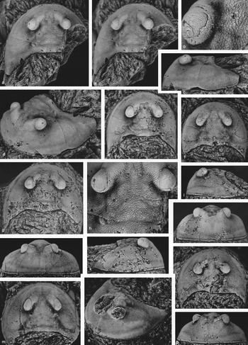
Gogglops ensifer gen. et sp. nov., Horizon Yt22-2, Yaoxian Formation, Taoqupo, 11 km NW of Yaoxian, Shaanxi Province, China. (a–d, h) Holotype, incomplete cephalon, dorsal stereo-pair, anterior-oblique, lateral views, ×5, dorsal view of eye, ×15, dorsal view of central part of cephalon, ×10; NIGPAS 160534; (e, l) cephalon, dorsal, anterior views, ×4.5; NIGPAS 160535; (f) dorsal view, ×6.0; NIGPAS 160536; (g, k) cephalon, dorsal, lateral views, ×4.5; NIGPAS 160537; (i, o, p) cephalon, lateral, dorsal, anterior views, ×4.7; NIGPAS 160538; (j, m) cephalon, anterior, dorsal views, ×5.0; NIGPAS 160539; and (n) cephalon lacking eyes, anterior-oblique view, ×6; NIGPAS 160540.

Other material. This includes, of prepared material, twelve almost complete to partial cephala, seven thoracic tergites and three tail spines, one of which has the most posterior thoracic tergite attached, NIGPAS 153620, 160535–160555, Figures 5e–g, i–p and 6a–v; these all represent paratype specimens from the type locality. In addition, there are probably at least 30 largely unprepared cephala and other body parts concentrated in small pieces of limestone, OUMNH BY.1334 - BY.1376/p; these all represent topotype specimens.
Locality and age. Upper Ordovician, Yaoxian Formation, Sandbian Stage, type locality for the Yaoxian Formation, about 15 km NNW of Yaoxian, Shaanxi Province, China.
Derivation of name. From the Latin ensifer, sword-bearing, referring to the shape of the tail spine.
Diagnosis. As for the genus.
Description. Cephalon is sub-semicircular in outline; at its widest point (generally opposite the occipital ring) it is 1.5 (Fig. 5f) to 1.9 (Fig. 5a) times as wide (trans.) as long (sag.). It is gently to moderately convex (trans. & sag.; Fig. 5c, i–l, p) over the central cephalic area, and (trans.) over the inner part of the cheek which descends moderately steeply abaxially, with the outer part of the cheek and the pre-occular cephalic area descending very steeply in a gentle to moderately convex arc to the cephalic margin (Fig. 5c, i–l, p). The occipital ring is 4.5–5.2 times as wide (trans.) as long (sag.), and 0.13–0.18 times the length of the cephalon. It is of similar length over its central one-third, narrowing abaxially to about three-quarters of this. On the posterior part of the occipital ring, very close to the posterior margin, there is a low node either side the median line (Figs 5a, e, 6e, 7). The occipital furrow is shallow (Figs 5a, 6a) to moderately well incised (Fig. 5m).
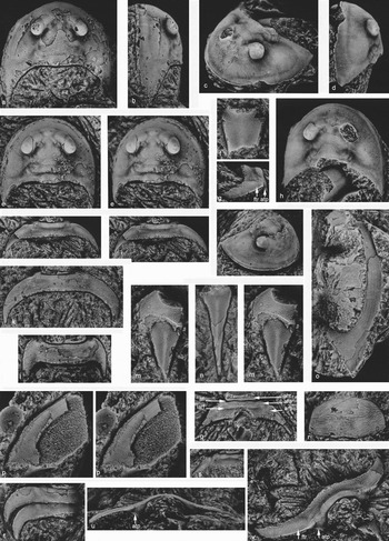
Gogglops ensifer gen. et sp. nov., Horizon Yt22-2, Yaoxian Formation, Taoqupo, 11 km NW of Yaoxian, Shaanxi Province, China. (a, b) Cephalon, dorsal, lateral views, ×4.8; NIGPAS 160541; (c, d, h) cephalon, anterior-oblique, lateral, dorsal views, ×4.5; NIGPAS 160542; (e) cephalon, dorsal stereo-pair, ×6.0; NIGPAS 160543; (f) incomplete tail-spine, dorsal view, ×5.5; NIGPAS 160544; (g, i, u, v) thoracic tergite, lateral, anterior, anterior-oblique views, ×4.5, dorsal stereo-pair, ×3.0; NIGPAS 160545; (j) cephalon, anterior-oblique view, ×2.8; NIGPAS 160546; (k) thoracic tergite, dorsal view, ×4.0; NIGPAS 160547; (l) thoracic tergite, dorsal view, ×7.4; NIGPAS 160548; (m) tail-spine and last thoracic tergite, dorsal stereo-pair, ×11.0; NIGPAS 160549; (n) tail-spine, dorsal view, ×6.0; NIGPAS 160550; (o) cheek showing doublure, dorsal view, ×3.5; NIGPAS 160551; (p) cheek showing doublure, ventral stereo-pair, ×7.5; NIGPAS 153620; (q) thoracic tergite, dorsal view, ×5.4; NIGPAS 160553; (r) last thoracic tergite, dorsal view, ×7.3; NIGPAS 160554; (s) incomplete thoracic tergite, dorsal view, ×4.0; NIGPAS 160555; and (t) two incomplete thoracic tergites, dorsal view, ×6.8; NIGPAS 160552. Abbreviations: af – articulating furrow; ahr – articulating half-ring; atp – anterior tergal process; ara – articulating ring anterior part; arp – anterior ring posterior part; ftr – fine transverse ridge; iarf – intra-axial ring furrow; mc – margin of cuticle.

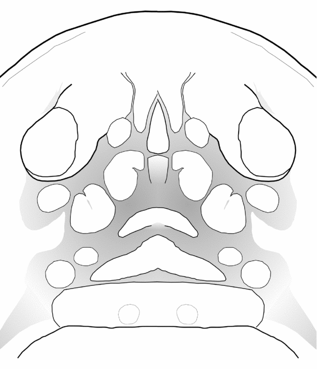
Diagrammatic representation of raised areas on the central cephalic area of Gogglops ensifer, based on the holotype NIGPAS 160534.

A shallow axial furrow defines the cheek from the central cephalic area; posteriorly, immediately anterior to the occipital ring, this furrow is interrupted by a swollen area, and at its mid-length by a low, broad, antero-medially directed ridge from the cheek. The central glabellar area narrows anteriorly, is subdivided by several shallow furrows that define gently raised sub-circular to suboval (? muscle attachment) areas (Figs 5a, h, 6e, 7). Immediately anterior to the occipital ring the posterior section of the central cephalic area is (bilaterally) defined by an inwards and slightly forwardly projecting furrow that bifurcates adaxially, the posterior branch the broader and shallower, and at the bifurcation close to the axial furrow there is a small oval raised area (Fig. 5h). Postero-laterally of this small oval area is a slightly larger sub-circular raised area, the axial furrow deflecting around it. On the central cephalic area postero-medially to the eye there are two moderately large gently swollen areas, each of which is partially subdivided by a weak furrow. The central cephalic area narrows between the eyes where it comprises a pair of posteriorly (Fig. 5a) to postero-laterally (Fig. 6e) pointing finger-like ridges, between which there is an elongate median raised ridge. Anterior to the finger-like ridges, the central cephalic area expands in width. In the axial furrow posterior to and beneath the visual surface of the eye, also at the posterior extremity of the finger-like ridge and adaxial of the basal part of the eye, there is a small sub-circular to sub-oval swelling.
The eye is sited anteriorly on the cephalon at about two-thirds its length; it projects postero-laterally from the broad, rounded (trans.), swollen basal part to the large, bulbous visual surface overhanging the cheek. The visual surface faces forwards, backwards, upwards and laterally, and, to a lesser degree, downwards (Fig. 5a–d).
The cheek area postero-laterally to the eye is gently independently convex of the pre-occular area (Figs 5d, n, 6c). The steeply descending outer cephalic area bears a shallow lateral furrow which is positioned fairly close to the cephalic margin medially, rising gradually postero-laterally towards the genal spine where it meets a similarly impressed posterior border furrow, so defining very gently convex lateral and posterior borders that are widest where they meet (Figs 5c, d, n, 6 c, d). These border furrows also appear to coincide with the upper margin of the anteriorly narrowing cheek doublure (and so might be termed paradoublural lines; Figs 5d, 6o, p). A marginal cephalic suture would appear to be present, and for some cephala to represent moults (e.g. Figs 5a, 6c, d appear to lack a doublure which is clearly present in other specimens, e.g. Fig. 6o, p). Immediately bounding the cephalic margin there is a very narrow cephalic rim which is bounded by an even narrower cephalic furrow (Figs 5d, i–k, n, 6c, j). Some specimens (Fig. 5n) show a fine median pre-occular ridge between the central cephalic area and the cephalic border furrow; others display here a very weak, narrow, median pre-occular furrow (Fig. 5j). The postero-lateral cephalic margin takes the form of a broadly based, robust, moderately long genal spine (Figs 5a, c, d, g, i–k, n, o and 6a, b, d, e, h, j), but in very small specimens this feature is scarcely represented (Fig. 5j).
Evidence of a hypostomal sclerite is lacking.
Isolated trunk tergites show differing overall shapes, according mainly to the posterior deflection of the pleurae. Those assumed to be more anterior in position (e.g. Fig. 6i, k) are larger and more abaxially deflected from the axis before curving backwards, compared with those presumed to have had a more posterior position (e.g. Fig. 6l, t) which are smaller and deflected more immediately backwards from the axis; all trunk tergites end distally in a sharp point. The main axial ring is weakly to moderately convex (trans.) and takes up a proportionately greater width of the tergite in the more posterior ones (cf. Fig. 6k with 6l). In some tergites (Fig. 6k, l, t) the axial ring is preceded by a narrow, well-defined articulating furrow and then more anteriorly by an articulating half-ring; in others (Fig. 6g, i, q, s, v) there is an extra furrow (here termed the intra-axial ring furrow) that parallels the articulating furrow and which occurs immediately posterior to it, to transversely divide the axial ring into a shorter (sag.) anterior and a longer posterior section. On the anterior margin of the tergite, at the extremely weak axial furrow, the cuticle extends ventrally into a well-developed tergal process (presumed apodeme; Fig. 6g, i, k, u, v; see also Section 7).
The tail spine is 3.1 times as wide (anteriorly; trans.) as long (sag.) in one essentially complete example (Fig. 6n) that also appears to show more rapid widening anteriorly; it bears a median ridge dorsally (Fig. 6f, n, m) that dies out before reaching the anterior margin. Attached to one of the tail spines (Fig. 6m) is a partial example of the last body tergite. Another example of the same tergite (Fig. 6r), isolated but more complete, shows a transversely elongate, biconvex outline; it is moderately dorsally convex (trans), more so anteriorly as it falls (sag.) to its anterior margin. Sclerites that might be regarded as post-ventral plates have not been recognized.
The cephalon is covered with very small, closely spaced scale-like features that are particularly well developed on the central cephalic area, occipital ring and posterior part of the cheek, where they are triangular-shaped and posteriorly pointing (Figs 5h, 6a). The outer face of the doublure of the cheek displays terrace line sculpture (Fig. 6o, p). The inner face also shows evidence of terrace lines anteriorly (Fig. 6o), as well as very subdued scale-like features posteriorly (Fig. 6p) near the genal angle. The tergites bear very small pustules and scales, especially on the axial area, extending laterally onto the pleurae, and in both being more concentrated near and at the posterior margin (Fig. 6g, i, k, l, q, s, t, v). All the isolated tergites show an extremely fine, transverse ‘ridge’ on the anterior part of the axis, which extends a short distance abaxially across the anterior part of the pleura before flexing very gently posteriorly to meet the more strongly posteriorly curving anterior pleural margin at a point about mid-way between the anterior tergal process and the tip of the pleural spine (e.g. Fig. 6g, k, v; see also Section 7). In places this fine ridge is seen to be made up of extremely fine pustules. The last body tergite bears wave-like lines of coalesced, posteriorly pointing scales (Fig. 6m, r), and the tail spine shows very small and subdued scales (Fig. 6f, m, n)
7. Discussion and relationships
The cephalon of G. ensifer is transversely strongly convex and it is dorso-ventrally high, the outer cephalic area descending steeply for some distance to the cephalic margin (e.g. Fig. 5c, i, j, l). This is clearly the original, undistorted carapace relief for the species. Most other aglaspidids, with reference here in particular to those in the finer clastics, are preserved as flattened carapaces, but nevertheless it is likely that they never had the high carapace relief of the Chinese species. For example, the various species described from the Cambrian St Lawrence Formation of Wisconsin (Hesselbo, Reference Hesselbo1992) are all preserved flattened, but if in life they had had any marked degree of relief of the cephalon, it is likely that this would be shown by clear, consistent, extensive fracturing (or crumpling), especially in the outer cephalic area. However, while some show carapace fracturing (Hesselbo, Reference Hesselbo1992, figs 2.5, 2.8), the majority have their cephala entire, uniformly rounded, and without fracturing (Hesselbo, Reference Hesselbo1992, figs 5.2, 8.2, 8.3, 10.2, 10.5, 12.1, 12.2, 12.11, 15.3, 16.2). We conclude that the Cambrian species had low convexity.
The morphology of some body tergites in G. ensifer, whereby they have an extra transverse furrow in front of the axial furrow in order to form an extra raised, transverse ridge (see description), is reminiscent of the morphology of thoracic segments in some proetid trilobites. In the latter, from anterior to posterior, there is respectively an articulating half-ring, articulating furrow, a raised transverse ridge (the so-called preannulus) then an intra-annular furrow, and most posteriorly the axial ring (see Owens, 1970, fig. 1). In this comparison, the anterior part of the axial ring in Gogglops would equate to a preannulus, and the intra-axial ring furrow (see description) behind this anterior part would compare with the proetid intra-annular furrow. This type of tergite morphology has not, as far as we are aware, been previously recognized in aglaspidids, although the comparison with proetids is merely morphological rather than any suggestive of true homology.
The tergal process in Gogglops, that is, the ventral extension of the anterior margin of the tergite at (in front of) the very shallow axial furrow (Fig. 6g, i, k, u, v), corresponds best to ‘anterior tergal process 2’ of Hesselbo (Reference Hesselbo1992) in the Cambrian aglaspidids of Wisconsin that he studied (e.g. in Flobertia kochi Hesselbo, Reference Hesselbo1992, figs 18.5 and 20.2). ‘Tergal process 1’ of Hesselbo (Reference Hesselbo1992) lies too far abaxially along the anterior tergal margin to equate with the tergal process in Gogglops, which shows only a single such process and not two. While the Wisconsin aglaspidid specimens show these processes preserved in an essentially flattened mode, in the Chinese specimens the process is preserved fully in its original form, projecting dorsoventrally (internally; Fig. 6g, u, v). It seems certain that this particular tergal process in Gogglops, and in other aglaspidids, represents an apodeme (i.e. a muscle attachment site) as concluded by Hesselbo (Reference Hesselbo1992), and as has been suggested for similar structures in other fossil and recent euarthropods, for example trilobites (e.g. Cisne, Reference Cisne1981).
The fine, transverse ridge on all tergites (e.g. Fig. 6v), on the anterior part of the axial ring and associated pleura, can be compared with a feature that Hesselbo (Reference Hesselbo1992) described in various aglaspidids from the Cambrian strata of Wisconsin. In some of his figures this feature, which is characterized (in parts at least) by a line of fine tubercles, is tagged as the ‘articulating ridge’ (with the figure notation ‘ar’; e.g. ?Flobertia kochi, figs 18.5, 19.2), or the ‘articulating step’ (denoted ‘as’; e.g. Cyclopites vulgaris, figs 22.8 and 23.5; also note that in Glypharthrus simplex, fig. 11.5, what is apparently the same feature is also marked ‘ts’ = tergite striae, which seems likely to be a notation error).
Prior to its formal description here, Gogglops had already been included in a phylogenetic analysis of aglaspidid relationships within Artiopoda by Ortega-Hernández, Legg & Braddy, Reference Ortega-Hernández, Legg and Braddy2013, where it was referred to as the ‘Chinese taxon’ (their table 1) or ‘Chinese arthropod’ (their Fig. 8). In their analysis, it was securely nested within Aglaspidida sensu stricto, as sister to Glypharthrus + Aglaspis itself. Our own assessment of the morphology of Gogglops has resulted in agreement with the character codings employed by those authors except for character 31 (visible ecdysial sutures: there is evidence of a marginal suture) and the associated character 32 (position of ecdysial sutures: marginal). However, a re-running of the cladistic analysis with our slightly modified coding did not fundamentally alter the topology of the similarly treated trees recovered by Ortega-Hernández, Legg & Braddy (Reference Ortega-Hernández, Legg and Braddy2013). We have therefore not republished our trees here in the main body of the text but include them as online Supplementary Material, together with comments on the two re-coded characters (available at http://journals.cambridge.org/geo). Despite its calcareous cuticle and the fact that the most important autapomorphy for aglaspidids – the possession of post-ventral plates – has not been identified for it, Gogglops is securely recovered as a member of Aglaspidida sensu stricto (see online Supplementary Material Figure S2 and Ortega-Hernández, Legg & Braddy, Reference Ortega-Hernández, Legg and Braddy2013, fig. 6b). Within Aglaspidida sensu stricto, two morphotype groups have been identified by Ortega-Hernández, Legg & Braddy (Reference Ortega-Hernández, Legg and Braddy2013, fig. 8): ‘Cambrian-type’ representatives; and ‘Ordovician-type’ taxa. Interestingly, Gogglops shows morphological elements deemed by those authors characteristic of both groups: the presence of genal spines, cephalic furrows and large eyes of the former, and the convex/vaulted cephalon of the latter. The tail spine of Gogglops was indicated by them as being long, which they associated with the Cambrian-type taxa. However, while this is the case in one specimen (Fig. 6n), in another, smaller specimen (Fig. 6m) it is relatively much shorter.
As mentioned above, Gogglops could be part of a group including the genera mentioned from the Siberian Platform. These, however, were excluded from the analysis of Ortega-Hernández, Legg & Braddy (Reference Ortega-Hernández, Legg and Braddy2013, table 2) as ‘aglaspidid-like genera excluded from analysis’. In light of the complete morphology of Gogglops described here, it now seems probable that many of these relatively poorly known Siberian genera are relevant to the composition of the important early euarthropod group Aglaspidida, and modern, detailed reappraisal of them should be a priority.
Acknowledgements
We would like to thank Owen Green (Department of Earth Sciences, University of Oxford) for thin-section imaging and discussion; Javier Ortega-Hernández (Department of Earth Sciences, University of Cambridge) and David Legg (Oxford University Museum) for discussion and running phylogenetic analyses, and the latter for drafting the line diagram of Gogglops; Monica Price (Oxford University Museum of Natural History) for SEM EDX analysis of the aglaspidid cuticle; Elena Mikhailova (National Mineral Resources University, St Petersburg) for assistance with Russian literature; and Paul Selden, Peter Van Roy and an anonymous reviewer for improving the manuscript through their perceptive reviews. DJS was supported in this study by the Leverhulme Trust (grant no. EM-2014-068) and X-JZ by the Natural Science Foundation of China (award nos 41290260, 41330101, 41521061 and 41672002).
Supplementary material
To view supplementary material for this article, please visit http://doi.org/10.1017/S0016756817000309