Introduction
The Kana (Japanese syllabogram) Pick-out Test (KPT) was developed to evaluate frontal lobe function easily and quickly at the bedside (Kaneko, Reference Kaneko1996). The test is said to be suitable for inspecting higher-order brain function and to be a good method for screening persons with mild or slight dementia (Inoue et al., Reference Inoue, Suyama, Kato, Urakami, Nakashima and Meshitsuka2003). Although the clinical usefulness of the KPT in dementia is well documented, the neural substrates involved remain unclear. There are few functional neuroimaging studies investigating brain regions related to the KPT performance of dementia patients (Nakatsuka et al., Reference Nakatsuka, Matsubara and Ohtani2003; Tachibana et al., Reference Tachibana2007). In the present study we investigated the relationship of KPT scores to regional cerebral blood flow (rCBF) in order to assess the neural substrates of KPT performance in patients with mild Alzheimer's disease (AD).
In most neuroimaging studies in which rCBF was evaluated, stereotaxic analysis of regions of interest (ROIs) was used. However, the reproducibility and objectivity of the results are controversial because small ROIs were manually placed on selected slices of single photon emission computed tomography (SPECT) images of the patient. Image analysis using ROIs has been criticized for the serious shortcoming that any information outside of the ROIs is not obtained. To overcome the above disadvantages, in the present study we conducted an evaluation using statistical parametric mapping (SPM) comparison which has the advantage of making no a prior anatomical hypotheses.
Methods
Kana pick-out test
In the KPT, the subjects are shown a short story written in Japanese kana characters. The original paper version on which a short story is written in Japanese kana characters was described previously (Inoue et al., Reference Inoue, Suyama, Kato, Urakami, Nakashima and Meshitsuka2003). The subjects are required to find as many vowel symbols as possible within two minutes, while understanding the meaning of the story (Kaneko, Reference Kaneko1996; Nakatsuka et al., Reference Nakatsuka, Matsubara and Ohtani2003). While reading the story, the subjects circle the vowel symbols with a pencil. If the subjects concentrate only on reading the story, they tend to become careless in finding the symbols. Inversely, if they concentrate on finding the symbols, they tend to forget the substance of the story. Thus, this test assigns two simultaneous tasks: find the vowel symbols and recognize the content of the story (Inoue et al., Reference Inoue, Suyama, Kato, Urakami, Nakashima and Meshitsuka2003). The Japanese kana character set consists of 66 phonetic symbols that include five vowels; the story consists of 406 symbols with 61 vowels, so the full score for this test is 61. The lower normal limits of KPT scores are 11 for those in their seventies, 10 for those in their eighties, and 9 for those in their nineties (Kaneko, Reference Kaneko1996). In this study, the subjects with low scores on the KPT did not reach the lower limits of normal, and the subjects with high scores on the KPT were required to score higher more than the mean score for their ages (20 for those in their 60s, 18 for those in their 70s, and 16 for those in their 80s) (Kaneko, Reference Kaneko1996).
In this study, KPT memory score was also recorded. Two questions to examine whether the subjects remembered the substance of the story were asked, and the memory score was determined according to the following criteria: 10 for two correct answers, 5 for one correct answer (full score was 10).
Subjects
Twenty AD patients with a dementia severity of 0.5 (possible) or 1 (mild) based on the Clinical Dementia Rating (CDR) (Hughes, et al., Reference Hughes, Berg, Danziger, Coben and Martin1982), whose score on the KPT did not reach the lower limits of normal, were selected from 227 consecutive Japanese patients who were fully examined at the Memory Clinic of Okayama University Hospital, between April 2004 and March 2008. Thereafter, 20 AD patients with high scores on the KPT, matched for age, sex ratio, education, and scores on the Addenbrooke's Cognitive Examination (ACE) (Mathuranath et al., Reference Mathuranath, Nestor, Berrios, Rakowicz and Hodges2000; Yoshida et al., Reference Yoshida2010), were also selected.
The inclusion criteria for all participants were: (i) they underwent general physical and neurological examinations and extensive laboratory testing, including thyroid function tests, serum vitamin B12, and syphilis serology; (ii) they took the KPT, Mini-mental State Examination (MMSE) (Folstein et al., Reference Folstein, Folstein and McHugh1975) and ACE; (iii) underwent brain SPECT as well as head CT and/or head MRI; (iv) they were diagnosed with probable AD according to the NINCDS-ADRDA criteria (McKhann et al., Reference McKhann, Drachman, Folstein, Katzman, Price and Stadlan1984); (v) they had a dementia severity of 0.5 or 1 based on the CDR (Hughes, et al., Reference Hughes, Berg, Danziger, Coben and Martin1982); and (vi) they or their nearest relatives gave informed written consent. The exclusion criteria were (i) complications from other neurological diseases or illnesses; (ii) history of mental illness or substance abuse prior to the onset of dementia; (iii) any evidence of focal brain lesions on head MRI; (iv) treatment with cholinesterase inhibitors, antipsychotics, antidepressants or anxiolytic drugs; and (v) left-handedness or ambidexterity. The profile of each participant (age, sex, years of education, and disease duration) was recorded, and the CDR score was rated by the chief clinician.
Other instruments
Addenbrooke's Cognitive Examination (ACE) was developed to provide a brief test sensitive to early stage dementia, and is capable of differentiating between dementia subtypes including AD, frontotemporal dementia, progressive supranuclear palsy and other parkinsonian syndromes (Mathuranath et al., Reference Mathuranath, Nestor, Berrios, Rakowicz and Hodges2000). ACE includes the MMSE but extends it to encompass important areas not covered by the MMSE, such as frontal-executive function and visuospatial skills. ACE has a comparable sensitivity to the Dementia Rating Scale (Bak et al., Reference Bak, Rogers, Crawford, Hearn, Mathuranath and Hodges2005), a well-established dementia screening tool, widely used in research but not in clinical practice because of its length and difficulty of administration. For this study, we used the Japanese version of ACE described by Yoshida et al. (Reference Yoshida2010). The reliability of the Japanese version of ACE is excellent, and its validity is, to some extent, established (Yoshida et al., Reference Yoshida2010).
The Physical Self-Maintenance Scale (PSMS) and the Instrumental Activities of Daily Living scale (IADL) are validated scales for the assessment of activities of daily living (ADL) (Lawton and Brody, Reference Lawton and Brody1969; Hokoishi et al., Reference Hokoishi2001). The PSMS is a six-item scale that rates self-care ability in toileting, feeding, dressing, personal hygiene and grooming, locomotion (physical ambulation), and bathing. The IADL scale assesses patients' ability to perform eight complex daily tasks: ability to use the telephone, shopping, food preparation, household tasks, laundering, mode of transportation, responsibility for medications, and ability to manage finances.
The Neuropsychiatric Inventory (NPI) is a valid and reliable instrument for measuring behavior in dementia (Cummings et al., Reference Cummings, Mega, Gray, Rosenberg-Thompson, Carusi and Gornbein1994; Hirono et al., Reference Hirono1997). It is a caregiver-based tool that assesses ten common behaviors in dementia.
Ethics
This study adhered to the 1975 Helsinki Declaration of Human Rights and the study protocol was approved by the Committee of Okayama University Hospital on Human Research. After providing a complete description of the study to the subjects and their relatives, written informed consent was obtained.
Brain perfusion SPECT imaging
All subjects were examined by brain perfusion SPECT. Patients were examined in a comfortable supine position with their eyes closed in quiet surroundings. First, the passage from the heart to the brain was monitored after intravenous administration of 99mTc-ethylcysteinate dimer (ECD, 600 MBq, Daiichi Radioisotope Laboratories Ltd., Tokyo, Japan). Ten minutes after the angiography, SPECT images were obtained using a triple-head, rotating gamma camera interfaced to a minicomputer (GCA9300A/ DI; Toshiba, Tokyo, Japan) equipped with a fanbeam, low-energy, high-resolution collimator. Sixty projection images over a 360° angle in a 128 × 128 matrix were acquired. All images were reconstructed using ramp-filtered back-projection and then three-dimensionally smoothed with a Butterworth filter (order 8, cutoff 0.12 cycles/cm). The reconstructed images were corrected for gamma ray attenuation using the Chang method (μ = 0.09).
Data analysis
Spatial reprocessing and statistical analysis of images was performed on a voxel-by-voxel basis using Statistical Parametric Mapping 2 (SPM2, Wellcome Department of Imaging Neuroscience, U.K.) running on MATLAB (The Mathworks, Inc). All SPECT images of each subject were normalized to the standard brain of the Montreal Neurological Institute (MNI), and the spatial normalization was performed with 12-parameter affine and non-linear transformations (Friston et al., Reference Friston1995a; Reference Friston1995b). The voxel sizes of the reslice option were (2 mm, 2 mm, 2 mm). The non-linear parameter was set at 25 mm cut-off basis functions and 16 iterations. All of the normalized SPECT images were then smoothed with an isotropic gaussian kernel filter (12-mm full-width at half-maximum). To examine the images for specific regions showing differences in perfusion, two sample t-tests were performed. Global normalization was performed by proportional scaling with the mean voxel value. Masking was applied using the threshold method (0.8 times the global value). The statistical height threshold was set at P < 0.005 uncorrected, and the extent threshold was set at P < 0.01 with correction for multiple non-independent comparisons.
Statistical analysis
Statistical analysis was performed using the SPSS 14.0J software program (SPSS Inc., Chicago, IL). Comparisons between the two groups were performed by independent sample t-tests. A value of p < 0.05 was accepted as significant.
Results
Age, years of education, duration of illness, neuropsychological test scores and ADL
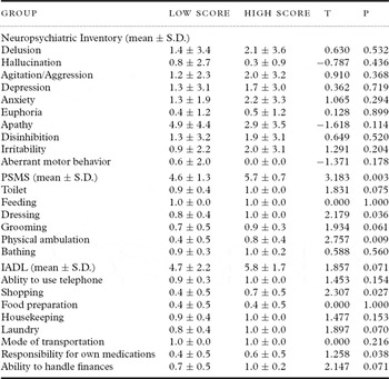
No significant differences were found between high- and low-score groups with respect to age, years of education, or duration of disease by Student's t-test (Table 1). With the exception of KPT scores, no significant differences were found between high- and low-score groups with respect to ACE scores, MMSE scores, KPT memory scores or NPI subscale scores (Tables 1 and 2).
Clinical characteristics and neuropsychological tests

Low score = Alzheimer's diseasae patients with low score on Kana Pick-out Test.
High score = Alzheimer's diseasae patients with high score on Kana Pick-out Test.
ACE = Addenbrook's cognitive examination; MMSE = Mini-mental State Examination.
KPT = Kana Pick-out Test; S.D. = Standard Deviation.
Activities of daily living (ADL) and instrumental ADL

Low score = Alzheimer's disease patients with low score on Kana Pick-out Test.
High score = Alzheimer's disease patients with high score on Kana Pick-out Test.
PSMS = Physical Self-Maintenance Scale; IADL = Instrumental Activities of Daily Living scale.
S.D. = Standard Deviation.
Comparison on ADL scales revealed that AD patients with a low KPT score got lower scores than those with a high KPT score, in the total scores of PSMS and the subscale scores of dressing, physical ambulation, shopping and responsibility for own medications among PSMS and IADL (Table 2).
rCBF
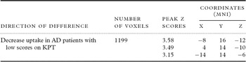
A group comparison of the SPM results between the high- and low-score groups were performed. Specific voxels with a significantly lower perfusion in the low-score group than in the high-score group are shown in Figure 1 and Table 3. A similar test for specific voxels with a significantly lower perfusion in the high-score group than in the low-score group found no cluster of voxels.
SPM (z) map of rCBF decrease in AD patients with low scores on the Kana Pick-out Test compared with AD patients with high scores. Three-way-glass view of the area of significant hypoperfusion.

Significant regional uptake differences between AD patients with low and high scores on Kana Pick-out Test (KPT)

AD = Alzheimer's disease; MNI = Montreal Neurological Institute.
Figure 1 shows the z-score for each voxel in this cluster superimposed onto a three-way-glass brain view. It shows a significant cluster of voxels in the left subgenual cingulate gyrus (SGC), extending to right SGC. Table 3 shows the probability results of the SPM analysis and the location of peak z scores in terms of the MNI coordinates.
Discussion
Kana Pick-out Test score and rCBF
The KPT is a popular test of attention in Japan. This task requires parallel processing of reading and picking out letters, and demands an appropriate allocation of attentional resources to the two activities. Therefore, the KPT is thought to be a suitable test of working memory and executive function (Tamura et al., Reference Tamura, Kikuchi, Otsuki, Kitagawa and Tashiro2003), and to reflect prefrontal area function (Tachibana et al., Reference Tachibana2007).
There have been only a few studies of the relationship between results of the KPT and rCBF (Nakatsuka et al., Reference Nakatsuka, Matsubara and Ohtani2003; Tachibana et al., Reference Tachibana2007). At a memory clinic, the mean rCBF among four outpatients whose MMSE score was normal but whose KPT scores were abnormal was reported to be decreased in the posterior cingulate gyrus and cinguloparietal transitional area, compared to the mean rCBF among four cases in which both the MMSE and the KPT scores were normal (Nakatsuka et al., Reference Nakatsuka, Matsubara and Ohtani2003). A functional MRI (fMRI) study of the four cases showed significant increases in blood oxygenation level-dependent signals in the prefrontal area, sensorimotor area, parietal association area, and visual cortex during performance of the computerized KPT (Tachibana et al., Reference Tachibana2007). Inconsistent with these two previous findings, we found rCBF in the bilateral SGC was decreased in AD patients with low scores on the KPT compared with AD patients with high scores.
The results of the three reports, including our own, were different from each other. What caused the differences? There are two major differences among the three studies. The first is a difference in the diseases. All subjects in the study by Tachibana et al. (Reference Tachibana2007) were cognitively normal subjects, whereas subjects in the other two studies were memory clinic outpatients. All eight cases in the study by Nakatsuka et al. (Reference Nakatsuka, Matsubara and Ohtani2003) were patients with subjective complaints of forgetfulness, but all eight cases showed normal MMSE scores. In our study, all 40 subjects were patients with AD, and the mean MMSE scores were 22.1 among patients with low KPT scores and 21.6 among patients with high KPT scores.
The second was the difference in the methods of evaluating brain function. Tachibana et al. (Reference Tachibana2007) used fMRI, whereas the other two studies used brain SPECT. In the study by Nakatsuka et al. (Reference Nakatsuka, Matsubara and Ohtani2003), 99mTc-hexamethyl-propyleneamine oxine was injected intravenously, and the z-value, which shows the distance from the mean, was calculated by three-dimensional stereotactic surface projections (3D-SSP). Thus, the exact rCBF was not calculated, and deep brain structures such as the basal ganglia and thalamus were not evaluated. In this study, 99mTc-ECD was used, and rCBF was compared using SPM. The difference of patients and methods among three studies, we suppose, might influence the difference in results.
In addition, the number of participants in the three studies was different. In Nakatsuka's study, four subjects with low KPT scores were compared with another four subjects with normal KPT scores (Nakatsuka et al., Reference Nakatsuka, Matsubara and Ohtani2003). In Tachibana's study, four cases were examined with fMRI (Tachibana et al., Reference Tachibana2007). In this study, 20 subjects with low KPT scores were compared with 20 subjects with high KPT scores. The number of the participants in this study was larger than those of previous studies, which we believe is an advantage of this study.
Subgenual cingulate gyrus
The extensive interconnections between the posterior SGC and the nucleus tractus solitarius of the vagus led to this region initially being termed the “visceromotor cortex” (Drevets et al., Reference Drevets, Savitz and Trimble2008), and the SGC is thought to be involved in autonomic conditioning functions (Vogt, Reference Vogt2005). At the same time, the SGC has been implicated in the modulation of emotional behavior on the basis of neuroimaging studies in humans (Drevets et al., Reference Drevets, Savitz and Trimble2008). The SGC is activated during sad events (Vogt, Reference Vogt2005), and the mean gray matter volume of the SGC is abnormally reduced in subjects with mood disorders, irrespective of mood state (Drevets et al., Reference Drevets, Savitz and Trimble2008). Moreover, a preliminary report in six patients suggested that deep brain stimulation of the SGC may provide a benefit in treatment-resistant depression (Lozano et al., Reference Lozano, Mayberg, Giacobbe, Hamani, Craddock and Kennedy2008).
The SGC is a very important area for mood disorder as stated above. Therefore, it is, of course, possible that a depressive mood affects the results of the KPT, and that a depressive mood among patients with AD causes both the low scores of KPT and hypoperfusion in the subcallosal area. However, in this study, depression scores in NPI did not differ between high- and low-score groups. Therefore, we believe that the relationship between low scores on the KPT and low perfusion in the SGC was not mediated by depressive mood.
The SGC has been implicated in the tracking of reward value (Rolls, Reference Rolls2000; Reference Rolls2004), and disinhibition in frontotemporal dementia showed unique associations with tissue loss in the SGC (Rosen et al., Reference Rosen, Allison, Schauer, Gorno-Tempini, Weiner and Miller2005). The SGC possesses significant connections with the orbitofrontal cortex and amygdala (Doyon et al., Reference Doyon, Owen, Petrides, Sziklas and Evans1996), with the ventral striatum (Baker et al., Reference Baker1996), and with other limbic structures (Dupont et al., Reference Dupont, Orban, De Bruyn, Verbruggen and Mortelmans1994). Lesion of the SGC was reported to lead to impaired decision-making based on future consequences (Bechara et al., Reference Bechara, Damasio, Damasio and Anderson1994). The alterations of rCBF in the SGC could affect those neuronal networks related to executive functions, and correlate with scores of the KPT in mild AD. Our results suggest that functional activity of the subcallosal area is closely related to the score on the KPT. KPT might be a promising strategy to use for detection of the early stages of AD with low SGC function.
Significant metabolic decrease in specific medial prefrontal areas, namely the SGC and the anterior cingulate cortex, are reported to be initiated early in the course of AD (Fouquet et al., Reference Fouquet2009). In support of this contention, the same two medial prefrontal areas showed specific perfusion decrease from the entorhinal to the limbic neuropathologic Braak stages (Braak and Braak, Reference Braak and Braak1991), corresponding to amnestic mild cognitive impairment and early AD, respectively (Bradley et al., Reference Bradley2002).
KPT and ADL
The KPT was developed to evaluate frontal executive function easily and quickly at the bedside (Kaneko, Reference Kaneko1996; Inoue et al., Reference Inoue, Suyama, Kato, Urakami, Nakashima and Meshitsuka2003). It has been reported that a range of deficits in executive functioning adversely affect the ability of AD patients to perform ADL (Njegovan et al., Reference Njegovan, Hing, Mitchell and Molnar2001; Feldman et al., Reference Feldman, Sauter, Donald, Gelinas, Gauthier, Torfs, Parys and Mehnert2001). In this study, AD patients with low scores on the KPT showed lower scores than those with high scores on the KPT in the total score of PSMS and the subscale scores of dressing, physical ambulation, shopping and responsibility for own medications among PSMS and IADL. Our results suggest that performance on the KPT is closely related to several ADL as well as rCBF in the SGC, independently of ACE and MMSE scores.
Limitation of this study
Our study has several limitations. The participants in this study consisted of patients with mild AD who were recruited at a university center. Thus, our results apply only to a clinic-based mild AD outpatient population. Further study is needed to clarify the neural substrates of the KPT among aged subjects with normal cognitive function or patients with dementias other than AD. Secondly, we compared rCBF between two groups with AD patients and found a difference in the SGC between two groups. It does not mean that the SGC is the only important area for performing the KPT. However, regardless of the above limitations, this is the first report to assess the neural substrates of performance on the KPT among patients with mild AD.
Conflict of interest
None.
Description of authors' roles
Y. Kishimoto was involved in data analysis and wrote the paper. S. Terada designed the study, analyzed the data and wrote the paper with Y. Kishimoto. S. Sato assisted in preparing the statistical design and supervised the statistical analysis. O. Yokota, H. Honda and N. Takeda collected the data. All authors discussed the results and conclusions. Y. Uchitomi supervised the study design, participated in data analysis and assisted with writing the paper.
Acknowledgments
We thank Ms. Ido, Ms. Horiuchi, Ms. Imai and Ms. Yabe for their skillful assistance with this study. This work was supported by a grant from the Japanese Ministry of Education, Culture, Sports, Science and Technology (21591517), and the Zikei Institute of Psychiatry.