I. INTRODUCTION
Superparamagnetic iron oxide nanoparticles (SPIO) are attractive materials as contrast agents for magnetic resonance imaging (MRI). SPIO are typically encapsulated with a surface coating to provide biocompatibility and to stabilize the iron-oxide core from chemical degradation. Common SPIO surface chemistries include polysaccharidesReference Anzai and Prince1, Reference Moore, Weissleder and Bogdanov2 and organic polymers.Reference Petri-Fink, Chastellain, Juillerat-Jeanneret, Ferrari and Hofmann3–Reference Moffat, Reddy, McConville, Hall, Chenevert, Kopelman, Philbert, Weissleder, Rehemtulla and Ross6 Alkoxysilanes are being increasingly used for SPIO surface chemistries as they form relatively strong Si–O–Fe covalent bonds with the iron oxide surface and the resulting silanized surface is both hydrophilic and biocompatible.Reference Zhang, Wängler, Morgenstern, Zentgraf, Eisenhut, Untenecker, Krüger, Huss, Seliger, Semmler and Kiessling7 Functionalized alkoxysilanes may be used to add to the SPIO surfaces functional groups such as amines,Reference Zhang, Wängler, Morgenstern, Zentgraf, Eisenhut, Untenecker, Krüger, Huss, Seliger, Semmler and Kiessling7–Reference Bruce and Sen14 aldehydes,Reference De Palma, Trekker, Peeters, Van Bael, Bonroy, Wirix-Speetjens, Reekmans, Laureyn, Borghs and Maes15 carboxyls, and thiols,Reference De Palma, Peeters, Van Bael, Van den Rul, Bonroy, Laureyn, Mullens, Borghs and Maes16 thus enabling crosslinking of additional molecules to the SPIO surface.
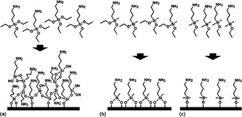
Aminopropyl triethoxysilane (APS3) is arguably the most commonly used functionalized alkoxysilane due to the versatile nature of the amine functionalization of the SPIO surface after silanization with APS3. As shown in Fig. 1(a), APS3 possesses three hydrolyzable alkoxy anchor groups and may form Fe–O–Si bonds with the SPIO surface but may also oligomerize by forming Si–O–Si bonds with other APS3 molecules. This oligomerization of APS3 is disadvantageous because it results in a thick silica shell due to the condensation of multiple molecular layers on the SPIO surface, which reduces the efficacy of the SPIO as a MRI contrast agent by decreasing the saturation magnetization of the nanoparticles.Reference Yamaura, Camilo, Sampaio, Macedo, Nakamura and Toma12 A further disadvantage of APS3 oligomerization is the occurrence of interparticle crosslinking, leading to the formation of undesired SPIO aggregates of uncontrolled sizes.Reference Bruce and Sen14 This undesired oligomerization is inherent to the trialkoxy functionality of APS3 and necessitates careful control of reaction parameters to minimize the silanization thickness and to avoid the crosslinking SPIO aggregates. Despite these disadvantages, APS3 is the de facto standard alkoxysilane for SPIO applications due to its widespread use as reported in the published literature, and this underscores the need to investigate new alkoxysilane SPIO surface chemistries to improve upon the deficiencies of APS3.
Chemical structure of APS3 (a), APS2 (b), and APS1(c) and diagrams representing surface bonding with SPIO. APS3 may form Si–O–Fe bonds to the SPIO surface but may also form Si–O–Si bonds to other APS3 molecules, resulting a thick disordered surface layer. In contrast, APS2 is less likely to oligomerize, forming a more uniform surface layer. Similarly, APS1 is unable to oligomerize and may only form a surface monolayer.

In this work, a dialkoxysilane (aminopropyl methyl diethoxysilane, APS2) and a monoalkoxysilane (aminopropyl dimethyl ethoxysilane, APS1) are studied as new SPIO surface chemistries and compared with APS3, the most commonly used tri-alkoxysilane. Due to the presence of fewer alkoxy functional groups in both APS2 and APS1, the previously mentioned undesired consequences of APS3 oligomerization may be avoided. As a dialkoxysilane, APS2 [Fig. 1(b)] possesses two hydrolyzable alkoxy anchor groups, thus reducing the propensity to form multiple molecular surface layers on the SPIO surface and reducing the ability to form undesired polydisperse SPIO aggregates. As a monoalkoxysilane, APS1 [Fig. 1(c)] possesses a single hydrolyzable alkoxy anchor group, limiting condensation of the silane to one monolayer on the SPIO surface and eliminating the possibility of crosslinking SPIO aggregates. A previous study on planar silica substrates has demonstrated that APS1 and APS2 form amine-functionalized surfaces with decreased surface thickness as compared with APS3.Reference Moon, Kim, Kim, Kang, Kim, Kim, Hahn and Park17
We report our development of APS1 and APS2 as new surface chemistries for the application of SPIO as an MRI contrast agent. We find that SPIO silanized with APS1 and APS2 (SPIO-APS1 and SPIO-APS2, respectively) have ligand densities consistent with surface monolayers while exhibiting excellent MRI contrast properties. Furthermore, we find that the chemical stability of SPIO-APS1 and SPIO-APS2 is equivalent to the multilayer SPIO surface coating formed by APS3 (SPIO-APS3), despite the single-layer thicknesses of APS1 and APS2. These results support the viability of both APS1 and APS2 as new minimal thickness hydrophilic surface coatings for the application of SPIO as an MRI contrast agent.
II. RESULTS AND DISCUSSIONS
A. Surface modification of SPIO with APS1, APS2, and APS3
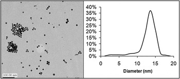
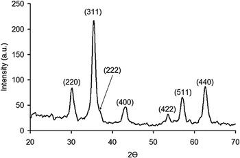
The SPIO used in this study was synthesized by a high-temperature solution-phase reaction of Fe(acac)3 in trioctylamine and heptanoic acid, yielding monodisperse nanoparticles with an average diameter of 13.5 nm, as shown in Fig. 2. The x-ray diffraction (XRD) peaks (Fig. 3) are consistent with a dominant magnetite (Fe3O4) phase with a minimal presence of hematite (Fe2O3).
Transmission electron microscopy (TEM) and particle size distribution of the as-synthesized SPIO.

The characteristic x-ray diffraction (XRD) reflections of magnetite are clearly observed and are labeled. The minor peaks at approximately 24, 40, 48, 59 and 66° suggest a minimal amount of hematite is present.

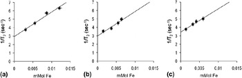
The SPIO was modified by each alkoxysilane, APS1, APS2, and APS3, by deposition from aqueous alcohol solutions. Because SPIO is a T2 MRI contrast agent, the R2 relaxivities of SPIO-APS1, SPIO-APS2, and SPIO-APS3 were determined to assess the MRI contrast agent performance of each preparation. For each sample, the T2 relaxation times of several SPIO-APS dilutions in deionized water were measured at room temperature from multispin multiecho (MSME) scans (TR = 3000 ms, TE = 12, 24, 36, and 48 ms) using a 4.7 T Bruker PharmaScan. The proportional relationship between the inverse of the measured T2 relaxation times (R2 = 1/T2) and the SPIO concentration was used to calculate the R2 relaxivity of each SPIO-APS preparation. From the results shown in Fig. 4, the R2 relaxivity values of SPIO-APS1, SPIO-APS2, and SPIO-APS3 at 4.7 T are 87.5 ± 9.9, 86.7 ± 14, and 82.4 ± 9.4 s−1 mM Fe−1, respectively. These R2 relaxivity values are similar to a recent study of an iron-oxide nanoparticle T2 contrast agent measured at 4.7 T18.
R2 molar relaxivity curves of SPIO-APS1 (A), SPIO-APS2 (B), and SPIO-APS3 (C). The R2 molar relaxivities are 87.5 ± 9.9, 86.7 ± 14, and 82.4 ± 9.4 s−1 mM Fe−1, respectively, as determined by the linear relationship between R2 and Fe molarity for each sample and the standard error of the least squares fit.

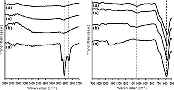
Each SPIO-APS preparation was further characterized by Fourier transform infrared (FTIR) spectroscopy, as shown in Fig. 5. The decreased intensity of CH2 stretching bands (2920 and 2850 cm−1) in SPIO-APS3 [Fig. 5(b)], SPIO-APS2 [Fig. 5(c)], and SPIO-APS1 [Fig. 5(d)] relative to as-synthesized SPIO [Fig. 5(a)] indicates the removal of carboxylic acid from the SPIO surface. Removal of the carboxylic acid is necessary to allow the formation of Fe–O–Si bonds between the SPIO surface and the APS ligands. The weak CH2 stretching bands observed in Figs. 5(b)–5(d) are attributed to the aminopropyl head groups of APS3, APS2, and APS1 (Fig. 1). The appearance of a Si–O stretching band (1030 cm−1) is observable in all three SPIO-APS spectra. The Si–O stretching bands indicate the presence of APS ligands on the SPIO surface as they are absent in the spectrum of the as-synthesized SPIO. The strong band observed at 588 cm−1 is characteristic of Fe–O stretching. The significant decrease of CH2 stretching bands and the appearance of a Si–O stretching band in all SPIO-APS spectra confirms the presence of APS on the surface of all three SPIO-APS materials.
FTIR of the as-synthesized SPIO (a), SPIO-APS3 (b), SPIO-APS2 (c), and SPIO-APS1 (d). CH2 stretching bands (2920 and 2850 cm−1) from carboxylic acid are present in the as-synthesized SPIO (a). The carboxylic acid is displaced in SPIO-APS (b–d) by the APS ligand, as evidenced by the decreased intensity of the samples CH2 stretching bands (2920 and 2850 cm−1) and the appearance of a Si–O stretching band (1030 cm−1). The strong band near 588 cm−1 is a Fe–O stretching vibration.

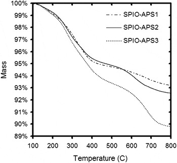
To characterize the surface thickness of the SPIO-APS1 and SPIO-APS2 preparations and to compare these new surface chemistries with SPIO-APS3, thermogravimetric analysis (TGA) was performed to quantify the mass loss of the alkoxysilane surface coating after thermal desorption. The mass loss determined by TGA (Fig. 6) was greatest for SPIO-APS3 (10.2%), less for SPIO-APS2 (7.70%), and least for SPIO-APS1 (6.88%). These results are consistent with a study reporting that SPIO-APS3 forms thicker surface films than SPIO-APS2 and SPIO-APS1.Reference Moon, Kim, Kim, Kang, Kim, Kim, Hahn and Park17 From the mass loss data, the surface ligand density of each SPIO-APS preparation may be estimated by dividing the thermally desorbed mass by the molecular weight of the appropriate APS molecule and the approximated surface area of 13.5 nm spherical nanoparticles. The approximated surface ligand densities are 5.2 × 10¹⁴ and 4.0 × 10¹⁴ ligands per cm2 for SPIO-APS3 and SPIO-APS2, respectively, which are in agreement with silane ligand densities in literature,Reference De Palma, Peeters, Van Bael, Van den Rul, Bonroy, Laureyn, Mullens, Borghs and Maes16, Reference Moon, Kim, Kim, Kang, Kim, Kim, Hahn and Park17 and 3.6 × 10¹⁴ ligands per cm2 for SPIO-APS1. Whereas the silane ligand density of SPIO-APS1 is higher than reported ligand densities of APS1 monolayers on planar silica,Reference Moon, Kim, Kim, Kang, Kim, Kim, Hahn and Park17 we attribute the increased density to decreased steric hindrance within the high curvature monolayer of the nanoparticle substrate.
TGA of SPIO-APS shows greatest mass loss due to ligand desorption for SPIO-APS3 and least mass loss for SPIO-APS1. The samples were heated at 20 °C per min under N2.

The ligand densities calculated from the TGA results are also supported by Si/Fe elemental ratios determined by inductively coupled plasma optical emission spectroscopy (ICP-OES) measurements of each SPIO-APS preparation: 3.1, 2.6, and 2.2% for SPIO-APS3, SPIO-APS2, and SPIO-APS1, respectively. Assuming stoichiometric Fe3O4 nanoparticles with an average diameter of 13.5 nm, the expected Si/Fe ratios based on the observed mass loss are 3.2, 2.4, 2.1% for SPIO-APS3, SPIO-APS2, and SPIO-APS1, respectively. These results demonstrate that both SPIO-APS1 and SPIO-APS2 form ultra thin surface coatings on SPIO with greatly reduced ligand density as compared with SPIO-APS3 and are viable surface chemistries for MRI contrast agent applications of SPIO.
B. Chemical stability of SPIO-APS1 and SPIO-APS2
To complement the materials characterization of SPIO-APS1 and SPIO-APS2, additional consideration is needed for application as an MRI contrast agent. Whereas minimization of the APS surface thickness of SPIO is desired for maximization of the SPIO MRI contrast, the chemical stability of the ultrathin surface coatings is an important consideration, particularly for long-term studies. A powerful application of SPIO is in vivo MRI tracking of SPIO-labeled cells for cell trafficking studies such as tracking stem and progenitor cells,Reference Miyoshi, Flexman, Cross, Maravilla, Kim, Anzai, Oshima and Minoshima18, Reference Sykova and Jendelova19 visualizing transplanted hepatocytes,Reference Shapiro, Sharer, Skrtic and Koretsky20 and visualizing tumor-targeting by cytotoxic lymphocytes.Reference Kircher, Allport, Graves, Love, Josephson, Lichtman and Weissleder21, Reference Valable, Barbiera, Bernaudinc, Roussel, Segebartha, Petit and Rémy22 The SPIO-labeled cells (i.e., cells with internalized SPIO) are distinguishable in vivo by their negative MRI contrast and this offers a noninvasive alternative to post-mortem histology. The long-term nature of cell-labeling studies requires the SPIO surface chemistry to provide adequate chemical stability in intracellular conditions, including the acidic environment of lysosomes, intracellular acidic vesicles at pH 4.5–5.Reference Mukherjee, Ghosh and Maxfield23 Stability in an acidic environment is particularly important because a recent study has observed that alkoxysilane-modified SPIO internalized in cells are sequestered in lysosomes.Reference Lu, Hung, Hsiao, Yao, Chung, Lin, Wu, Hsu, Liu, Mou, Yang, Huang and Chen24 To support their viability for MRI cell-labeling applications, the chemical stability of both SPIO-APS1 and SPIO-APS2 in acidic conditions must be evaluated.
Since APS3 is a well-established SPIO surface chemistry for in vivo applications, SPIO-APS1 and SPIO-APS2 were compared with SPIO-APS3 to provide a relative measure of the chemical stability of these new surface chemistries. All SPIO-APS materials were incubated in neutral and acidic media to simulate intracellular conditions using methods similar to chemical stability studies of dextran-modified SPIO.Reference Arbab, Wilson, Ashari, Jordan, Lewis and Frank25, Reference Skotland, Sontum and Oulie26 The three mediums used in this study were a neutral medium (RPMI) and acetic and citric acid media (RPMI-acetic and RPMI-citric, respectively). These three media provide a useful range from neutral to acidic conditions to assess the chemical stability of APS1 and APS2 relative to APS3. It is important to emphasize that this evaluation of the three SPIO-APS materials in the different media is intended to be a comparison of the new APS1 and APS2 surface chemistries relative to APS3. In the context of this study, it is appropriate to compare both SPIO-APS1 and SPIO-APS2 with SPIO-APS3 due to the previous studies abundantly demonstrating APS3 as an effective SPIO surface chemistry for MRI contrast agent applications.
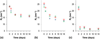
To assess the ability of the APS1 and APS2 surface chemistries to adequately stabilize the SPIO core in a long-term MRI study, the R2 relaxivity of each SPIO-APS preparation was measured at several time points during incubation in the neutral and acidic media. Each of the three SPIO-APS materials was incubated in each media for 14 days at 37 °C, and during the incubation, MRI phantoms were prepared using aliquots of each SPIO-APS from each media and the R2 relaxation was measured for each sample. As shown in Fig. 7, a decreased R2 is observed by all three SPIO-APS materials in each of the media; there is no observable difference between the SPIO-APS1, SPIO-APS2, or SPIO-APS3 preparations. These results indicate that APS1 and APS2 possess comparable chemical stability to APS3, despite the decreased surface layer thickness of APS1 and APS2.
Fe normalized R2 of SPIO-APS1 (diamond), SPIO-APS2 (square), and SPIO-APS3 (circle) incubated in RPMI (a), RPMI-Acetic (b), and RPMI-Citric (c) at 0, 1, 4, 8, and 14 days.

Whereas there are no observable differences in chemical stability between the APS1, APS2, or APS3 surface chemistries, large differences are observed in the different media.Reference Arbab, Wilson, Ashari, Jordan, Lewis and Frank25 After the 14 day in vitro incubation, the average change of R2 is approximately −30% in RPMI, −47% in RPMI-acetic media, and −73% in RPMI-citric. The significantly greater R2 reduction observed in the RPMI-citric medium compared with RPMI-acetic is attributed to the greater capacity of citrate to solubilize iron than acetate, as citrate possesses two additional carboxylate anions.Reference Arbab, Wilson, Ashari, Jordan, Lewis and Frank25 The R2 decrease of SPIO-APS in neutral RPMI is slightly less than in RPMI-acetic, which is consistent with reported results of dextran-coated SPIO (SPIO-dextran) degradation.Reference Arbab, Wilson, Ashari, Jordan, Lewis and Frank25, Reference Skotland, Sontum and Oulie26 It is important to note that the significant degradation of SPIO-APS in RPMI-citric does not preclude APS1 or APS2 as viable SPIO surface chemistries: previous studies of SPIO-dextran indicates that RPMI-acetic medium is a more realistic simulation of intracellular degradation than the RPMI-citric acid medium, which is overly aggressive.Reference Arbab, Wilson, Ashari, Jordan, Lewis and Frank25, Reference Skotland, Sontum and Oulie26
These results demonstrate that the new APS1 and APS2 offer comparable chemical stability to the well-established APS3 surface chemistry for applications of SPIO as a contrast agent. Despite the ultrathin nature of these new SPIO surface coatings, both APS1 and APS2 stabilize the SPIO equivalently to APS3 in neutral and acidic media.
III. CONCLUSIONS
In this study, two new hydrophilic amine-functionalized SPIO surface coatings were developed using mono- and di- alkoxysilane analogs of the commonly used aminopropyl-triethoxysilane. New APS1 and APS2 surface chemistries were used to modify SPIO, creating surface layers with surface ligand densities consistent with molecular monolayers. The SPIO-APS1 and SPIO-APS2 preparations both exhibit comparable molar T2 relaxivity to previously reported SPIO contrast agents, demonstrating its efficacy as an MRI contrast agent. The results of this study establish APS1 and APS2 as viable alternatives to the commonly used APS3 for SPIO applications requiring minimal surface. Furthermore, the new SPIO-APS1 and SPIO-APS2 surface chemistries demonstrate adequate chemical stability in both neutral and acidic media, exhibiting comparable stability to SPIO-APS3.
IV. EXPERIMENTAL METHODS
APS1, APS2, and APS3 were purchased from Gelest. All other materials were purchased from Sigma Aldrich.
A. Synthesis of SPIO-APS
A previously reported method was used to synthesize SPIO.Reference Barker, Cage, Russek and Stoldt27, Reference Larsen, Haag, Serkova, Shroyer and Stoldt28 Briefly, 18.6 mL of trioctylamine and 1.4 mL of heptanoic acid were heated to 260 °C with magnetic stirring and .045 M Iron(III) Acetylacetonate was added. The reaction was stopped 3 h after nucleation (observed as a change in solution color from dark red to black) and after cooling to ambient temperature, the SPIO was precipitated with ethanol. The as-synthesized SPIO was magnetically precipitated and washed twice with ethanol, then resuspended in 10 mL toluene.
To modify SPIO using APS3, 20 mg of SPIO was magnetically precipitated and washed with butanol, then resuspended in 20 mL of 30% vol/vol NH4OH in butanol and heated to reflux with magnetic stirring for 2 h. After cooling to ambient temperature, the SPIO was magnetically precipitated and washed twice with butanol and resuspended in 20 mL of butanol. Two hundred μL of APS3, 100 μL of deionized water, and 10 μL of acetic acid were added to the SPIO in butanol and the solution was reacted with mechanical (nonmagnetic) stirring for 24 h at 70 °C. After cooling to ambient temperature, the SPIO-APS3 was magnetically precipitated and washed twice with acetone, twice with ethanol, twice with deionized water, and resuspended at 5 mg/mL in deionized water. The procedures for APS1 and APS2 modification were performed in the exact same manner.
B. Chemical stability of SPIO-APS in neutral and acidic media
Simulated lysosomal degradation of the three SPIO-APS types was evaluated by incubating the APS-SPIO in neutral and acidic solutions, a method adapted from previous studies investigating the chemical stability of dextran-modified SPIO.Reference Arbab, Wilson, Ashari, Jordan, Lewis and Frank25, Reference Skotland, Sontum and Oulie26 The three solutions used as incubation media to evaluate the simulated lysosomal degradation of SPIO were RPMI-1640 medium at pH 7.0 (RPMI), RPMI 1640 with 20 mM sodium citrate at pH 4.0 (RPMI-citric), and RPMI 1640 with 20 mM sodium acetate at pH 4.0 (RPMI-acetic). RPMI-citric and RPMI-acetic were adjusted to pH 4.0 with citric acid and acetic acid, respectively. SPIO-APS1, SPIO-APS2, and SPIO-APS3 were each added to 12 mL of RPMI, RPMI-citric, and RPMI-acetic at a concentration of 40 μg per mL, as determined by ICP-OES, yielding nine samples which were incubated in a 37 °C water bath for 14 days. Aliquots were extracted after 1 h, 1 day, 2 days, 4 days, 8 days, and 14 days to prepare phantoms for MRI. The MRI phantoms were prepared by vortex mixing a 0.5 mL aliquot of each sample with 0.5 mL of 16% gelatin in a 1.5 mL centrifuge tube. The gelatin phantoms were immediately immersed in an ice bath to rapidly set the gelatin, solidifying the SPIO-APS/incubation medium sample and effectively quenching the solution from further acidic degradation. The phantoms were stored at 4 °C and T2 measurements of the phantoms were performed using a 4.7 T Bruker PharmaScan MRI using a multispin multiecho (MSME) pulse sequence using a 68-mm Bruker volume coil, which was used as an RF receiver and transmitter (field of view FOV = 6.4 cm, repetition time TR = 3000 ms, echo times TE = 12, 24, 36, and 48 ms for T2 mapping). For the series of 4 echo times in this MR protocol, the Bruker t2vtr-fitting function based on the equations below was applied in order to calculate T2 relaxation time as a function of signal intensity and TE values of each image:


where  is a constant which gets fitted. All calculated T2 times were given in milliseconds (ms). All T2 relaxivity vales were calculated as R 2 = 1/T2 and given as (s−1).