




No CrossRef data available.
Article contents
Syntactic language processing: ERP lesion data on the role of the basal ganglia
Published online by Cambridge University Press: 12 February 2004
Abstract
The role of the basal ganglia in syntactic language processing was investigated with event-related brain potentials in fourteen neurologically impaired patients. Seven of these patients had basal ganglia lesions while 7 other patients primarily had lesions of the left temporo–parietal region excluding the basal ganglia. All patients listened to sentences that were either correct or included a verb argument structure violation. In previous experiments this type of violation elicited a biphasic pattern of an N400–P600 complex in young healthy participants. While the N400 may result from incorrect semantic-thematic role assignment, the P600 reflects the fact that verb information does not license the syntactic structure at present. Results of the patient experiment revealed a double dissociation: patients with left temporo–parietal lesions only show a P600, whereas patients with lesions of the basal ganglia showed no P600, but a negativity with extended duration that resembled an N400. The latter pattern not only confirms previous reports that the basal ganglia modulate the P600 but extends these results by showing that the N400 as a late semantic–thematic integration process appears partially modulated by the basal ganglia. (JINS, 2003, 9, 1053–1060.)
Information
- Type
- THEMATIC ARTICLES
- Information
- Journal of the International Neuropsychological Society , Volume 9 , Issue 7 , November 2003 , pp. 1053 - 1060
- Copyright
- © 2003 The International Neuropsychological Society
INTRODUCTION
While the role of cortical structures in language processing has been confirmed by numerous investigations (see Friederici, 1999; Goodglass, 1993, for reviews), the participation of subcortical structures, such as the basal ganglia, in language is highly controversial. In particular, functional implications of subcortical structures are at stake. Nadeau and Crosson (1997) and Crosson (1999) postulate that the thalamus rather than the basal ganglia is engaged during lexical–semantic processing. This argument is supported by data that do not reveal lexical–semantic deficits in patients with basal ganglia lesions (Gotham et al., 1988; Mortimer et al., 1982; Tyler & Marslen-Wilson, 1986). However, other empirical data suggest that lexical–semantic and prosodic processes do engage the basal ganglia (e.g., Lieberman, 2001). Neuroimaging studies with healthy subjects and Parkinson patients (PD), who suffer from a neurodegenerative disorder of the basal ganglia, have confirmed a correlation of the basal ganglia function with the perception of emotional prosody (e.g., Breitenstein et al., 1998, 2001; Kotz et al., in press; Pell, 2002), as well as with lexical–semantic processes (e.g., Cappa & Abutalebi, 1999; Lieberman, 2001; Wallesch & Papagno, 1988).
In addition, the basal ganglia have been linked to language production in general (Alexander et al., 1987; Robin & Schienberg, 1990) or to processing of syntactic information in both language production and comprehension (e.g., Grossman et al., 1993; Lieberman et al., 1992). Three general cognitive processes have been implicated in syntactic comprehension of sentences: (1) regulation of attention, (2) working memory and (3) speed of information processing. Several authors investigated syntactic complexity (e.g., subject–object relative clauses) during sentence comprehension in basal ganglia patients (e.g., Grossman et al., 1991, 1992, 1993; Lieberman et al., 1990, 1992; Natsopoulos et al., 1993; Pickett et al., 1998). While Grossman et al. (1993) initially argued that syntactic comprehension deficits result from attentional rather than syntactic deficits, Lieberman et al. (1990, 1992) proposed that repeated errors on syntactically complex sentences cannot be attributed to an attention deficit, but to a working memory deficit. Recently, Grossman et al. (2002) attributed syntactic comprehension deficits in PD patients to slowed lexical access.
Finally, Ullman (2001) and Ullman et al. (1997) proposed that a fronto–striatal network engages in the computation of procedural knowledge, which reflects the implicit rules and operations of syntax. Utilizing a verb participle production paradigm that allowed one to separate regular verb forms (rule-based; e.g., walk, -ed) and irregular verb forms (lexically based; e.g., teach, taught), the authors reported that patients with anterior lesions and PD patients at a late stage of their disease progression show a selective deficit for regular verb forms, while patients with posterior lesions or Alzheimer's disease cannot produce irregular verb forms. This dissociation of verb-specific deficits was taken as evidence that the whole fronto–striatal loop plays a role in implicit rule based syntactic processing.
In summary, while it is clear from the literature that the basal ganglia are engaged during language processing, a clear functional specification of the role of the basal ganglia in language remains open. In particular, the claim that the basal ganglia play a specific role during syntactic processing is controversial as the mental operations proposed to underlie or correlate with syntactic processing are diverse. Furthermore, there is no clarity as to whether lexical–semantic processes engage the basal ganglia or not. Most authors consider that the deficit arising from basal ganglia damage is not one that is solely “automatic” in nature (but see Ullman et al., 1997). The latter statement can be best clarified by analogy to the motor control hypothesis introduced by Marsden and Obeso (1994). These authors suggested that the primary role of the basal ganglia is a controlled response to changes in cortically regulated automatic behavior. If one applies this proposition to syntactic processes in language comprehension one could speculate that the basal ganglia engage in the controlled reordering or altering of cortically driven automatic syntactic processes.
This dissociation of automatic and controlled syntactic processing is also made explicit in a recent model on auditory sentence processing by Friederici (2002). The model describes that in a first step, a simple syntactic structure is built on the basis of word-category information (e.g., noun, verb). As will be described below, this first processing phase is highly automatic. In a second phase, which is controlled, lexical–semantic information is processed to realize thematic role assignment. If initial syntactic information and lexical–semantic information do not map onto each other, as in the case of some syntactic violations, the sentence structure needs to be reanalyzed in a third phase which is also a controlled process.
A number of event-related brain potential (ERP) studies investigating syntactic processes in healthy participants have shown that automatic and controlled syntactic processes can be separated (Friederici, 1995; Friederici, 2002; Hahne & Friederici, 1999). For example, phrase structure violations (e.g., violating the expectancy of a word class as in, *The fish was in the_ caught rather than The fish was in the pond caught; literal German translation) elicit an early anterior negativity (E(L)AN), followed by a late positivity (P600). Adhering to the sentence processing model described above, the early anterior negativity has been correlated with automatic syntactic processes, as the component does not vary as a function of manipulations that implicate control. This was shown by manipulating the proportion of violations that do not modulate the E(L)AN component (Hahne & Friederici, 1999). Furthermore, the E(L)AN is not influenced by additional violations of lexical-semantic information (Frisch et al., 2000; Hahne & Friederici, 1999). On the other hand, the P600 has been linked to controlled syntactic processes (e.g., Frisch et al., 2002; Kaan et al., 2000). Finally, a third component, the N400 with a maximal centro-parietal distribution has been linked to the processing of lexical–semantic information. At the sentence level, the N400 is discussed as a component reflecting controlled, integrative processing of lexical–semantic information.
In three investigations with patients we explored the role of automatic versus controlled syntactic processes in lesion patients and PD patients. Friederici et al. (1999) reported that patients with anterior lesions show no early anterior negativity, but a P600 elicited by phrase structure violations, while patients with basal ganglia lesions show an early anterior negativity, but a strongly reduced P600. The authors take this evidence in support of the hypothesis that anterior cortical areas, but not subcortical regions such as the basal ganglia, are engaged in automatic syntactic processes, while the basal ganglia seem to modulate controlled syntactic processes. In a study with early PD patients a similar pattern emerged: PD patients showed an early anterior negativity, but barely any P600 effect (Friederici et al., 2003). Thus, unilateral focal vascular lesions as well as PD patients with unilateral functional deficits result in a comparable syntactic deficit as evidenced in the reduction of the P600 effect. Furthermore, these data support a functional as well as a structural separation of the two syntactic processes. While automatic syntactic processes seem to be regulated in anterior cortical regions, controlled late syntactic processes appear to be modulated by the basal ganglia. One question that these studies left open was to clarify whether the P600 reflects late syntactic processes or rather varies as a function of attentional demands. This question is also reflected in an ongoing debate whether the P600 is language-specific or just a P300-like effect, indicating the attention driven detection of an unexpected, task-relevant target (Coulson et al., 1998; Gunter et al., 1997). To test this question, Frisch et al. (2003) tested patients with focal vascular basal ganglia lesions and patients without basal ganglia lesions. They were presented with correct and incorrect sentences that included a morphosyntactic violation (e.g., *In the house it was often to paint rather than, In the house it was often painted; literal German translation) as well as in a classic non-linguistic oddball paradigm (e.g., counting deviant tones in a series of standard tones). The prediction was that patients with basal ganglia lesions should show no P600, while patients without basal ganglia lesions should show a P600. If the basal ganglia indeed modulate late controlled syntactic processes rather than general attentional processes, both patient groups should show a P300 elicited in the non-linguistic oddball paradigm. Both predictions were confirmed. While both patient groups displayed a P300 in the oddball task, no P600 was elicited by morphosyntactic violations in the patients with basal ganglia lesions.
In summary, ERP lesion data and data collected from patients with Parkinson's disease show that the functional significance of the basal ganglia in syntactic language processing can be described in the following way: The basal ganglia do not seem to play a role during automatic syntactic processing, but during controlled syntactic processing. What remains to be investigated is whether the latter syntactic processing deficit can be replicated in other syntactic paradigms that typically elicit a P600, and whether the basal ganglia also engage in lexical–semantic integration processes (as reflected by an N400) or not.
Following this brief review on syntactic processes investigated with ERPs in healthy subjects and patient populations of diverse etiology we would like to specify the hypothesis that the basal ganglia are engaged in controlled late syntactic processes that result from the lack of mapping between lexical–semantic and initial syntactic information as proposed by Friederici (2002). This hypothesis is not in full agreement with the hypothesis proposed by Ullman et al. (1997) as they claim both cortical and subcortial areas to be involved in the processing of “procedural” knowledge.
Therefore, the goal of the current ERP study was to address syntactic comprehension in the same two patient groups as tested in Frisch et al. (2003). Here we were interested in investigating the temporal and functional dissociation of two controlled processes, the N400 which can reflect lexical–semantic information processing relevant for thematic role assignment and the P600 within one syntactic structure, the verb–argument structure, which adheres to both syntactic and semantic–thematic restrictions of the verb. Previous evidence from healthy young participants revealed a biphasic pattern of an N400 followed by a P600 elicited by verb–argument structure violations with a centro-parietal distribution (Friederici & Frisch, 2000; Frisch et al., 2000; Osterhout et al., 1994). We predicted that patients with focal basal ganglia lesions should show an N400 but no P600, while patients with primarily temporo–parietal lesions should potentially show a P600 but no N400.
EXPERIMENT
It is central for language processing research that verbs can be classified with respect to the number and type of constituents that they take as (syntactic and semantic) arguments. For example, an intransitive verb such as to grin can only take a subject argument expressing who is doing the grinning (e.g., The little boy grins). In contrast to a transitive verb which allows or demands an object besides the subject, adding a direct object to a verb such as to grin would render a sentence ungrammatical as well as semantically anomalous (see, *The little boy grins the old man). In the experiment, participants listened to grammatical and ungrammatical sentences. Ungrammatical sentences contained violations of the verb-argument structure (see above). In a number of studies that explored this syntactic violation type a biphasic ERP pattern of a negativity, resembling an N400, followed by a positivity (P600) was reported in healthy young subjects (Friederici & Frisch, 2000; Frisch et al., 2000; Osterhout et al., 1994).
METHODS
Research Participants
Fourteen brain damaged patients (4 female, all right-handed) in a chronic state participated in the current study after giving informed consent. Lesions primarily resulted from left hemisphere ischemic (n = 12) strokes, but 2 patients of the basal ganglia group had suffered a left-hemisphere hemorrhage. The average time since lesion in the basal ganglia group was: 2.85 years (range: 2–4 years) and in the patient group without basal ganglia lesions: 5.71 years (range: 3–9 years). Lesion sites were determined by (T1- and T2-weigthed) anatomical MRI datasets from a 3.0 T system (Bruker 30/100 Medspec) and evaluated by an experienced neuroanatomist. The individual patient information is listed in Table 1.
Patient history: Descriptions of lesions determined by MRI scans for each individual patient in both groups (abbreviations: caud = caudatum, put = putamen, pall = globus pallidus). The severity of the language comprehension disorder is indicated by the number of mistakes in the Token Test: no/very mild disorder (0–6); mild (7–23); moderate (24–39); severe (>40). In addition, the auditory comprehension scores of the Aachen Aphasia Test (AAT) are listed for each patient (only patients with a Token Test score greater than zero were tested with the AAT). The degree of the comprehension disorder is evaluated based on a total of 60 points.

Materials
All sentences were German passive constructions. In contrast to English, German allows passivization of intransitive verbs (such as arbeiten/to work). In this case, however, the sentence initial position can only be filled with an expletive (such as es/there), a prepositional phrase (such as im Zimmer/in the room) or an adverb (such as gestern/yesterday). Filling the initial position with a subject argument (such as das Zimmer/the room) creates an argument-structure violation since the subject can neither be syntactically nor semantically integrated. Thus, we realized an argument-structure violation by using sentences with an intransitive verb and a subject NP (Das Zimmer wurde gearbeitet/The room was worked). In the correct conditions, the sentence initial element was a prepositional phrase (Im Zimmer wurde gearbeitet/In the room it was worked). This allowed us to keep the critical word (verb participle) identical across correct and incorrect conditions.
In order to exclude possible confounds with a sentence final wrap up effect (e.g., Friederici & Frisch, 2000; Osterhout, 1997), the critical verb participle was always followed by a conjunction ‘and’ and a second verb participle which was transitive and therefore always correct. Forty sentences per condition, resulting in 80 critical sentences were created on the basis of 80 noun-(intransitive) verb sets. In addition, 80 filler sentences (half of them ungrammatical) with a similar sentence structure were created.
A female native speaker of German spoke the sentences at a normal speech rate. The sentences were recorded onto digital audio tape and digitized at a sampling rate of 44.1 KHz. In order to ensure a precise time locking of the ERP in each individual sentence, the onset of the critical word was marked by way of a careful visual and auditory inspection of the auditory speech signal.
Procedure
Patients listened to all 160 sentences that were presented via loudspeakers in a pseudorandomized order. A visual cue on the center of a computer screen indicated the onset of each sentence. 800 ms after the offset of the sentence, subjects judged whether the sentence was acceptable or not by pressing one of two response buttons. The next trial started 1000 ms after the subject's button press. In the non-linguistic task patients heard standard tones (600 Hz) with a probability of .8 and deviants (660 Hz), with a probability of .2. The two-tone block contained a total of 500 auditory stimuli. All stimuli had a duration of 200 ms (including 10-ms rise and 40-ms fall time; sound pressure level (SPL) 75 dB) and were presented with a constant offset-to-onset interval of 600 ms.
ERPs were recorded from 19 scalp sites by means of Ag/AgCl electrodes with a NEUOSCAN 4.1 amplifier. C2 served as ground electrode. Recordings were referenced to the left mastoid and were re-referenced to linked mastoids off-line. Electrode impedances were kept below 5 kΩ. In order to control for eye movement artifacts, a horizontal and a vertical electro-oculogram (EOG) were recorded. Eye artifact control measures were applied to the raw data of each patient to increase the number of critical trials in each condition (Pfeifer et al., 1995). Then individual EEG recordings were scanned for additional artifacts on the basis of visual inspection. The average percentage of trials rejected due to behavioral performance and additional artifacts was 24.9%.
Data analysis
Accuracy in the behavioral task was calculated as the percentage of incorrectly performed trials in one condition relative to all trials in that condition. An ANOVA with lesion as a between-subjects factor and grammaticality as a within-subjects factor was conducted. ERPs were computed for each of the critical conditions for each electrode and each subject. All ERP averages were aligned to a 200 ms baseline relative to the onset of the auxiliary verb preceding the critical verb. Only trials with correct responses and without movement and amplifier saturation artefacts entered the averages. Separate repeated-measures ANOVAs were conducted separately for midline electrodes (FZ, CZ, PZ), for anterior lateral electrodes (FC3/4, F3/4, F7/8) and for posterior lateral electrodes (P3/4, P7/8, O1/O2) in order to capture potential distributional differences. The ANOVA for the midline analysis included lesion as the between-subjects factor (lesions including vs. excluding the basal ganglia) and two within-subjects factors grammaticality (grammatical vs. ungrammatical) and electrode (FZ vs. CZ vs. PZ). The ANOVA for the two lateral regions of interest were calculated with lesion as the between-subjects factor, and with two within-subjects factors grammaticality, and hemisphere (left vs. right), respectively. The statistical analyses were computed in two time windows relative to the critical word (verb), selected on the basis of visual inspection: 300 to 700 ms for the N400 and 800 to 1200 for the P600. Main effects of grammaticality in the respective time windows will reflect a N400 effect and a P600 effect, respectively. Results will be reported as statistically significant for p values .05 or less. Furthermore, to ensure that any modulation of the patient data was of linguistic nature, the P300 oddball paradigm was applied as a non-linguistic test. The statistical analyses followed the same ANOVA design as presented for the linguistic experiment with a within-subjects factor probability (rare vs. often) and a between-subjects factor lesion on the averages in a time window between 300 and 600 ms.
RESULTS
Accuracy
We found a main effect of lesion [F(1,12) = 13.55, p < .01] due to more errors made by the basal ganglia group (33.2%) as compared to the group without a basal ganglia lesion (16.6%) and of grammaticality [F(1,12) = 8.76, p < .05] showing that on average patients made more errors in the violation condition (35.0%; SD = 17.88) than in the correct condition (14.8%; SD = 9.64). There was no grammaticality × lesion interaction (p > .1).
ERPs
Figure 1 displays the ERP patterns from the onset of the critical verb up to 1500 ms for each of the two lesion groups at selected electrode-sites. It is apparent that the patients without basal ganglia lesions show a clear P600 effect for verb-argument violations, but no apparent N400 effect (B). On the other hand, patients with basal ganglia lesions do not show a P600 effect, but an extended negativity resembling an N400 (A).

ERP effects for patients with basal ganglia lesions (A) and patients without basal ganglia lesions (B) at selected electrode-sites. Marked on a schematic head are the electrodes that are graphically displayed showing anterior (e.g., F3, FZ, F4), central (e.g., C3, CZ, C4) and posterior (e.g., P3, PZ, P4) regions. For orientation L = left sites, M = midline sites and R = right sites. The correct condition is displayed in a solid line, the incorrect condition in a dotted line. Negativity is plotted upwards and each tick on the x-axis indicates 500 ms. ERPs from the onset of the critical verb (onset at zero ms/vertical bar) up to 1500 ms show that patients with basal ganglia lesions display an extended N400-like negativity effect, but no P600 effect, while patients without basal ganglia lesions show no N400-like negativity effect, but a clear P600 effect.
N400-like Negativity
The global ANOVA for the N400-like negativity in the selected time window (300–700 ms) at midline sites revealed no main effects of lesion nor grammaticality (all Fs < 1), but a marginal interaction of grammaticality × lesion [F(1,12) = 3.98, p = .07]. Follow-up analyses by lesion group did not confirm a statistically significant N400-like negativity in either patient group at midline sites (Fs < 1).
Analyses of lateral sites showed no significant effects for lesion, grammaticality, hemisphere nor an interaction of any of the three factors (all Fs < 1) in the anterior region. In the posterior region we found an interaction of grammaticality × lesion [F(1,12) = 7.77, p < .01], but no main effects of lesion nor grammaticality. Follow-up analyses by lesion group revealed that patients with basal ganglia lesions show a N400-like negativity at posterior electrode sites [F(1,6) = 7.37, p < .05], but the patients without basal ganglia lesions did not (p > .1).
Analyses of the N400-like negativity preceding the P600 time window resulting from verb–argument structure violations indicate that patients with unilateral basal ganglia lesions show a bilaterally distributed extended N400-like negativity effect at posterior sites, while patients without basal ganglia lesions do not show such an N400-like negativity effect.
P600
Analyses of the midline sites revealed a main effect of grammaticality [F(1,12) = 4.42, p < .05], but not of lesion (F < 1). However, an interaction of grammaticality × lesion [F(1,12) = 9.99, p < .01] as well as an interaction of grammaticality × lesion × electrode [F(2,24) = 3.91, p < .03] can be reported. These interactions resulted from the fact that patients without basal ganglia lesions display a P600 effect [F(1,6) = 15.24, p < .001] and an interaction of grammaticality × electrode [F(2,12) = 5.39, p < .05] at midline sites, but patients with basal ganglia lesions do not show comparable effects (all effects: F < 1). Grammaticality was significant at all three electrode sites for patients without basal ganglia lesions: Fz [F(1,6) = 6.67, p < .05]; Cz [F(1,6) = 15.24, p < .001]; Pz [F(1,6) = 24.63, p < .001].
A similar picture emerged for the analyses of lateral sites. Analyses of anterior sites showed no main effects nor any critical interactions for any factor (all Fs < 1). Analyses of posterior sites displayed a main effect of grammaticality [F(1,12) = 8.00, p < .01], but not of lesion (F < 1). However, a significant interaction of grammaticality × lesion [F(1,12) = 12.82, p < .001] was found. Follow-up analyses by patient group revealed that patients without lesions of the basal ganglia showed a P600 effect at posterior sites [F(1,6) = 14.66, p < .001], but not at anterior sites (F < 1). This effect was not qualified by hemisphere (F < 1). Patients with basal ganglia lesions showed no main effect of grammaticality nor any interaction at either anterior or posterior electrode sites (all Fs < 1).
The data show that patients with basal ganglia lesions show no P600 effect following the preceding extended N400-like negativity effect, while patients without basal ganglia lesions show only a P600 effect.
Statistical analyses of repeated-measures ANOVA on the P300 effect did not reveal any significant differences as a function of lesion, but a main effect of probability indicating that patients of both groups showed a normal P300 effect. Detailed data analyses of the P300 effect were reported elsewhere (Frisch et al., 2003).
In summary, while patients with unilateral basal ganglia lesions show an extended negativity comparable to an N400 effect at posterior electrode sites, but no P600 effect as a result of verb–argument violations, patients without lesions of the basal ganglia show no N400-like negativity effect, but a P600 effect to this type of violation. These results are in contrast to the biphasic pattern of an N400 followed by a P600 in younger healthy participants (see Frisch et al., 2000).1
In a pilot study with a sample of 14 age-, gender- and education-matched controls for the patients tested in the current experiment we also found a biphasic pattern of an N400 followed by a P600.
DISCUSSION
Taken together, the data from the current experiment further support the role of the basal ganglia in controlled syntactic processing by replicating the lack of a P600 effect in patients with focal unilateral basal ganglia lesions (Friederici et al., 1999; Frisch et al., 2003) and extending it by reporting evidence on the role of the basal ganglia in a second controlled process that relates to semantic–thematic processing.
As it was hypothesized, a P600 effect due to verb–argument structure violations was only found in patients without basal ganglia lesions. However, these patients showed an extended N400-like negativity effect preceding the P600 time window which resembles the N400 effect in young healthy participants (see Frisch et al., 2000), while patients without basal ganglia lesions did not show such an N400-like negativity effect. Furthermore, a P300 effect in response to rarely occurring auditory stimuli was shown in both groups of patients and was comparable to healthy controls (see Frisch et al., 2003).
With respect to our hypothesis that the basal ganglia only regulate controlled syntactic processes, the current results support the fact that the basal ganglia play a necessary role in the mediation of the P600 effect. Thus, the present results are in agreement with recent findings that the P600 effect is strongly reduced in Parkinson patients (Friederici et al., 2003) and in patients with unilateral lesions of the basal ganglia (Friederici et al., 1999; Frisch et al., 2003; Kotz & Friederici, 2003).
The question of whether there is a functional correlation of the basal ganglia and lexical–semantic processes can be partially answered. The fact the basal ganglia group show an N400-like negativity effect, but with an extended duration points to the modulatory role of the basal ganglia in lexical–semantic processing such as thematic role assignment. This extended N400-like negativity adds to controversial previous evidence as the data imply that speed of information processing affecting lexical–semantic information might be modulated by the basal ganglia (e.g., Crosson, 1999; Nadeau & Crosson, 1997; but see Cappa & Abutalebi, 1999; Wallesch & Papagno, 1988).
In particular, it needs to be noted that in comparison to the N400 seen in younger and older healthy participants (e.g., Frisch et al., 2000) the duration of the N400-like negativity effect in the basal ganglia patients differs. While the negativity in the patients shows a similar onset to the one seen in healthy controls, the duration of the N400-like negativity effect in this study extends up to 700 ms post-stimulus onset in the patients. We would like to argue that this effect is due to global cognitive slowing that impairs lexical–semantic processing during language comprehension. As this duration difference only occurred for the N400-like negativity effect that reflects semantic–thematic role assignment, but was not visible in the non-linguistic P300 effect (see Frisch et al., 2003), it is plausible that the rate of lexical-semantic information processing in the broader sense is changed as a result of unilateral basal ganglia lesions. In a recent paper, Grossman et al. (2002) discussed evidence that the striatum may play a critical role in information processing speed (see also Rao et al., 2001; Schubotz et al., 2000). In accordance, it appears that while the time course of semantic–thematic processing is hampered by basal ganglia lesions, the process is still realized in an extended time window. This is clearly not the case for controlled syntactic processes reflected in the P600 effect which is absent in the basal ganglia patients.
In conclusion, our study demonstrates that patients with focal lesions of the basal ganglia show a selective deficit of controlled syntactic processes as reflected in the P600 effect, while controlled semantic–thematic processes as reflected in a preceding N400-like negativity effect are present but its duration was extended. These results show that the basal ganglia play a mediating role in controlled syntactic processes during comprehension and may also play a role in the rate of controlled semantic processes.
ACKNOWLEDGMENTS
We want to thank Anja Hahne, Korinna Eckstein, and Angelika Wolf for their assistance in preparing the sentence materials, Claudia Misch for speaking the sentences, Ina Koch for recording the ERP data and Juliane Hofmann for statistical support. Thanks also to Bruce Crosson, Arturo Hernandez and Natalie Phillips for helpful comments on an earlier version of the paper.
References
REFERENCES
Alexander, M.P.,
Naeser, M.A., &
Palumbo, C.L.
(1987).
Correlations of subcortical CT lesion sites and aphasic profiles.
Brain,
110,
961–991.CrossRefGoogle Scholar
Breitenstein, C.,
Daum, I., &
Ackermann, H.
(1998).
Emotional processing following cortical and subcortical brain
damage: Contribution of the fronto–striatal circuitry.
Behavioural Neurology,
11,
29–42.CrossRefGoogle Scholar
Breitenstein, C.,
Van Lancker, D.,
Daum, I., &
Waters, C.
(2001).
Impaired perception of vocal emotions in Parkinson's disease:
Influence of speech time processing and executive functioning.
Brain and Cognition,
45,
277–314.Google Scholar
Cappa, S.F. &
Abutalebi, J.
(1999).
Subcortical aphasia. In
F. Fabbro (Ed.),
The concise encyclopedia of language pathology (pp.
319–327).
Oxford, UK:
Pergamon Press
Coulson, S.,
King, J., &
Kutas, M.
(1998).
Expect the unexpected: Event-related brain response to
morphosyntactic violations.
Language and Cognitive Processes,
13,
21–58.Google Scholar
Crosson, B.
(1999).
Subcortical mechanisms in language: Lexical–semantic
mechanisms and the thalamus.
Brain and Cognition,
40,
414–438.Google Scholar
Friederici, A.D.
(1995).
The time course of syntactic activation during language processing:
A model based on neuropsychological and neurophysiological data.
Brain and Language,
50,
259–281.CrossRefGoogle Scholar
Friederici, A.D.
(1999).
The neurobiology of language comprehension. In
A.D. Friederici (Ed.),
Language comprehension: A biological perspective
(2nd ed., pp.
265–304).
Berlin:
Springer.
Friederici, A.D.
(2002).
Towards a neural basis of auditory sentence processing.
Trends in Cognitive Sciences,
6,
78–84.CrossRefGoogle Scholar
Friederici, A.D., &
Frisch, S.
(2000).
Verb-argument structure processing: The role of verb-specific and
argument-specific information.
Journal of Memory and Language,
43,
476–507.CrossRefGoogle Scholar
Friederici, A.D.,
Kotz, S.A.,
Werheid, K.,
Hein, G., &
von Cramon, D.Y.
(2003).
Syntactic comprehension in Parkinson's disease: Investigating
early and late integrational processes using ERPs.
Neuropsychology,
17,
133–142.CrossRefGoogle Scholar
Friederici, A.D.,
von Cramon, D.Y., &
Kotz, S.A.
(1999).
Language related brain potentials in patients with cortical and
subcortical left hemisphere lesions.
Brain,
122,
1033–1047.CrossRefGoogle Scholar
Frisch, S.,
Hahne, A., &
Friederici, A.D.
(2000).
ERP evidence for the priority of phrase structure information over
argument structure information in sentence processing.
Journal of Cognitive Neuroscience
(Suppl. 12)
51.Google Scholar
Frisch, S.,
Kotz, S.A..,
Friederici, A.D., &
von Cramon, D.Y.
(2003).
Why the P600 is not just a P300.
Electroencephalography and Clinical Neurophysiology,
114,
336–340.Google Scholar
Frisch, S.,
Schlesewsky, M.,
Saddy, D., &
Alpermann, A.
(2002).
The P600 as an indicator of syntactic ambiguity.
Cognition,
85,
B83–B92.CrossRefGoogle Scholar
Goodglass, H.
(1993).
Understanding aphasia.
San Diego, CA:
Academic Press.
Gotham, A.M.,
Brown, R.G., &
Marsden, C.D.
(1988).
“Frontal” cognitive function in patients with
Parkinson's disease “on” and “off”
levodopa.
Brain,
111,
299–321.Google Scholar
Grossman, M.,
Carvell, S.,
Gollomp, S.,
Stern, M.B.,
Reivich, M.,
Morrison, D.,
Alavi, A., &
Hurtig, H.I.
(1993).
Cognitive and physiological substrates of impaired sentence
processing in Parkinson's disease.
Journal of Cognitive Neuroscience,
5,
480–498.CrossRefGoogle Scholar
Grossman, M.,
Carvell, S.,
Gollomp, S.,
Stern, M.B.,
Vernon, R.N., &
Hurtig, H.I.
(1991).
Sentence comprehension and praxis deficits in Parkinson's
disease.
Neurology,
41,
1620–1626.Google Scholar
Grossman, M.,
Carvell, S.,
Stern, M.B.,
Gollomp, S., &
Hurtig, H.I.
(1992).
Sentence comprehension in Parkinson's disease: The role of
attention and memory.
Brain and Language,
42,
347–384.Google Scholar
Grossman, M.,
Zurif, E.,
Lee, C.,
Prather, P.,
Kalmanson, J.,
Stern, M.B., &
Hurtig, H.I.
(2002).
Information processing speed and sentence comprehension in
Parkinson's disease.
Neuropsychology,
16,
174–181.CrossRefGoogle Scholar
Gunter, T.C.,
Stowe, L.A., &
Mulder, G.
(1997).
When syntax meets semantics.
Psychophysiology,
34,
660–676.CrossRefGoogle Scholar
Hahne, A. &
Friederici, A.D.
(1999).
Electrophysiological evidence for two steps in syntactic analysis:
Early automatic and late controlled processes.
Journal of Cognitive Neuroscience,
11,
194–205.CrossRefGoogle Scholar
Kaan, E.,
Harris, A.,
Gibson, G., &
Holcomb, P.J.
(2000).
The P600 as an index of syntactic integration difficulty.
Language and Cognitive Processes,
15,
159–201.CrossRefGoogle Scholar
Kotz, S.A. &
Friederici, A.D.
(2003).
Electrophysiology of normal and pathological language processing.
Journal of Neurolinguistics,
16,
43–58.CrossRefGoogle Scholar
Kotz, S.A.,
Meyer, M.,
Alter, K.,
Besson, M.,
von Cramon, D.Y., &
Friederici, A.
(in press).
On the lateralization of emotional prosody: An fMRI investigation.
Brain and Language.
Lieberman, P.
(2001).
Human language and our reptilian brain: The subcortical bases of
speech, syntax, and thought.
Perspectives in Biology and Medicine,
4,
32–51.Google Scholar
Lieberman, P.,
Friedman, J., &
Feldman, L.S.
(1990).
Syntactic deficits in Parkinson's disease.
Journal of Nervous and Mental Disorders,
178,
360–365.CrossRefGoogle Scholar
Lieberman, P.,
Kako, E.,
Friedman, J.,
Tajchman, G.,
Feldman, L.S., &
Jiminez, E.B.
(1992).
Speech production, syntax comprehension, and cognitive deficits in
Parkinson's disease.
Brain and Language,
43,
169–189.CrossRefGoogle Scholar
Marsden, C.D. &
Obeso, J.A.
(1994).
The functions of the basal ganglia and the paradox of sterotaxic
surgery in Parkinson's disease.
Brain,
117,
877–897.CrossRefGoogle Scholar
Mortimer, J.A.,
Pirozzolo, F.J.,
Hansch, E.C., &
Webster, D.D.
(1982).
Relationship of motor symptoms to intellectual deficits in
Parkinson disease.
Neurology,
32,
133–137.Google Scholar
Nadeau, S.E. &
Crosson, B.
(1997).
Subcortical aphasia.
Brain and Language,
58,
403–409.Google Scholar
Natsopoulos, D.,
Grouios, G.,
Bostantzopoulou, G.,
Mentenopoulos, G.,
Katsarou, Z., &
Logothetis, J.
(1993).
Algorithmic and heuristic strategies in comprehension of complement
clauses by patients with Parkinson's disease.
Neuropsychology,
31,
951–964.Google Scholar
Osterhout, L.,
Holcomb, P.J., &
Swinney, D.A.
(1994).
Brain potentials elicited by garden-path sentences: Evidence of the
application of verb information during parsing.
Journal of Experimental Psychology: Learning, Memory and Cognition,
20,
786–803.Google Scholar
Osterhout, L.
(1997).
On the brain response to syntactic anomalies—manipulations of
word position and word class reveal individual differences.
Brain & Language,
59,
494–522.CrossRefGoogle Scholar
Pell, M.D.
(2002).
Surveying emotional prosody in the brain: Some key landmarks and
new directions. In
B. Bel and
I. Marlein (Eds.),
Speech prosody, Proceedings of the 1st International Conference on
Speech Prosody, Aix-en-Provence, France (pp.
77–82).
Pfeifer, E.,
Novagk, R., &
Maess, B.
(1995).
Software for EEG/ERP evaluation. In
A.D. Friederici (Ed.),
Max Planck Institute of Cognitive Neuroscience Leipzig annual
report (pp.
51–55).
Leipzig, Germany:
Max Planck Institute of Cognitive Neuroscience.
Pickett, E.R.,
Kuniholm, E.,
Protopapas, A.,
Friedman, J., &
Lieberman, P.
(1998).
Selective motor, syntax and cognitive deficits associated with
bilateral damage to the head of the caudate nucleus and the putamen: A
single case study.
Neuropsychologia,
36,
173–188.CrossRefGoogle Scholar
Rao, S.M.,
Mayes, A.R., &
Harrington, D.L
(2001).
The evolution of brain activation during temporal processing.
Nature Neuroscience,
4,
317–323.Google Scholar
Robin, D.A. &
Schienberg, S.
(1990).
Subcortical lesions and aphasia.
Journal of Speech and Hearing Disorders,
55,
90–100.Google Scholar
Schubotz, R.I.,
Friederici, A.D., &
von Cramon, D.Y.
(2000).
Time perception and motor timing: A common cortical and subcortical
basis revealed by fMRI.
Neuroimage,
11,
1–12.Google Scholar
Tyler, L.K. &
Marslen-Wilson, W.D.
(1986).
The effects of context on the recognition of polymorphemic words.
Journal of Memory and Language,
25,
741–752.Google Scholar
Ullman, M.T.
(2001).
A neurocognitive perspective on language: The
declarative/procedural model.
Neuroscience,
2,
717–726.Google Scholar
Ullman, M.T.,
Corkin, S.,
Coppola, M.,
Hickok, G.,
Growdon, J.H.,
Koroshetz, W.J., &
Pinker, S.
(1997).
A neural dissociation within language: Evidence that the mental
dictionary is part of declarative memory, and that grammatical rules
are processed by the procedural system.
Journal of Cognitive Neuroscience,
9,
266–276.CrossRefGoogle Scholar
Wallesch, C.W. &
Papagno, C.
(1988).
Subcortical aphasia. In
F.C. Rose,
R. Whurr, &
M.A. Wyke (Eds.),
Aphasia (pp.
256–287)
London:
Whurr Publishers.
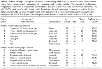
Patient history: Descriptions of lesions determined by MRI scans for each individual patient in both groups (abbreviations: caud = caudatum, put = putamen, pall = globus pallidus). The severity of the language comprehension disorder is indicated by the number of mistakes in the Token Test: no/very mild disorder (0–6); mild (7–23); moderate (24–39); severe (>40). In addition, the auditory comprehension scores of the Aachen Aphasia Test (AAT) are listed for each patient (only patients with a Token Test score greater than zero were tested with the AAT). The degree of the comprehension disorder is evaluated based on a total of 60 points.
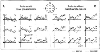
ERP effects for patients with basal ganglia lesions (A) and patients without basal ganglia lesions (B) at selected electrode-sites. Marked on a schematic head are the electrodes that are graphically displayed showing anterior (e.g., F3, FZ, F4), central (e.g., C3, CZ, C4) and posterior (e.g., P3, PZ, P4) regions. For orientation L = left sites, M = midline sites and R = right sites. The correct condition is displayed in a solid line, the incorrect condition in a dotted line. Negativity is plotted upwards and each tick on the x-axis indicates 500 ms. ERPs from the onset of the critical verb (onset at zero ms/vertical bar) up to 1500 ms show that patients with basal ganglia lesions display an extended N400-like negativity effect, but no P600 effect, while patients without basal ganglia lesions show no N400-like negativity effect, but a clear P600 effect.
 You have
Access
- 112
- Cited by