I. INTRODUCTION
Bioprosthetic heart valves (BHVs) are used for the replacement of dysfunctional heart valves and usually made by porcine or bovine pericardium.Reference Manji, Menkis, Ekser and Cooper1,Reference Rapoport, Connolly, Fulmer, Dai, Murti, Gorman, Gorman, Alferiev and Levy2 BHVs could be used in mini-invasive surgical treatments and reduce the use of anticoagulants, thus making it become the main trend for heart valve replacements.Reference Hedayat, Asgharzadeh and Borazjani3
Commercially available BHVs are usually crosslinked with glutaraldehyde (GLUT) to make them less-immunogenic and have better resistance to enzymatic degradation.Reference Human and Zilla4,Reference Zilla, Brink, Human and Bezuidenhout5 However, GLUT crosslinked BHVs might have failures within ten years.Reference Vyavahare, Ogle, Schoen, Zand, Gloeckner, Sacks and Levy6 Furthermore, mechanical damage and calcification seem to accelerate in young patients most likely due to increased metabolism. Pericardium or aortic valve leaflets have similar extracellular matrix (ECM) components: collagen and elastin. Collagen provides mechanical strength to sustain high cyclic pressure environment.Reference Sacks and Schoen7 Elastin withstands the load at low strains and provides the tissue with elasticity.Reference Levy, Vyavahare, Ashworth, Bianco and Schoen8,Reference Paule, Bernick, Strates and Nimni9 The soluble tropoelastin molecules bound by desmosine and isodesmosine forms insoluble elastin.Reference Anwar10 Previous studies have shown that GLUT crosslinked BHVs could crosslink collagen but not elastin, which can lead to the loss of elastin. This is mainly because elastin has very few free amine groups which are needed for GLUT crosslinking.Reference Tripi and Vyavahare11 Elastin-associated degradation may lead to calcification and structural damage of BHVs. Loss of elastin potentially leads to the reduction of elastic recoil and elasticity of tissues. Therefore, it is hypothesized that improvement of elastin stabilization will decrease calcification and increase the durability of BHVs. There are many challenges for chemical stabilization of elastin. Elastin is difficult to be crosslinked by GLUT since it has a small quantity of lysine-derived amino groups (∼0.3% of total amino acids).Reference Bowes and Kenten12
Amino reagents have been used in previous studies for detoxification procedure in GLUT-fixed BHVs. Zilla et al. reported that efficient detoxification of thick aortic wall tissue crosslinked by GLUT was possible by one-week incubation of acetic acid buffer-based amino reagents.Reference Zilla, Fullard, Trescony, Meinhart, Bezuidenhout, Gorlitzer, Human and Von13 Zilla et al. also verified the concept that diamine extension of GLUT crosslinks by an interim step decreased bioprosthetic aortic wall calcification.Reference Zilla, Bezuidenhout, Weissenstein, van der Walt and Human14 Grimm et al. showed that L-glutamic acid treatment for GLUT-fixed pericardium improved biocompatibility of BHVs by decreasing reactive aldehyde groups.Reference Grimm, Grabenwöger, Eybl, Moritz, Böck, Müller and Wolner15 Jee et al. used L-arginine to modify bovine pericardium by the reaction of residual aldehyde and amine group of L-arginine.Reference Jee, Kim, Park and Kim16 Girardot et al. reported the “Ultifix” method, by using carbodiimide crosslinking, and tissues were fixed by linking the amine and the carboxyl moieties through amide bonds. The amide bonds were more stable than the Schiff-base bonds formed by GLUT.Reference Girardot and Girardot17
Being innovated by previous amino agent’s treatments and carbodiimide crosslinking methods, in the current study, we developed a new tissue fixation method that utilizes exogenous amino donor arginine or lysine and carbodiimide chemistry to better stabilize elastin of porcine pericardium. Unlike previous purpose for amino agents used as detoxification agents to react with residual aldehyde groups, arginine or lysine used in the current study would be served as exogenous amino donor to provide more amino groups and crosslinking sites to couple with carboxyl moieties of elastin through carbodiimide chemistry. This strategy would better maintain BHVs’ structural integrity and stability.
II. MATERIALS AND METHODS
A. Materials
If not specially mentioned, reagents were purchased from local chemical company with analytical grade. Fresh porcine pericardium was provided by VENUS MEDTECH (Hangzhou, China) and transferred on ice. 1-Ethyl-3-(3-dimethylaminopropyl) carbodiimide (EDC), N-hydroxysuccinimide (NHS), Toluidine Blue-O (TBO), and porcine pancreatic elastase (≥8 units/mg protein) were bought from Best-Reagent (Chengdu, China). Type I collagenase [≥125 collagen digestion unit (CDU)/mg solid] and hyaluronidase (Type II, lyophilized powder, ≥300 units/mg solid) were purchased from Sigma-Aldrich (St. Louis, Missouri).
B. Heart valve biomaterial crosslinking treatments
Fresh porcine pericardiums were rinsed three times with sterile saline for 1 h. Fat tissues were removed gently with scissors and forceps. Whole pericardiums were treated with four different methods: (i) GLUT; (ii) GLUT plus EDC/NHS; (iii) GLUT plus arginine plus EDC/NHS; (iv) GLUT plus lysine plus EDC/NHS.
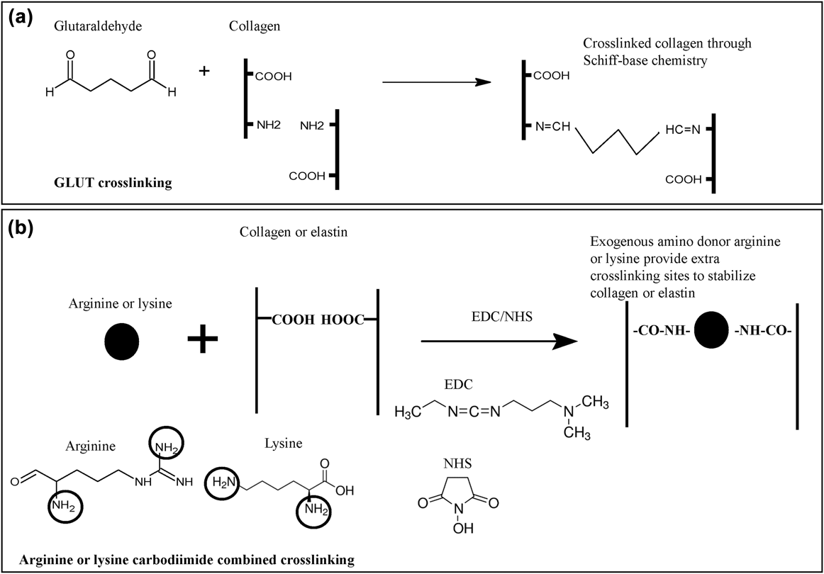
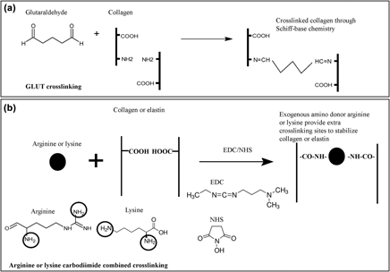
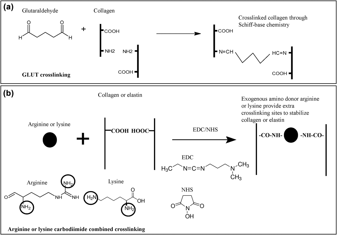
For the GLUT control, fresh pericardiums were incubated with 0.25% GLUT with shaking at 150 rpm on an orbital shaker (SK-O180-E, DRAGONLAB, Beijing, China) for 24 h at room temperature.Reference Dewanjee18 For GLUT plus EDC/NHS control, fresh pericardiums were first treated with 0.25% GLUT for 24 h and then 0.2 M EDC and 0.1 M NHS mixture for 4 h.Reference Girardot and Girardot17,Reference Tam, Zhang, Feaver, Parchment, Sacks and Vyavahare19 For the GLUT plus arginine or lysine plus EDC/NHS treatments, fresh pericardiums were first crosslinked with 0.25% GLUT for 24 h then incubated with 10% arginine or 10% lysine at room temperature with shaking at 150 rpm on an orbital shaker (SK-O180-E, DRAGONLAB, Beijing, China) for 24 h. Then the tissues were incubated in 0.2 M EDC and 0.1 M NHS mixture for 4 h. Figure 1 illustrates the fundamental differences between these chemical crosslinking methods.
Crosslinking chemistry of pericardium. (a) GLUT crosslinking mechanism. (b) Arginine or lysine carbodiimide combined crosslinking. Black circles highlight amino groups in arginine and lysine.
C. TBO staining for carboxyl group quantification
The concentration of the –COOH groups was quantified by the TBO method.Reference Chen, Li, Yang, Zhou, Luo, Maitz, Zhao, Wang, Xiong and Huang20 50 µL TBO solution (5 × 10⁻³ M, pH = 10) was put on the top of each tissue in a 96-well plate and incubated for 12 h. Then 0.2 mL NaOH solutions (0.1 mM, pH = 10) were used each time in each well to wash samples three times. 1 mL of 50 wt% acetic acid solutions were used to elute the TBO dye for 1 h. Finally, 0.2 mL of the TBO elution from each sample well was transferred into a 96-well plate and the absorbance was colorimetrically determined with a microplate reader (Varioskan Flash, Thermo Scientific, North Shore City, NZ, US) at 631 nm. A standard curve for TBO in 50 wt% acetic acid solutions was made to quantify the COOH concentration.
D. Ninhydrin assay for amine content quantification
The ninhydrin assay was used to quantitatively assess the number of free amines present in the biomaterial.Reference Chan, Burugapalli, Naik, Kelly and Pandit21 Briefly, treated pericardiums were cut into small pieces (about 1 cm × 1 cm). Each sample was placed in a centrifuge tube and 2 mL ninhydrin solution was added (1% w/v ninhydrin, 0.1 M sodium citrate, pH = 5.0). Then the tubes were heated at 95 °C in water bath. After 60 min incubation, the absorbance value (optical density, OD) of the supernatant at 567 nm was determined by UV-Vis spectrometry (UV-3200, MAPADA Instruments, Shanghai, China). The tissues were washed with deionized water three times before being dried at 45 °C for 6 h, and the weight of samples was recorded. The percentage of amine content was normalized with the GLUT control sample as follows: percentage of amine content = (ODsample − ODblank/ODGLUT − ODblank) × 100%.
E. Differential scanning calorimetry
The tissue shrinking temperature (Td) was determined by differential scanning calorimetry (DSC).Reference Weng and Khor22 Samples (1–5 mg) were cut from the pericardium. Each group of three patches was soaked in PBS to prepare the samples of DSC. Then the samples were maintained flat in sealed DSC pans. The samples were tested using a DSC (Mettler-Toledo, Schwerzenbach, Switzerland). The samples were heated at 10 °C/min in the range of 20–120 °C. The temperature value at the endothermic peak was recorded as tissue shrinking temperature.
F. Enzymatic degradation in vitro
Each treated pericardium (about 10 mg each) was cut in 1 cm × 1 cm, rinsed with deionized water, freeze-dried, and weighed. Then the tissue was incubated in 0.5 mL of 50 CDU/mL Type I collagenase or 30 unit/mL porcine pancreatic elastase or glycosaminoglycans (GAGs) degrading enzyme solution (5 U/mL hyaluronidase) for 48 h at 37 °C with shaking at 100 rpm on an orbital shaker (SK-O180-E, DRAGONLAB, Beijing, China).Reference Tripi and Vyavahare11,Reference Tam, Zhang, Feaver, Parchment, Sacks and Vyavahare19 The digested tissue was washed with deionized water, freeze-dried, and weighed. Percent weight loss was calculated as follows: percent weight loss = (weight before enzymatic degradation − weight after enzymatic degradation)/weight before enzymatic degradation × 100%. Another digested tissue was saved for histology.
G. Rat subdermal implantation model
The conducted animal experiments have been approved by institutional review from Medical Ethics Committee of Sichuan University. All treated tissues (1 cm × 1 cm each) were washed thoroughly with 50 mL sterile distilled water three times, 1 h each time. Then the tissues were soaked in 75% ethanol for 24 h for sterilization. Then the tissues were washed with 50 mL sterile PBS buffer and made ready for implantation. Male Sprague–Dawley rats, 200–300 g each, were anesthetized by intraperitoneal injection with 10% chloral hydrate (1 mL/300 g rat). Two dorsal surgical incisions were made, and two subdermal pockets were created on either side. One pericardium was placed in each pocket. Six specimens in each group (GLUT, GLUT/EDC, GLUT/Arg/EDC, GLUT/Lys/EDC; 6 × 4 = 24 specimens; 12 rats in total) were implanted. Incisions were closed with surgical staples and the samples together with fibrous capsule were explanted after 30 days’ implantation. Half of the explanted tissues were saved for histology. The other half was frozen for calcium content analysis.
H. Histology
Pericardium (about 1 cm × 1 cm) was fixed in 10% buffered formalin (pH 7.4) at room temperature for 24 h prior to infiltration, embedded in paraffin, and sectioned at 8 µm. Picrosirius red and Victoria blue were utilized to stain collagen and elastin.Reference Tripi and Vyavahare11,Reference Leong, Munnelly, Liberio, Cochrane and Vyavahare23 The serous side and fibrous side of the pericardium were indicated. Alcian blue staining was used to visualize GAGs. The blue color is indicative of the presence of GAGs.Reference Tripi and Vyavahare11 Alizarin red staining was used to stain calcium deposition; calcium deposits appear red. Images were captured by using a digital slide scanner (MIRAX Desk, Carl Zeiss Microimaging GmbH, Göttingen, Germany).
I. Calcium content analysis
The tissues (about 10 mg each) were freeze-dried, weighed, and acid hydrolyzed in 1 mL of 6 N HCl for 2 h at 60 °C. The samples were centrifuged at 1000 rpm for 5 min and supernatants were collected and diluted 20-fold in distilled water. Calcium content was analyzed using atomic adsorption spectrometry (VARIAN, SpectrAA-220Fs, Palo Alto, California) and normalized to dry tissue weight. Dilution ratios were used for calculation of calcium element contents.
J. Scanning electron microscopy
The tissue was dehydrated by 70% ethanol and 100% ethanol for 1 h each. Then the tissue was sputtered with gold, and then images were taken by scanning electron microscopy (SEM; JSM-7500F, JEOL, Tokyo, Japan). Only the serous side of the pericardium was prepared for imaging to visualize the digested collagen or elastin fibers.
K. Uniaxial tensile testing
Uniaxial tensile testing was followed with previous reported methods with minor changes.Reference Tam, Zhang, Feaver, Parchment, Sacks and Vyavahare19 The Instron 4310 machine (Instron, Norwood, Massachusetts) was used for uniaxial tensile testing. Samples were cut from each tissue utilizing a dog-boned tissue stamp (2 mm × 35 mm). Then the samples were saved in PBS buffer and made ready for the testing. Tissue sample thickness was measured utilizing a digital micrometer. To prevent the tissues from slipping, sandpaper was used at both ends of the clamps. Using a 100 N load cell, the samples were preloaded (about 0.1 N) and stretched at an extension speed of 10 mm/min. Tensile force was applied to the tissue sample until the tissue failed. Concurrently, a stress–strain curve was recorded. Six specimens in each group were tested for statistical analysis.
L. Cytotoxicity test
To investigate the cytotoxicity of the samples, L929 fibroblasts were cultured in Rosewell Park Memorial Institute (RPMI) 1640 Medium (HyClone, Logan, Utah), 100 U/mL penicillin, and 100 μg/mL streptomycin and supplemented with 10% Fetal Bovine Serum at 37 °C, 5% CO2. The pericardium was washed three times with 50 mL sterile distilled water and immersed in 75% ethanol overnight for sterilization and washed with sterile PBS buffer to remove the residual ethanol. The extracts were made by soaking the pericardium in the culture medium at 37 °C for 48 h in a tissue-weight to volume ratio of 0.2 g/mL (according to the standard cytotoxicity test ISO 10993-5). The cells were detached by 0.25% trypsin to obtain cell suspension and seeded in the 96-well culture plate for 24 h, and then the culture medium in the 96-well culture plate was abandoned, and the extracts of the samples were added to the wells. After being incubated for another 24 h, the extracts were removed and the fresh culture medium supplemented with 10% Cell Counting Kit-8 (CCK-8, Beyotime, Shanghai, China) was added and incubated for 60 min at 37 °C according to the instruction. The absorbance at 450 nm was measured on a microplate reader (Varioskan Flash, Thermo Scientific, North Shore City, NZ, US).
M. Platelet adhesion test
The thrombogenic properties of the treated tissues were tested by a platelet adhesion assay.Reference Jiang, Suen, Wertheim and Ameer24 Platelet-rich plasma (PRP) was prepared by centrifugation from pig citrated whole blood (1:9 volume ratio of 1% sodium citrate: whole blood). Each tissue was cut as 0.5 cm diameter round sample and put in the 96-well plate with 0.2 mL PRP for 1 h at 37 °C under static conditions. The number of adherent platelets was quantified by detecting the amount of lactate dehydrogenase (LDH) present after cell lysis. The suspended PRP was aspirated and each well was washed three times with PBS. Then 0.2 mL 0.05 wt% Triton X-100 was added to lyse the adherent platelets. The LDH activity in the lysed platelet suspensions was measured using an LDH cytotoxic assay kit (Beyotime, Shanghai, China). The result of platelet adhesion was also observed using SEM. Briefly, adherent platelets were fixed using 10% buffered formalin (pH 7.4) in PBS for 1 h and dehydrated in 70% ethanol and 100% ethanol for 1 h each and freeze-dried. The tissues were then sputtercoated with a layer of gold and imaged using SEM.
N. Statistical analysis
Results are showed as means ± standard error of the mean (SEM). Single-factor analysis of variance was performed for statistical analyses of the data. Asterisks represent P-values smaller than 0.05 and were considered as statistically significant.
III. RESULTS
A. Carboxyl groups and amine content quantification
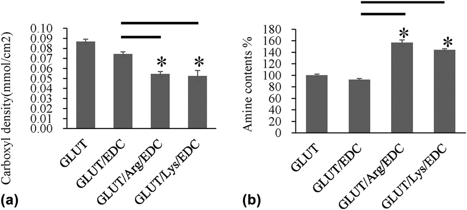
Based on EDC/NHS crosslinking chemistry, each mole arginine or lysine would consume one mole carboxyl group. We determined that carboxyl groups were decreased in arginine (GLUT/Arg/EDC) or lysine (GLUT/Lys/EDC)-treated tissues [Fig. 2(a)]. These results demonstrated that the amino groups on arginine or lysine effectively consumed carboxyl groups on pericardium. We further evaluated amine contents by ninhydrin assay. Our results showed that the amine contents in arginine or lysine-treated tissues were 157 ± 5% and 144 ± 2%, respectively, by normalizing to GLUT control tissues [Fig. 2(b)]. These results further indicated the effective binding of arginine or lysine since additional amino groups might be introduced by arginine or lysine.
(a) Carboxyl group density quantification. GLUT: control 0.25% GLUT treatment. GLUT/EDC: 0.25% GLUT plus 0.2 M EDC/0.1 M NHS. GLUT/Arg/EDC: 0.25% GLUT plus 10% arginine plus 0.2 M EDC/0.1 M NHS. GLUT/Lys/EDC: 0.25% GLUT plus 10% lysine plus 0.2 M EDC/0.1 M NHS. (b) Amine content quantification. The asterisk (*) means significant difference (P < 0.05) between the compared two groups. N = 6 (six independent experiments).

B. Thermal stability
Thermal shrinking temperatures (Td) have been shown to be effectively related to collagen stability.Reference Tripi and Vyavahare25 Td was taken at the point of the minimum value within this endothermic peaks (The DSC curve data were not shown). All groups exhibited a sufficiently high degree of crosslinking as the Td of arginine or lysine-treated pericardium were higher than that of GLUT control. The Td of GLUT and GLUT/EDC control was measured to be 93.92 ± 0.58 and 95.42 ± 0.67 °C, while arginine or lysine treated tissues were measured as 105.49 ± 0.27 °C and 105.07 ± 0.13 °C, respectively (Table I). These results indicated the increase of thermal stability.
Tissues thermal shrink temperature.

C. Enzyme stability studies and calcification for crosslinked pericardium
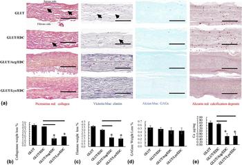
When each tissue was challenged with type 1 collagenase, broken collagen fibers (Picrosirius red staining, collagen fiber was visualized as red) were noticed in GLUT and GLUT/EDC control tissue but not in arginine or lysine-treated tissue [Fig. 3(a)]. GLUT and GLUT/EDC control group had 4.43 ± 0.26% and 4.11 ± 0.07% mass loss, while arginine or lysine-treated tissue had 1.50 ± 0.31% and 1.94 ± 0.05%, which was significantly less than GLUT/EDC control (P < 0.05) [Fig. 3(b)]. These results demonstrated the improvement of integrity and stability of collagen.
(a) Collagen fiber by Picrosirius red staining, elastic fiber by Victoria blue staining, GAGs by Alcian blue staining, and calcification deposits by Alizarin red staining. Arrows highlight broken collagen fibers and broken elastic fibers. Scale bar: 100 µm. (b) Collagen stability of pericardium against collagenase degradation. (c) Elastin stability of pericardium against elastase degradation. (d) GAGs stability of pericardium against GAGase degradation. (e) Calcium content of each explant. The asterisk (*) means significant difference (P < 0.05) between the compared two groups. N = 6 (six independent experiments).
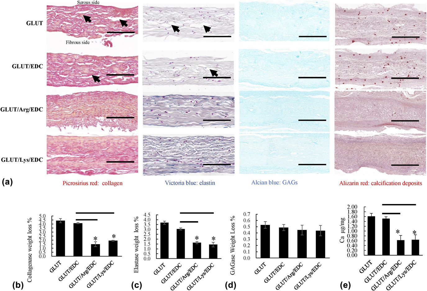
When each tissue was challenged with porcine pancreatic elastase, broken elastic fibers (Victoria blue staining, elastic fiber was visualized as blue) were noticed in GLUT and GLUT/EDC control tissue but not in arginine or lysine-treated tissue [Fig. 3(a)]. GLUT and GLUT/EDC control group had 3.68 ± 0.16% and 3.04 ± 0.11% mass loss, while arginine or lysine-treated tissue had 1.63 ± 0.11% and 1.44 ± 0.24%, which was significantly less than GLUT/EDC control (P < 0.05) [Fig. 3(c)]. These results demonstrated the improvement of integrity and stability of elastin. We further tested GAGs stability by challenging with GAGase (hyaluronidase) and results did not show significant difference [Fig. 3(d)], which might be due to few amount of GAGs in fresh pericardiums.Reference Tam, Zhang, Infante, Parchment, Sacks and Vyavahare26
Alizarin red staining on both arginine and lysine-treated tissues showed no visible calcification in each treated tissue [Fig. 3(a)]. Calcium content analysis of each explanted pericardial tissue showed that arginine or lysine-treated tissue had 0.624 ± 0.193 and 0.637 ± 0.213 Ca µg/mg tissue dry weight versus 1.610 ± 0.124 and 1.512 ± 0.075 Ca µg/mg tissue dry weight in GLUT and GLUT/EDC control [Fig. 3(e)]. These results indicated that the strategy combined arginine or lysine and carbodiimide crosslinking decreased significantly more than 50% calcifications in BHVs.
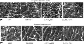
We further examined the surface appearance by using a SEM for each tissue after collagenase and elastase degradation in vitro. Consistent with histology observation, broken collagen fibers and elastic fibers were noticed in GLUT and GLUT/EDC control tissues but not in arginine or lysine-treated tissues (Fig. 4). These results further indicated the improvement of stability of collagen and elastin.
Surface appearance by using a SEM in each group after collagenase (a) and elastase (b) degradation in vitro. Arrows highlight broken elastic fibers and collagen fibers. Scale bar: 10 µm. N = 3 (three independent experiments).

D. Mechanical tests and biocompatibility
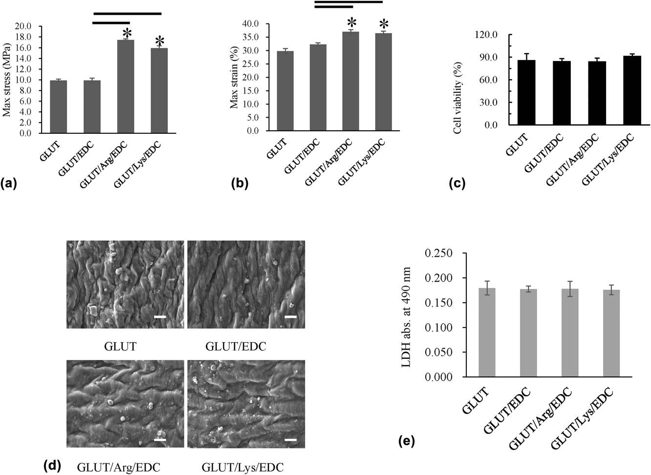
Arginine or lysine-treated tissues had 17.46 ± 0.18 and 15.91 ± 0.42 MPa maximum stress versus 9.88 ± 0.23 and 9.86 ± 0.46 MPa in GLUT and GLUT/EDC control, 37.05 ± 0.79% and 36.49 ± 0.73% maximum strain versus 29.78 ± 0.93% and 32.25.78 ± 0.60% in GLUT and GLUT/EDC control [Figs. 5(a) and 5(b)]. These results demonstrated the improved maximum strength after arginine or lysine treatments. The cytotoxicity test showed no statistical difference between the arginine or lysine-treated tissues and GLUT or GLUT/EDC control tissues [Fig. 5(c)]. Similar to the cytotoxicity test, there was no statistical difference between the arginine or lysine-treated tissues and GLUT or GLUT/EDC control tissues for platelet adhesion tests [Figs. 5(d) and 5(e)]. These results indicated that arginine or lysine-treated tissues had similar biocompatibility comparing to traditional GLUT treatments.
(a) and (b) Maximum stress and maximum strain for each group. (c) Cytotoxicity of the treated tissues in each group. (d) Platelet adhesion test by using a SEM. Scale bar: 10 µm. (e) LDH quantification to determine number of adherent platelets. The asterisk (*) means significant difference (P < 0.05) between the compared two groups. N = 6 (six independent experiments).

IV. DISCUSSION
A. Deficiency of the GLUT crosslinking method
For the past fifty years, GLUT has been used to crosslink BHVs. The GLUT crosslinking method is quite efficient to stabilize collagen and relatively simple to perform.Reference Nimni, Cheung, Strates, Kodama and Sheikh27 However, GLUT only actively crosslinks collagen and leaves elastin unstabilized because elastin does not have enough active amine groups.Reference Tam, Zhang, Feaver, Parchment, Sacks and Vyavahare28 Elastin exposed to enzymatic degradation would accelerate BHVs’ failure.Reference Leong, Munnelly, Liberio, Cochrane and Vyavahare23 Slow enzymatic degradation of elastin leads to BHVs’ structural loss, increasing risk of calcification, and eventually decreasing the lifetime of BHVs.
To better stabilize elastin, Vyavahare’s lab reported that polyphenols such as tannic acid or pentagalloyl glucose could stabilize elastin in biological cardiovascular implants.Reference Tam, Zhang, Infante, Parchment, Sacks and Vyavahare26,Reference Isenburg, Simionescu and Vyavahare29 However, polyphenol–elastin binding is mainly hydrogen binding and might not have long time stability.Reference Luck, Liao, Murray, Grimmer, Warminski, Williamson, Lilley and Haslam30
Being innovated by previous reports of carbodiimide crosslinking strategies and amino reagents introduced as the detoxification procedure in GLUT-fixed BHVs,Reference Zilla, Fullard, Trescony, Meinhart, Bezuidenhout, Gorlitzer, Human and Von13,Reference Jee, Kim, Park and Kim16,Reference Girardot and Girardot17 in this current study, we used arginine or lysine carbodiimide combined crosslinking to overcome the deficiency of GLUT crosslinking of BHVs. Carbodiimide chemistry has been used for modification of polymers.Reference Grabarek and Gergely31 Carbodiimide could link carboxyl and amine groups in proteins to create amide bonds. Carbodiimide has previously been demonstrated to stabilize BHVs and decrease calcification.Reference Girardot and Girardot17 However, carbodiimide treatment alone is insufficient to crosslink collagen in BHVs.Reference Tam, Zhang, Feaver, Parchment, Sacks and Vyavahare19
Unlike previous purpose for amino agents used as detoxification agents to react with residual aldehyde groups, arginine or lysine used in the current study would be served as exogenous amino donor to provide more amino groups and crosslinking sites to couple with carboxyl moieties of elastin through carbodiimide chemistry. Both arginine and lysine had two amino groups. The major ECM components, collagen and elastin, had carboxyl groups. By carbodiimide crosslinking chemistry, arginine or lysine could be chemically linked to elastin and maintain the structural stability.
B. Improvements of arginine or lysine carbodiimide combined crosslinking
We first demonstrated the effective binding of arginine or lysine to pericardium by carboxyl group density and amine content quantification [Figs. 2(a) and 2(b)]. The GLUT crosslinking agent mainly uses amino groups from pericardium. In our studies, we make use of carboxyl groups for introducing extra crosslinking sites. Previous studies showed that natural crosslinking agents like genipin, tannic acid, and procyanidins mainly use carboxyl groups in ECM-derived scaffolds by hydrogen bond.Reference Ma, Wang, Wu and Chang32 Then we showed that thermal stability was improved after arginine or lysine treatments (Table I). By collagenase and elastase degradation test in vitro, our results showed improved elastin integrity and stability after arginine or lysine treatments (Figs. 3 and 4). The thermal shrinking temperature in the arginine or lysine carbodiimide combined crosslinking was higher than previous GLUT or carbodiimide fixed BHVs.Reference Weng and Khor22,Reference Tam, Zhang, Infante, Parchment, Sacks and Vyavahare26 This might be due to the introduced extra crosslinking by amino donors, and the tissue thermal stability and structure integrity could increase. By the rat subdermal implantation calcification model, calcification deposits were decreased significantly in the arginine or lysine-treated samples [Figs. 3(a) and 3(d)]. Previously reported studies showed that the stability of collagen and elastin greatly determined BHVs’ calcification.Reference Tam, Zhang, Feaver, Parchment, Sacks and Vyavahare19,Reference Isenburg, Simionescu and Vyavahare29 The increased collagen and elastin stability would be beneficial to BHVs’ anticalcification. Besides calcification, the lifetime of BHVs is mainly determined by mechanical properties. Our results demonstrated the improved maximum stress and strain after arginine or lysine treatments [Figs. 5(a) and 5(b)]. The maximum stress indicates biggest tearing stress to cause rupture for the crosslinked tissue.Reference Caballero, Sulejmani, Martin, Pham and Sun33 We noticed that the amino donor treated BHVs have similar nonlinear behavior in strain–stress curve comparing to GLUT and GLUT/EDC control (Data not shown). This indicates that arginine or lysine combined treatment would have similar stress during physiological conditions.Reference Tripi and Vyavahare11 The arginine or lysine carbodiimide combined treated tissues had similar biocompatibility comparing to GLUT and GLUT/EDC control determined by the cytotoxicity test and platelet adhesion test [Figs. 5(c)–5(e)]. These results were reasonable since there were no noticed biocompatibility issues in previous amino reagents’ studies in BHVs.Reference Zilla, Fullard, Trescony, Meinhart, Bezuidenhout, Gorlitzer, Human and Von13,Reference Jee, Kim, Park and Kim16
C. Limitation of this study
Although we showed improvements of arginine or lysine carbodiimide combined crosslinking in turns of enzymatic degradation and calcification in 30 days’ rat model, more studies are needed to investigate the tissue stability, tissue-induced coagulation, and calcification for long time in vivo tests. These valves needed to be tested in animal studies to guarantee their safety and function under blood environment.
V. CONCLUSION
Our results showed that arginine or lysine carbodiimide combined crosslinking had sufficiently high degree of crosslinking as determined by the increase of tissue shrinking temperatures, less collagenase and elastase enzymatic degradation, and less calcification. This arginine or lysine carbodiimide combined crosslinking technique would be a promising method to make BHVs have better structural stability and anticalcification properties.
LIST OF ABBREVIATIONS

ACKNOWLEDGMENTS
This work is supported by National Natural Science Foundation of China (31700833), Young Elite Scientists Sponsorship Program by CAST (2017QNRC001), the Fundamental Research Funds for the Central Universities (YJ201641), National Key Research and Development Programs (2017YFC1104200 and 2016YFC1102200), and the Program of Introducing Talents of Discipline to Universities (111 Project, No. B16033).