Introduction
Genera Andracantha Schmidt, 1975 and Pseudocorynosoma Aznar, Pérez Ponce de León & Raga, 2006 were separated from the genus Corynosoma Lühe, 1904 based on morphological, molecular and ecological data by Aznar et al. (Reference Aznar, Pérez-Ponce de León and Raga2006) and García-Varela et al. (Reference García-Varela, Pérez-Ponce de León, Aznar and Nadler2009, Reference García-Varela, Pinacho-Pinacho, Sereno-Uribe and Mendoza-Garfías2013). Thus, the genus Pseudocorynosoma was included in the Acanthocephala classification proposed by Amin (Reference Amin2013), which also recognizes the following five species: Pseudocorynosoma anatarium (Van Cleave, Reference Van Cleave1945), Pseudocorynosoma constrictum (Van Cleave, Reference Van Cleave1918), Pseudocorynosoma iheringi (Machado Filho, 1961), Pseudocorynosoma peposacae (Porta, Reference Porta1914), and Pseudocorynosoma enrietti (Molfi & Freitas Fernandes, Reference Molfi and Freitas Fernandes1953), which was synonymized with Corynosoma molfifernandesi Machado Filho, Reference Machado Filho1962 (Aznar et al., Reference Aznar, Pérez-Ponce de León and Raga2006). Posteriorly, Pseudocorynosoma tepehuanesi García-Varela, Henández-Orts & Pinacho-Pinacho, 2017 was also described within this genus (see García-Varela et al., Reference García-Varela, Hernández-Orts and Pinacho-Pinacho2017).
All species of Pseudocorynosoma inhabit freshwater environments of the American continent: P. anatarium, P. constrictum, and P. tepehuanesi in North America, and P. enrietti, P. iheringi and P. peposacae in South America (Aznar et al., Reference Aznar, Pérez-Ponce de León and Raga2006; García-Varela et al., Reference García-Varela, Hernández-Orts and Pinacho-Pinacho2017). Pseudocorynosoma enrietti was originally described from specimens of the anatids Cairina moschata (Linnaeus) and Anas platyrynchos (Linnaeus) collected in Brazil (Molfi & Freitas Fernandes, Reference Molfi and Freitas Fernandes1953). In Argentina the only species recorded is P. peposacae, which parasitizes the ducks Netta peposaca (Vieillot) (syn. Metopiana peposacae) and Anas georgica spinicauda Vieillot (syn. Paecilonitta spinicauda) (Boero & Led, Reference Boero and Led1968). Little is known about Pseudocorynosoma life cycles, and intermediate hosts remain unknown for most species (Van Cleave, Reference Van Cleave1918, Reference Van Cleave1945; Van Cleave & Starrett, Reference Van Cleave and Starrett1940; Schmidt, Reference Schmidt1965; Aznar et al., Reference Aznar, Pérez-Ponce de León and Raga2006). Pseudocorynosoma constrictum is the only species with a known intermediate host, the amphipod Hyallela azteca (Saussure) (Podesta & Holmes, Reference Podesta and Holmes1970; García-Varela et al., Reference García-Varela, Pinacho-Pinacho, Sereno-Uribe and Mendoza-Garfías2013). This work aims to describe adults and cystacanths of P. enrietti collected from natural and experimental hosts in Patagonia to elucidate its life cycle.
Materials and methods
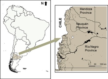
Adult worms were recovered from one freshly dead specimen of duck, A. platyrhynchos, from Lake Hess in Nahuel Huapi National Park (41°22′S; 71°43′W), found in spring 2002, and one freshly dead specimen of swan, Coscoroba coscoroba Molina, from Lake Verde in Laguna Blanca National Park (39°01′S; 70°24′W), found in spring 2008 (fig. 1).
Map of sampling localities: Coscoroba coscoroba from Lake Verde (white circle); Anas platyrhynchos from Lake Hess (white diamond); and Hyalella patagonica from Lake Mascardi (white triangle).

A total of 2000 amphipods, Hyalella patagonica Ortmann, were collected from Lake Mascardi in Nahuel Huapi National Park (41°17′S; 71°38′W) in spring 2002 (fig. 1). The amphipods were brought to the laboratory alive and dissected under stereomicroscope to obtain cystacanths. Eighty cystacanths were incubated in tap water for 12 h and then fixed in 2% formalin. Seventy-three were invaginated and cleared in Aman's lactophenol. The evaginated seven were stained in Mayer's acid carmine, destained in 1% hydrochloric acid in 70% ethanol, dehydrated in ascending concentrations of ethanol, cleared in creosote and mounted in Canada balsam. Another 250 live cystacanths were immediately used to generate infection. Three one-day-old Gallus gallus domesticus (Linnaeus) and two one-week-old A. platyrhynchos were experimentally infected with 50 cystancanths each in spring 2002. One chick was necropsied at 14 days post-infection (dpi) and the remaining two at 21 dpi; the ducklings were examined at seven and 14 dpi.
The complete intestinal tracts of the birds were examined under stereomicroscope (Olympus SZH10, Japan). Worms were collected, counted and fixed in 2% formalin. Specimens for morphological studies were then cleared in Aman's lactophenol before microscopic examination. From natural infection of A. platyrhynchos, body measurements were taken of 10 male and 10 gravid female P. enrietti specimens, the number of hooks rows on the transversal microdissected proboscis was counted under a stereomicroscope in eight males and ten females and measurements of the bell complex and eggs were taken in three microdissected females. Additionally, five males and five females obtained from experimental infection of A. platyrhynchos were measured at seven dpi. All measurements are given in micrometres, with the range followed by their mean in parentheses. Drawings were made using a camera lucida (Zeiss model 474620–9900, Germany). Some specimens were dried using the critical point method described by Amin et al. (Reference Amin, Heckmann, Wilson, Keele and Khan2015), examined under a scanning electron microscope (SEM Philips 515) operating at 20 kV in the Laboratory of Material Characterization of the Bariloche Atomic Centre, and photographed.
The specimens were deposited in the Colección Nacional de Parasitología, Museo Argentino de Ciencias Naturales Bernardino Rivadavia, Buenos Aires, Argentina (MACN–Pa).
Results
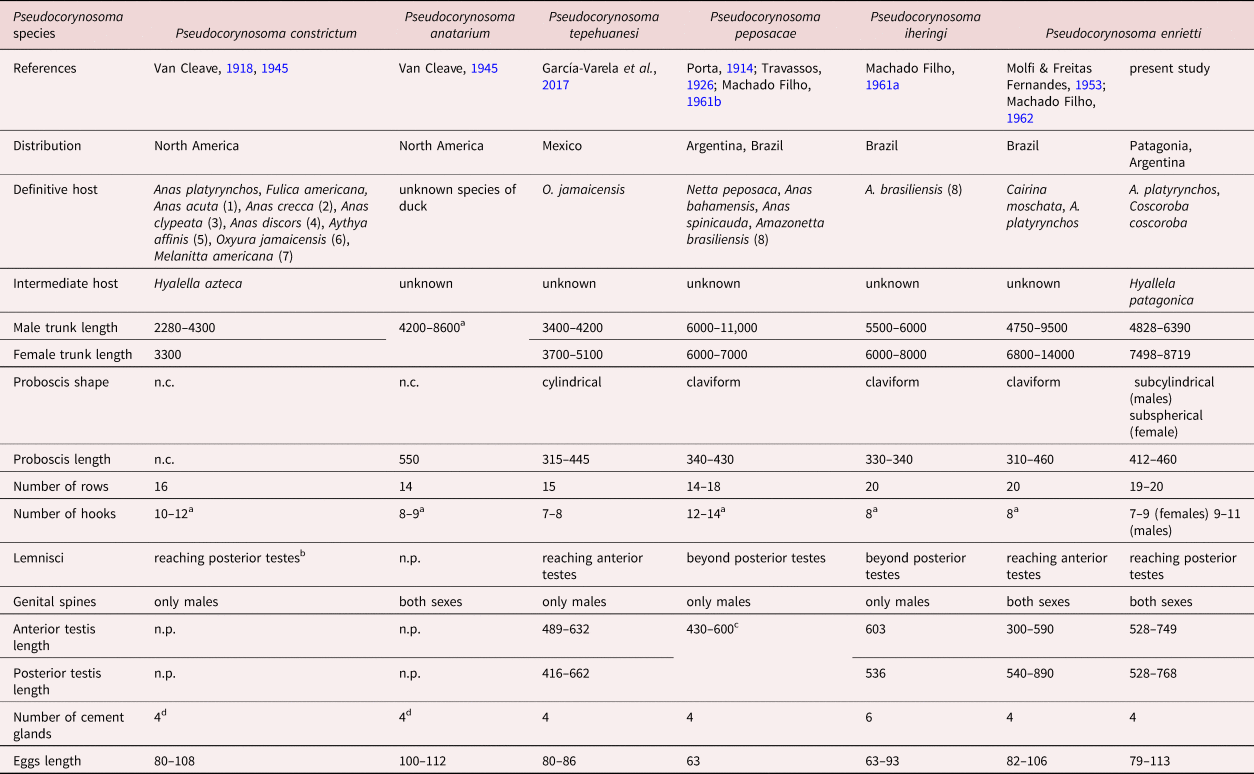
The specimens collected in the present study were assigned to P. enrietti due to the presence of the following characteristics: spines symmetrically distributed in the fore-trunk covering the anterior third; and genital spines in an isolated field in both sexes. The proboscis has 19–20 hook rows with 9–11 hooks per row in males and 7–9 hooks in females. Males have four cement glands similar in size. Eggs elongated, with filaments. All the body measurements analysed lay within the range given for the Brazilian specimens (see table 1). The only difference between Patagonian and Brazilian P. enrietti specimens was the length of the lemnisci, the former reaching only the anterior testes and the latter the posterior testis (Molfi & Freitas Fernandes, Reference Molfi and Freitas Fernandes1953; Machado Filho, Reference Machado Filho1962). Keys to the species of the genus Pseudocorynosoma given below.
Distribution, hosts and morphometric characters of natural specimens of Pseudocorynosoma from North and South America. Measurements given in micrometres.

Cited as: (1) Dafila acuta; (2) Nettion carolinense; (3) Spatula clypeata; (4) Querquedula americana; (5) Nyroca affinis; (6) Erismatura jamaicensis; (7) Oidemia americana; and (8) Nettion brasiliensis.
n.p., not provided.
a measures based on both sexes without discrimination
b according to Plate 1, fig. 2 (Van Cleave, Reference Van Cleave1945).
c measure not discriminated for each testis.
d according to fig. 4a, c of García-Varela et al. (Reference García-Varela, Hernández-Orts and Pinacho-Pinacho2017).
Keys
Key to species of the genus Pseudocorynosoma
1a. 4 cement glands 2
1b. 6 cement glands. Proboscis with 20 rows of 8 hooks, genital spines present only in males, egg size 63–93 μm P. iheringi
2a. Up to 18 hook rows on proboscis 3
2b. 19–20 hook rows on proboscis, dimorphism in number of hooks (7–9 in females; 9–11 in males), genital spines present in both sexes, egg size 79–113 μm. P. enrietti
3a. Up to 9 hooks in each row 4
3b. 10 or more hooks in each row 5
4a. Genital spines only in males, proboscis less than 550 μm long, egg size 80–86 μm P. tepehuanesi
4b. Genital spines in both sexes, proboscis 550 long, egg size 100–112 μm P. anatarium
5a. Trunk length less than 5000 μm, proboscis with 16 rows of 10–12 hooks, egg size 80–108 μm P. constrictum
5b. Trunk length more than 5000 μm, proboscis with 14–18 rows of 12–14 hooks, egg size 63 μm P. peposacae
Description of P. enrietti (Molfi & Freitas Fernandes, Reference Molfi and Freitas Fernandes1953)
General: Polymorphidae, with characteristics of genus Pseudocorynosoma: trunk cylindrical, slightly swollen anteriorly, often with constriction separating fore-trunk and hind-trunk. Nineteen to 20 rows of proboscis hooks in both sexes. Apical organ not observed. Proboscis receptacle double-walled, with ellipsoidal cerebral ganglion near to its posterior end. Trunk spines symmetrically covering the most anterior part of fore-trunk ventrally and dorsally. Genital spines in both sexes, surrounding genital pore in a separate field from trunk spines, males with quincuncial pattern in four rows, and females with irregular pattern or occasionally absent.
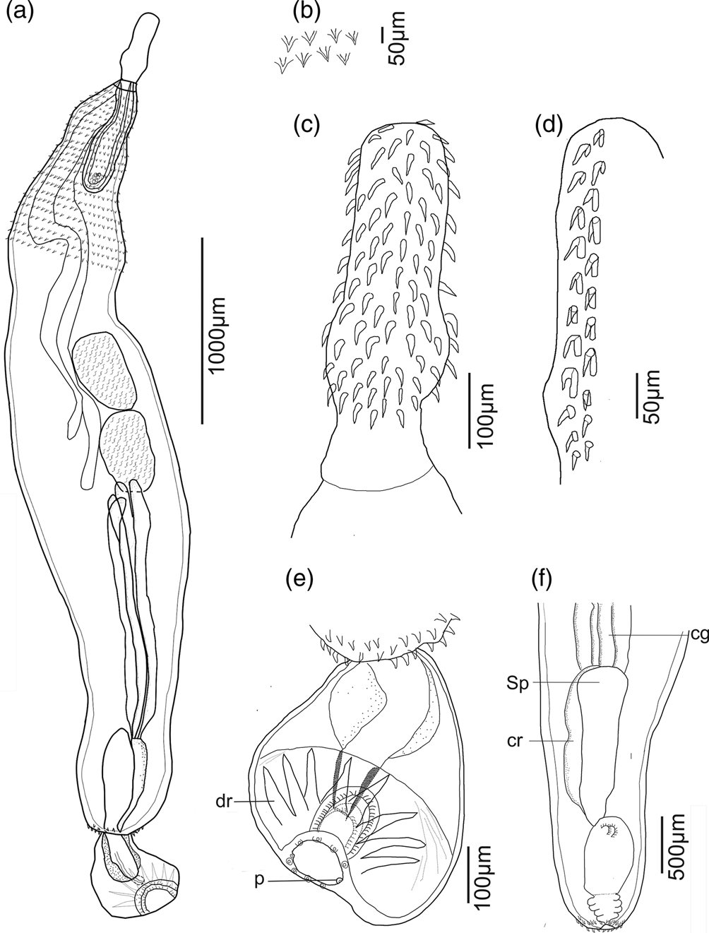
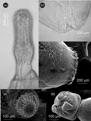
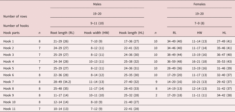
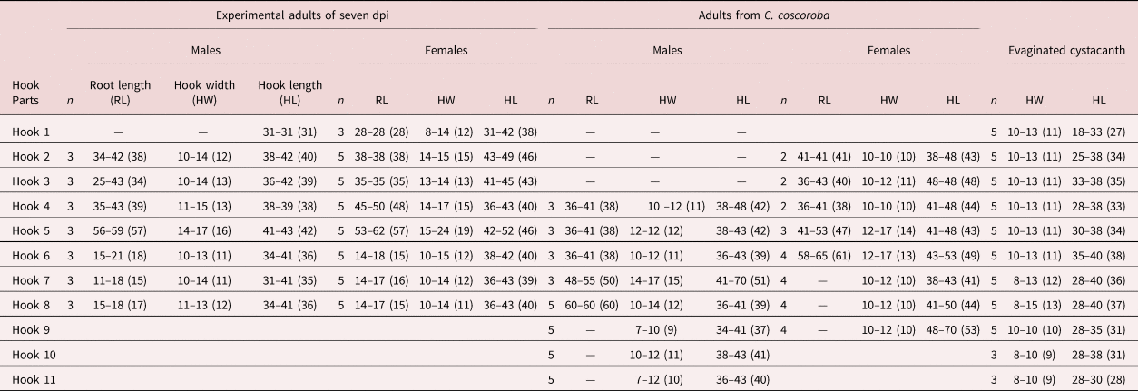
Male (based on 10 males obtained from naturally infected A. platyrhynchos): Trunk 4828–6390 (5314) long and 710–1221 (975) wide (fig. 2a). Trunk spines symmetrically covering anterior part of fore-trunk, 25–31 (27) long (fig. 2a, b). Proboscis subcylindrical 422–441 (432) long and 144–288 (193) wide (fig. 2a, c). Hooks per row 9–11 (10), with three recognizable groups: apical (1–6) hooks with intermediate values of root and hook length; central (7–8) with highest values for root length and hook width; and basal (9–11) with smallest root values. Hook and root length, and hook width are given in table 2 and shown in figs 2c, d and 3a, b. Neck 154–240 (202) long and 96–182 (138) wide. Proboscis 749–931 (845) long and 173–221 (190) wide. Cerebral ganglion 260 long and 106 wide. Lemnisci subequal, right lemniscus reaching posterior testis, 1344–2496 (1694) long, 48–202 (119) wide; left lemniscus 1200–2208 (1533) long, 48–202 (106) wide (fig. 2a). Reproductive system represents 73%–85% (80%) of trunk length in invaginated acanthocephalans and 64%–76% (70%) of trunk length in evaginated specimens. Testes two, oval, equatorial, nearly equal, in tandem (six of 10), or overlapped (four of 10); anterior testis 528–749 (588) long, 288–451 (350) wide, posterior testis 528–768 (642) long, 336–432 (373) wide (fig. 2a). Four cement glands, nearly equal, 1104–1536 (1288) long, 77–173 (138) wide (fig. 2a). Cement reservoir 230–432 (318) long and 96–278 (159) wide. Saefftigen's pouch 384–835 (627) long and 192–403 (272) wide. Glandular cellular cluster observed around junction of cement ducts and Saefftigen's pouch (fig. 2f). Invaginated bursa 816–1085 (931) long and 355 wide. Everted bursa with 8 digital rays and numerous papillae, 451–624 (526) long, 384–672 (538) wide, penis conspicuous (fig. 2e). Genital pore terminal. All specimens with 20–63 (35) genital spines, 22–32 (28) long (figs 2a, f and 3c–e).
Line drawings of Pseudocorynosoma enrietti males from Anas platyrhynchos. (a) Lateral view of entire worm; (b) spines of fore-trunk; (c) proboscis; (d) two rows of proboscis hooks; (e) detail of male genitalia with everted bursa; and (f) detail of male genitalia with inverted bursa. Abbreviations: cg – cement glands; cr – cement reservoir; dr – digital rays; p – papillae; Sp – Saefftigen's pouch.

Microphotographs of Pseudocorynosoma enrietti males from Anas platyrhynchos. (a) Optical microphotograph (OM) of proboscis armature, lateral view; (b) scanning electron microphotograph (SEM) of apical view of proboscis, showing 20 hook rows; (c) SEM of evaginated copulatory bursa; (d) OM of genital spines; and (e) SEM of genital spines.

Number of rows, hooks, measurements of hook length and width, and root length of natural males and females of Pseudocorynosoma enrietti from Anas platyrhynchos. Measurements given in micrometres (n = sample size).

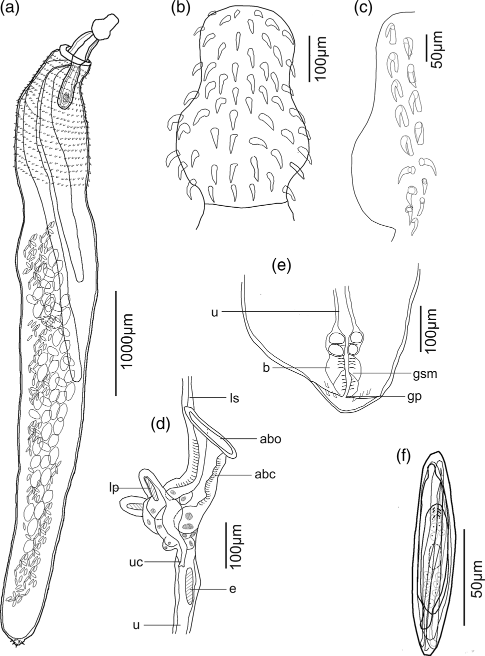
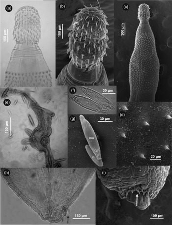
Female (based on 10 gravid females obtained from naturally infected A. platyrhynchos): Trunk 7498–8719 (8021) long and 1193–1619 (1331) wide (fig. 4a). Trunk spines in fore-trunk 25–28 (27) long (figs 4a and 5c, d). Proboscis subspherical 413–460 (436) long and 269–307 (283) wide (figs 4a, b and 5a, b). Hooks per row 7–9 (8) (fig. 4b, c), with three recognizable groups, apical (1–2) hooks with intermediate values of root and hook length, central (3–5) with biggest hook width and basal (6–9) with smallest root (see table 2 for hook and root length, and hook width). Neck 269–624 (413) long and 288–288 (288) wide. Proboscis receptacle 1008–1459 (1248) long and 240–307 (263) wide. Lemnisci digitiform, subequal, right lemniscus 2688–4320 (3312) long and 96–173 (152) wide; left lemniscus 1920–2928 (2340) long and 77–192 (161) wide (fig. 4a). Uterus not observed, covered by eggs. Bell complex 307–394 (336) long and 144–163 (154) wide, with two lateral pockets (figs 4d and 5e), vagina 163–192 (182) long and 77–115 (102) wide, with muscular sphincter surrounding vagina and four bulbs connected to vagina (figs 4e and 5h). Genital pore dorso-subterminal (figs 4e and 5h, i). Sixteen out of 20 specimens with 1–12 (4) genital spines and 9–15 (11) long (fig. 5h, i). Eggs (n = 20) elongated with filaments, outer membrane 79–113 (97) long, 19–23 (21) wide; fertilization membrane with polar prolongations, acanthor 50–59 (55) long and 14–17 (15) wide; six larval hooks, entoblast and central nuclear mass (figs 4f and 5f, g).
Line drawings of Pseudocorynosoma enrietti females from Anas platyrhynchos. (a) Lateral view of entire worm; (b) proboscis; (c) two rows of proboscis hooks; (d) bell complex, lateral view; (e) terminal genitalia; and (f) egg. Abbreviations: abc – anterior bell chamber; abo anterior bell opening; b – bulb; e – egg; gp – gonopore; gsm – genital sphincter muscle; lp – lateral pocket; ls – ligamental sac; u – uterus; uc – urogenital canal.

Microphotographs of Pseudocorynosoma enrietti females from Anas platyrhynchos. (a) Optical microphotograph (OM) of proboscis armature, lateral view; (b) scanning electron microphotograph (SEM) of proboscis, latero-apical view; (c) SEM of proboscis and distribution of spines in fore-trunk, lateral view; (d) SEM of fore-trunk spines; (e) OM of uterine bell; (f) OM; (g) SEM of eggs (imm. immature, and ma. mature); (h) OM of posterior end (genital spines indicated by arrow); and (i) SEM of posterior end (dorso-subterminal genital pore indicated by arrow).

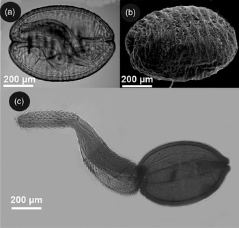
Cystacanth (based on 73 encapsulated specimens and seven evaginated specimens): Encapsulated cystacanth lemon-shaped, orange, enclosed in loose capsule, 539–966 (738) long and 340–568 (477) wide (fig. 6a, b). Evaginated cystacanth 1350–1750 (1525) long and 480–566 (514) wide (fig. 6c). Proboscis cylindrical 353–404 (384) long and 151–152 (124) wide, armed with 19–20 hooks rows. Hook measurements are given in table 3. Hooks per row 9–11 (10), in three groups as in adults. Proboscis receptacle 384–455 (420) long and 144–173 (152) wide. Fore-trunk with 18–20 (19) spines per row, symmetrically distributed on ventral and dorsal sides, 19–26 (21) long. Gonads in the hind-body observed in one male: two testes, anterior 303 long, 202 wide, posterior 353 long, 212 wide, other genital structures rudimentary. Hind-body smooth, lemon-shaped, 677–909 (754) long and 424–515 (471) wide. Genital spines not observed.
Microphotographs of Pseudocorynosoma enrietti cystancanths from Hyalella patagonica. (a) Optical microphotograph (OM) (invaginated proboscis indicated by arrow); (b) scanning electron microphotograph; and (c) OM of everted specimen.

Measurements of hook length and width, and root length of experimental males and females of Pseudocorynosoma enrietti from Anas platyrhynchos at seven days post-infection (dpi), of natural males and females from Coscoroba coscoroba and of evaginated cystacanths. Measurements given in micrometres (n = sample size).

Taxonomic summary
Definitive hosts and localities: A. platyrhynchos (Linnaeus) from Lake Hess (41°22′S; 71°43′W), Nahuel Huapi National Park; and C. coscoroba Molina from Lake Verde (39°01′S; 70°24′W), Laguna Blanca National Park.
Site of infection: intestine.
Infection values: one infected of one revised A. platyrhynchos, with 969 worms; one infected of one revised C. coscoroba, with 277 worms.
Intermediate host and locality: H. patagonica (Ortmann) from Lake Mascardi (41°17′S; 71°38′W), Nahuel Huapi National Park.
Site of infection: haemocoel.
Prevalence and mean intensity: 17.7%, 1.1.
Specimens deposited: 10 natural males (N° 766/2) and eight natural females (N° 766/1) of P. enrietti collected from A. platyrhynchos, four natural males (N°767/2) and four natural females (N° 767/1) collected from C. coscoroba, five experimental males (N° 768/2) and four experimental females (N° 768/1) collected from A. platyrhynchos, and three cystacanths collected from H. patagonica (N°769 1–3) were deposited in the MACN–Pa.
Remarks
The specimens collected in Patagonia presented a similar number of rows (19–20) to P. iheringi (20), but differed from P. anatarium and P. peposacae (both with 14), P. tepehuanesi (15) and P. constrictum (16) (Porta, Reference Porta1914; Van Cleave, Reference Van Cleave1918, Reference Van Cleave1945; Molfi & Freitas Fernandes, Reference Molfi and Freitas Fernandes1953; Machado Filho, Reference Machado Filho1961a; García-Varela et al., Reference García-Varela, Hernández-Orts and Pinacho-Pinacho2017). In addition, specimens of P. enrietti from Patagonia showed a different number of hooks per row in males and females, 9–11 vs. 7–9, respectively. Comparisons cannot be made with other species because no sex discrimination of number of hooks per row is included in their descriptions (table 1). Pseudocorynosoma enrietti from Patagonia showed a higher number of hooks per row than P. anatarium (8–9), P. iheringi (8) and P. tepehuanesi (7–8), but fewer than P. constrictum (10–12) and P. peposacae (12–14) (Porta, Reference Porta1914; Van Cleave, Reference Van Cleave1918, Reference Van Cleave1945; Molfi & Freitas Fernandes, Reference Molfi and Freitas Fernandes1953; Machado Filho, Reference Machado Filho1961a; García-Varela et al., Reference García-Varela, Hernández-Orts and Pinacho-Pinacho2017). The males of P. enrietti (from Brazil and Patagonia) had four cement glands whereas those of P. iheringi had six cement glands. Patagonian specimens presented genital spines in both sexes, like P. anatarium, but differed from P. constrictum and P. iheringi, which present them only in males (Van Cleave, Reference Van Cleave1918, Reference Van Cleave1945; Molfi & Freitas Fernandes, Reference Molfi and Freitas Fernandes1953). The eggs of the Patagonian specimens showed an intermediate size (79–113), similar to P. anatarium (100–112) and P. constrictum (80–108), but were larger than those of P. peposacae (63–68), P. iheringi (63–93) and P. tepehuanesi (80–86) (Porta, Reference Porta1914; Van Cleave, Reference Van Cleave1918, Reference Van Cleave1945; Molfi & Freitas Fernandes, Reference Molfi and Freitas Fernandes1953; García-Varela et al., Reference García-Varela, Hernández-Orts and Pinacho-Pinacho2017). Specimens of P. enrietti from Patagonia were characterized by marked sexual dimorphism in the shape of the proboscis, the number of hooks per row and the number of genital hooks. These differences between the sexes have not been reported for other species of the genus.
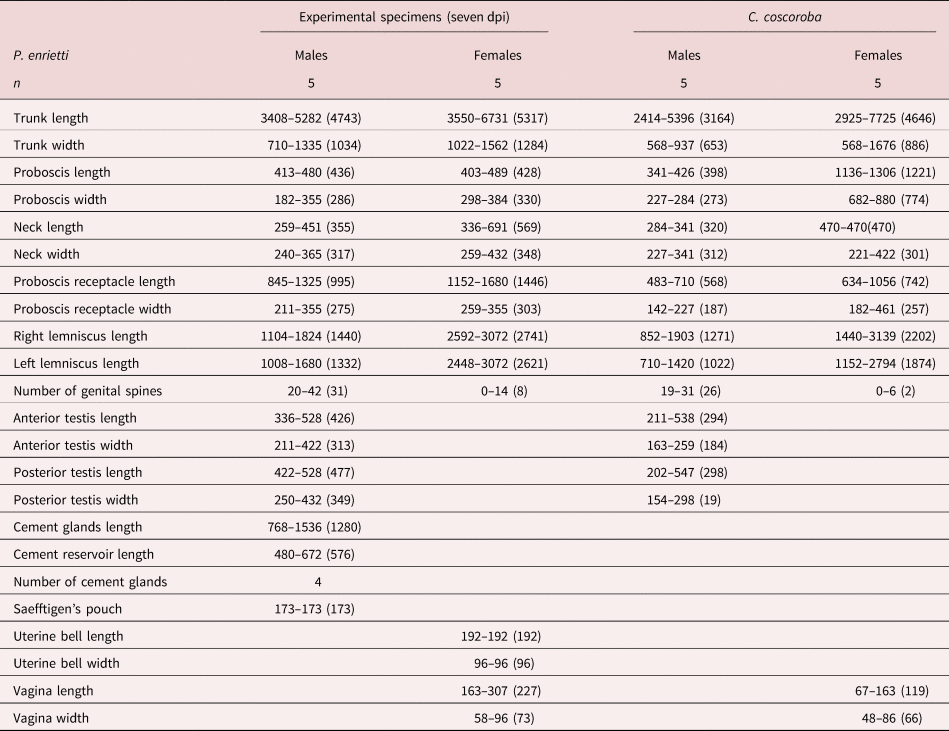
Most of the natural specimens obtained from C. coscoroba were immature; few females had ovarian balls and none were mature. The body measurements of these specimens were slightly smaller than experimental ones at seven dpi. The number of rows and hooks per row was similar in the natural and experimental specimens of both definitive hosts. Body measurements of experimental males and females, and those from natural C. coscoroba infection are given in table 4 and hook measurements in table 3.
Measurements of experimental males and females of Pseudocorynosoma enrietti from Anas platyrhynchos, seven days post-infection (dpi), and of natural males and females from Coscoroba coscoroba. Measurements given in micrometres (n = sample size).

Experimental infections and ecological notes:
Ten specimens of P. enrietti were recovered from A. platyrhynchos at seven dpi, and 11 specimens at 14 dpi, representing a transmission rate of 20% (10 of 50) and 22% (11 of 50), respectively. Both males and females were obtained and all specimens were found alive and attached to the posterior third of the duck intestine; some females at seven dpi showed ovarian balls and copulatory caps. No infections were found in the chicks.
Discussion
The specimens collected during this study were assigned to the genus Pseudorynosoma as they possessed 4 cement glands, genital spines in males and females, and a freshwater life cycle. The experimental and natural specimens showed a similar number of rows, hooks per row and morphometrical values, the former presenting lower values for lemnisci and testes lengths in males and for trunk length in females. Length of proboscis hooks was similar in experimental adults and evaginated cystacanths. In addition, in experimental specimens the distribution of the three groups of proboscis hooks (apical, central and basal) were similar to that of adults from natural infections. The minor differences can be attributed to the maturity of experimental worms. Both sexes presented conspicuous genital spines, with females showing lower numbers than males.
Despite amphipods representing an important food item for waterfowl in North America (Anteau & Afton, Reference Anteau and Afton2009), to our knowledge no studies in Patagonia have focused on the diet of these birds. The prevalence of P. enrietti in amphipods collected in Lake Mascardi was 17.7%; this very high infection value for an intermediate crustacean host could explain the high infection intensity recorded in the natural A. platyrhynchos examined (969 acanthocephalans). Although amphipods were not all collected from the same freshwater environment, both Lakes Hess and Mascardi are connected to the same Pacific watershed. This high parasite load could have a deleterious effect on birds; for example, the attachment structures of P. constrictum have been shown to cause substantial swelling, haemorrhaging and necrosis in the gut tissue of birds (Caballero-Viñas & Sánchez-Nava, Reference Caballero-Viñas and Sánchez-Nava2020). At least in the experimental hosts, the sexual development of females was very rapid: ovarian balls and copulatory caps were present at seven dpi.
The only life cycle known up to now for species of the genus Pseudocorynosoma is that of P. constrictum from North America, which includes the amphipod H. azteca as intermediate host, the waterfowl Anas acuta (Linnaeus), Melanitta deglandi (Bonaparte) and Aythya affinis (Eyton) as natural definitive hosts, and Anas strepera (Linnaeus) and different species of Aythya as experimental hosts (Schmidt, Reference Schmidt1965; Podesta & Holmes, Reference Podesta and Holmes1970). The present study is the first to describe the life cycle of a species of Pseudocorynosoma from South America, involving the anatids A. platyrhynchos and C. coscoroba as natural definitive hosts, A. platyrhynchos as experimental definitive host and H. patagonica as intermediate host. Pseudocorynosoma enrietti has been described in anatids from Curitiba, in the south of Brazil, so its distribution range can be extended to Argentinean Patagonia, considering specimens were found in Lakes Verde, Hess and Mascardi. Anas platyrhynchos and C. coscoroba are migratory birds that breed in the south of the continent (Argentina and Chile) and overwinter in northern Argentina and southern Brazil, where their distribution overlaps with C. moschata (del Hoyo et al., Reference Del Hoyo, Elliott and Sargatal1992). Superposition of the distribution range of the two originally described definitive hosts, A. platyrhynchos and C. moschata, with the new host, C. coscoroba (del Hoyo et al., Reference Del Hoyo, Elliott and Sargatal1992), may indicate the continuous presence of P. enrietti from the south of Brazil to the south of Argentina. The distribution range of P. enrietti is sustained by the presence of its intermediate hosts, among them H. patagonica, a freshwater amphipod widely distributed in Patagonia. Species of the genus Hyalella have been reported as infected by Pseudocorynosoma sp. in 23 environments of Argentinean Patagonia, from Laguna Blanca National Park in Neuquén Province (38°58′S; 70°24′W) to Fagnano Lake in Tierra del Fuego Province (54°30′S; 68°38′W) (Rauque, Reference Rauque2013). Thus, the distribution range of this acanthocephalan species may be wider than currently reported.
Acknowledgements
The authorities of Laguna Blanca and Nahuel Huapi National Parks granted permission for sampling. Thanks are given to two anonymous reviewers for suggestions that improved this manuscript.
Financial support
This study was funded by the following projects: Invasiones ocultas: Los parásitos y la introducción de peces en la cuenca del Río Negro, CONICET PIP 2015-2017 GI.; and Parasitismo en ecosistemas patagónicos: invasiones y zoonosis. Universidad Nacional del Comahue, UNCo B/225.
Conflicts of interest
None.
Ethical standards
The authors assert that all procedures contributing to this work comply with the ethical standards of the relevant national and institutional guides on the care and use of laboratory animals.