Introduction
Superior semicircular canal dehiscence syndrome is a relatively new syndrome described in 1998 by Minor et al. Reference Minor, Solomon, Zinreich and Zee1 The syndrome unifies the symptoms, signs and high-resolution computed tomography (CT) findings in patients presenting with vestibular symptoms or hearing loss caused by sound or pressure changes. Superior semicircular canal dehiscence syndrome has a robust pathology which is readily demonstrated radiologically, and it can be considered for surgical correction. A careful history and good quality radiological images are prerequisite. The incidence of superior semicircular canal dehiscence syndrome is estimated from autopsy studies at 0.7 per cent in the general populationReference Carey, Minor and Nager2 and at 1 per cent from in vivo imaging.Reference Krombach, DiMartino, Schmitz-Rode, Prescher, Haage and Kinzel3 Using high resolution temporal CT, a physical thickness of ≤0.1 mm of bone covering the superior semicircular canal appears completely dehiscent. The incidence may be as high as 1.3%; however, this incidence is far higher than the anecdotal incidence observed in our clinics, suggesting to us that radiological dehiscence may not always result in clinical sequelae.
We present here a unique clinical case with a fascinating anatomical aetiology; we also discuss more common aetiologies.
Case report
A 58-year-old woman initially presented in January 2006 with a history of right-sided autophony, otalgia and intermittently ‘muffled’ hearing. She had a 30-year history of Ménière's disease on the left, resulting in poor hearing in that ear, making her right ear her only functional ear at presentation. She also described occasional nasal obstruction, improved in the past with BeconaseTM spray.
Examination revealed normal tympanic membranes, with thickened nasal mucosa on nasendoscopy. Pure tone audiometry showed normal thresholds on the right but severe sensorineural hearing loss on the left, in keeping with the patient's Ménière's disease.
In view of these new problems in the patient's only functioning ear, a CT scan of the temporal bones was arranged. The working diagnosis at this stage was eustachian tube dysfunction. The patient was initially treated with medical therapy for rhinitis.
A CT scan of the patient's temporal bones (not shown) was performed with a slice thickness of >1 mm, rendering the image quality suboptimal for evaluation of the superior semicircular canal. The radiologist's report noted a ‘possible defect in the posterior aspect of the right superior semicircular canal’, but noted further that this was ‘a slightly unusual site for a defect’. All other middle-ear structures were normal.
The patient was reviewed in June 2006, six months after initial presentation, at which time nasendoscopy revealed rhinitic nasal passages with mucus. She was prescribed antihistamines and Flixonase (GlaxoSmithKlein, Brentford, UK).
Following a clinic review three months later, the patient was discharged to primary care with no further active management plan and a diagnosis of eustachian tube dysfunction.
The patient presented for a second opinion six months after her discharge from clinic, at which point her history was retaken. In addition to right-sided autophony, she had now developed momentary dizziness and imbalance on shouting. Her CT scans were reviewed by a neuroradiologist with an interest in temporal bone imaging (the second author). There appeared to be a defect in the right superior semicircular canal, possibly due to an unusually deep groove for the left superior petrosal sinus.
To confirm the diagnosis and delineate the venous anatomy, a repeat CT scan with intravenous contrast material was performed (using an Aquilion 64™ multidetector CT scanner; Toshiba, Crawley, UK). Contrast material is not usually administered during CT examinations of the temporal bone for suspected superior semicircular canal dehiscence syndrome. However, it was considered justified in this case due to the possibility of an abnormality of intracranial venous drainage.
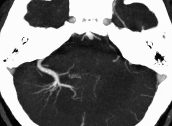
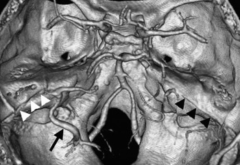
Image reconstruction was performed using bone and soft tissue algorithms. Data reconstructed on the bone algorithm were used to perform multiplanar reconstructions to show bony detail (Figure 1). Data reconstructed on the soft tissue algorithm were used to show vascular anatomy (Figures 2 and 3). The images confirmed a dehiscent right superior semicircular canal (Figure 1). The dehiscence was clearly due to an abnormally deep groove for the superior petrosal sinus (Figure 3). The scan also demonstrated a large developmental venous anomaly within the right cerebellar hemisphere, draining into the right superior petrosal sinus (Figures 2 and 3).
Oblique sagittal reconstructions of high resolution (0.5 mm slice thickness) temporal bone computed tomography images performed in the plane of the (a) right and (b) left superior semicircular canals, demonstrating a 2.5 mm defect in the bony covering at the postero-superior aspect of the right superior semicircular canal (arrow). The appearance on the left is normal.

A thin slab (8 mm) maximum intensity projection reconstruction of the computed tomography angiography examination, demonstrating the characteristic appearance of a right-sided posterior fossa developmental venous anomaly. A ‘Medusa head’ of radially oriented, dilated medullary veins joins to form a single large ‘collector vein’ which drains transcortically into the superior petrosal sinus.

A volume-rendering technique was used to produce this three-dimensional, surface-shaded display of the high resolution computed tomography angiography examination. The large developmental venous anomaly (black arrow) in the posterior fossa drains into the right superior petrosal sinus, which occupies an abnormally deep groove in the petrous ridge (white arrowheads). The left superior petrosal sinus has a more usual configuration (black arrowheads).

The patient was reassured that the condition was benign. In view of her non-functioning left ear, the surgical risk of canal occlusion was deemed too great, and no further treatment was offered after a full explanation.
Discussion
Superior semicircular canal dehiscence syndrome is caused by abnormal connections between the superior semicircular canal and the middle fossa. The present case details an unusual anatomical aetiology. This section reviews briefly the clinical features and management of superior semicircular canal dehiscence syndrome in the context of this case.
Superior semicircular canal dehiscence syndrome presents with pressure- or sound-related vertigo, and altered or impaired hearing. The symptoms result from dehiscence of bone covering the superior semicircular canals, exposing the vestibular system to variations in intracranial pressure and to bone-transmitted sound, resulting in so-called ‘third window’ conduction.Reference Minor4 These symptoms are underpinned by experimental work; fenestration of the semicircular canals in anaesthetised chinchillas lowered the threshold for auditory stimulation in the superior vestibular nerve afferentsReference Carey, Hirvonen, Hullar and Minor5 and increased the sensitivity of the superior canals to changes in pressure at the external auditory meatus.Reference Hirvonen, Carey, Liang and Minor6 Therefore, the classic symptoms are pressure-related imbalance, described by Tullio in 1929,Reference Tullio7 and autophony. Our experience is that patients present initially with auditory disturbance, developing vestibular features later. In a recent study, only five out of 65 patients presented solely with auditory symptoms,Reference Minor4 suggesting that vestibular dysfunction is a hallmark abnormality. Most patients report noise- or pressure-related vertigo only on direct questioning, highlighting the importance of good history-taking in diagnosing superior semicircular canal dehiscence syndrome.Reference da Cuhna and de Melo Tavares de8
Examination usually reveals normal tympanic membranes, with no membrane movement with respiration, a sign that would suggest a patulous eustachian tube, the other condition commonly associated with autophony. Therefore, a normal otological examination should point clinicians away from eustachian tube conditions and suggest superior semicircular canal dehiscence. There may be involuntary torsional eye movements and nystagmus relating to the affected canal. Investigating superior semicircular canal dehiscence syndrome requires audiometry and high resolution (<1 mm slice thickness) CT scanning to visualise the dehiscence.
Anecdotally, idiopathic dehiscences appear most commonly in our clinics. In other published groups, the commonest cause appears to be head trauma; around 50 per cent of patients in the Johns Hopkins series described a history of head trauma.Reference Minor9 Other pathologies resulting in dehiscence are infection and neoplasm – for example, vestibular schwannoma.Reference Carey, Minor and Nager2 Carey et al. studied 1000 temporal bones from 596 adult cadavers, including histology showing such erosions. The study also included eight instances of dehiscence of the superior semicircular canal from erosion specifically by the superior petrosal sinus; however, it lacked the matching symptomatology, as reported in the present case. One case, a 27-year-old with a left-sided vestibular schwannoma, showed bilateral dehiscences that were aetiologically different: tumour erosion on the left; superior petrosal sinus erosion on the right. The histology in this individual showed stratified lamellae running in parallel to the course of the vein, leading the authors to postulate that this defect was not in fact an erosive process but rather that mature bone had been laid down around the extended sinus as it developed in utero.Reference Carey, Minor and Nager2
The aetiology in our case is likely to be a similar developmental variation; the presence of the large, right-sided cerebellar developmental venous anomaly draining into the right superior petrosal sinus possibly explains the unusual configuration of this sinus. However, it is unclear what precipitated these symptoms in the sixth decade of life.
• The semicircular canal dehiscence syndrome is a recently described disorder giving rise to vestibular and auditory symptoms
• Dehiscence of bone overlying the superior semicircular canal generates a ‘third window’ for abnormal sound and pressure conduction
• This reported describes, for the first time, superior semicircular canal dehiscence syndrome caused by erosion from an enlarged superior petrosal sinus
• Superior semicircular canal dehiscence syndrome may result from abnormal vascular anatomy, and may present late in life without further evidence of precipitation
• Superior semicircular canal dehiscence syndrome is aetiologically varied; an unusual aetiology is presented here for the first time
The management of superior semicircular canal dehiscence syndrome involves conservative and surgical approaches. Surgical options for patients with disabling vestibular symptoms include a middle fossa craniotomy to plug or resurface the dehiscence. Data on the relative efficacy of these two approaches are as yet underpowered, but the initial suggestion from small cohorts is that canal plugging is superior to resurfacing.Reference Minor4 In a recent study, two patients out of 19 developed failure of all three ipsilateral canals following surgery, but this study did suggest that plugging of the dehiscence does lead to improvement or resolution of pressure- or sound-related vertigo.Reference Carey, Migliaccio and Minor10 In our case, the presence of a contralateral sensorineural hearing loss led us directly to a conservative approach.
Conclusion
Superior semicircular canal dehiscence syndrome is a relatively new condition in the lexicon of vestibular disorders, with various aetiologies. This report describes for the first time a clinical incidence of superior semicircular canal dehiscence syndrome caused by an unusually deep groove for the superior petrosal sinus. Computed tomography angiography showed that the sinus receives drainage from a large cerebellar developmental venous anomaly. Carey et al. have observed dehiscences at the superior petrosal sinus in their temporal bone survey but have ‘not observed symptoms of the superior canal dehiscence syndrome’ relating to it; they hypothesise that dehiscence at this site requires an additional aetiological event. The case presented here, however, with no history of head trauma or raised intracranial pressure, and a full thickness defect of the canal wall, suggests that symptoms can occur without a second precipitating event.