Introduction
The role of the eustachian tube in middle-ear homeostasis has long been recognised. Eustachian tube dysfunction has been implicated in most cases of middle-ear disease. In addition, normal eustachian tube function is a good prognostic indicator in the management of various middle-ear pathologies.
Studies of the eustachian tube are difficult due to its inaccessible location and the multiplicity of factors involved in normal function and dysfunction. Over the years, many tests have been devised to assess the functional status of the eustachian tube.
A clinically practical procedure for assessing eustachian tube function in individuals with an intact tympanic membrane is the eustachian tube swallow test, which was originally proposed by Williams,Reference Williams1 and subsequently by Bluestone and Cantekin.Reference Bluestone and Cantekin2 A baseline tympanogram is initially recorded. Positive and/or negative pressure is then generated in the external auditory canal; the individual is instructed to swallow thrice, and the tympanometry is repeated. A positive or negative shift in the peak of the tympanogram is considered indirect evidence of normal eustachian tube function. Absence of such a shift indicates eustachian tube dysfunction.
With the advent of diagnostic endoscopic techniques, attempts have been made to study eustachian tube function using endoscopes. For instance, endoscopic assessments of the pharyngeal orifice and the lumen of the eustachian tube have been carried out using fibre-optic endoscopes introduced from the pharyngeal or tympanic orifice.Reference Di Martino, Walther and Westhofen3, Reference Linstrom, Silverman, Rosen and Meiteles4 Although these tests provided information on the anatomic patency of the eustachian tube, they failed to assess its functional status. Navigation of the eustachian tube lumen using a fibre-optic endoscope requires significant skill and expertise. This can also prove to be a traumatic procedure for the patient, thereby defeating its purpose.
Dynamic slow motion video endoscopy of the eustachian tube has shown promising results as a method of assessing eustachian tube function. Poe et al. (2000) described the technique of eustachian tube video endoscopy and the normal pattern of eustachian tube dilatation on video endoscopy in their initial study, which comprised 17 normal individuals and 17 individuals with eustachian tube dysfunction.Reference Poe, Pyykkö, Valtonen and Silvola5 In a subsequent study of 64 ears with evidence of middle-ear disease, Poe et al. (2001) described the various pathological changes that contributed to eustachian tube dysfunction. The authors concluded that slow motion video endoscopy was a potentially useful tool for evaluating the pathophysiology of eustachian tube dysfunction.Reference Poe, Abou-Halawa and Abdel-Razek6
In 2007, Mathew et al. studied 61 normal ears and 63 ears with middle-ear disease and classified patterns of eustachian tube function on video endoscopy as follows.Reference Mathew, Kuruvilla and Job7 Grade 0 is representative of a normal eustachian tube, with no mucosal oedema or congestion. Medial cartilaginous lamina and lateral wall movements are normal. The tubal lumen opens well on swallowing. Grade 1 reflects oedema and congestion of mucosa limited to the pharyngeal orifice of the eustachian tube. There is normal lateral wall motion and the tubal lumen opens with swallowing. Grade 2a is associated with reduced lateral wall motion secondary to oedema and congestion involving the lumen. The tubal lumen opens partly with swallowing. Grade 2b is representative of reduced lateral wall motion secondary to abnormal tubal muscle contraction. The tubal lumen opens partly with swallowing. Grade 3a is descriptive of cases in which the tubal lumen fails to open with swallowing, secondary to gross oedema. Grade 3b concerns cases where the tubal lumen fails to open with swallowing, secondary to abnormal tubal muscle contraction. In cases classified as patulous (P), there is a noticeable concavity in the superior portion of the lateral wall of the lumen, with persistent patency of the lumen that extends towards the isthmus. The medial and lateral cartilaginous walls remain separate even at rest.
The study by Mathews et al. showed a significantly higher prevalence of eustachian tube dysfunction among individuals with middle-ear disease compared with those in the control group. In addition, there was a significant correlation between eustachian tube video endoscopy and the findings of manometric studies, which included the positive pressure equalisation test and tympanometry.
Our study aimed to assess the efficacy of dynamic slow motion video endoscopy as a test of eustachian tube function. The reliability of the test was assessed in terms of its correlation with the eustachian tube swallow test. We also studied the variations of eustachian tube function that may coexist with normal middle ears.
Materials and methods
The study by Mathew et al. showed that the prevalence of eustachian tube dysfunction among individuals with normal middle ears was 39 per cent, and the agreement between dynamic slow motion video endoscopy and the positive pressure equalisation test results was 0.3.Reference Mathew, Kuruvilla and Job7 For a null hypothesis agreement of 0, using a power of 80 per cent and 5 per cent level of significance, the required sample size for our study was calculated to be 84 ears.
The study comprised 100 ears of adults undergoing rigid nasal endoscopy for various indications (in the ENT out-patient department). All individuals included in the study satisfied the following inclusion criteria: no ear symptoms; normal otoscopy findings; pure tone audiogram thresholds of less than 20 dBHL at 0.25, 0.5, 1, 2 and 4 kHz; and a normal type A tympanogram. The exclusion criteria were as follows: a gross deviated nasal septum; gross nasal polyposis; nasopharyngeal pathology (e.g. adenoid hypertrophy or nasopharyngeal tumour); evidence of middle-ear disease on otoscopy or tympanometry; and very ill or debilitated individuals.
Individuals who satisfied the above criteria were then subjected to the reference test (the eustachian tube swallow test) and the index test (dynamic slow motion video endoscopy) as described below. The study design was scrutinised and approved by the institutional review board of our institution.
Eustachian tube swallow testReference Williams8
For this reference test, the subject was seated comfortably and instructed to avoid jaw movements, throat clearing or swallowing while the probe tip was in place. A baseline tympanogram was obtained using the GSI TympStar middle-ear analyser (Grason-Stadler, Eden Prairie, Minnesota, USA). The external auditory canal air pressure was then increased to +400 mm H2O and the subject was instructed to take three hard swallows of water, after which a second tympanogram was recorded. Canal pressure was returned to 0 mm H2O and the subject once again swallowed, thereby returning middle-ear pressure to the baseline condition. Finally, pressure in the ear canal was decreased to −400 mm H2O and the subject was again instructed to swallow thrice, after which a third tympanogram was obtained.
Positive pressure in the external auditory canal produces a shift of the tympanometric peak in the negative direction, while negative pressure in the ear canal causes the tympanometric peak to shift in the positive direction. A reverse shift may be encountered; this was considered a normal variant.
The shift in peak amplitude, or function change, was also used for the quantification of eustachian tube function. As suggested by Seidemann and Givens, function change data were reported as percentages according to the equation shown below.Reference Seidemann and Givens9 ‘FD1’ represents the difference between the peak amplitude and the +200 mm point on the baseline tympanogram. ‘FD2’ is the difference in amplitude between the peak pressure and +200 mm in the positive or negative external auditory canal pressure conditions.
 $$\percnt FC = \displaystyle{{FD2 - FD1} \over {FD1}}*100$$
Six criteria, based on those proposed by Schuchman and Joachims, were used to interpret the results of the eustachian tube swallow test and for the comparison with the dynamic slow motion video endoscopy grading of eustachian tubes.Reference Schuchman and Joachims10 These criteria are defined by: (1) a pressure change of ≥5 dPa in the positive or negative external auditory canal pressure situation; (2) a pressure change of ≥5 dPa in the positive and negative external auditory canal pressure situation; (3) a pressure change of ≥15 dPa in the positive or negative external auditory canal pressure situation; (4) a pressure change of ≥15 dPa in the positive and negative external auditory canal pressure situation; (5) a functional change of ≥5 per cent in the positive or negative external auditory canal pressure situation; and (6) a functional change of ≥5 per cent in the positive and negative external auditory canal pressure situation.
Dynamic slow motion video endoscopyReference Mathew, Kuruvilla and Job7
For this index test, a cottonoid soaked in 4 per cent lignocaine was placed along the floor of the nasal cavity for 5 minutes. A rigid Hopkins rod endoscope (Karl Storz, Tuttlingen, Germany) was used, which has a 45-degree angle and is 4 mm in diameter. The endoscopes were fitted with a Serwell video camera head and processor (Serwell Medi-Equip, Chennai, India). The images were viewed on a BenQ 17-inch colour liquid crystal display video monitor (BenQ India, Haryana, India), and were recorded using Imimo version 7 software (Avanttec Medical Systems, Chennai, India). The images obtained were then analysed using PowerDVD software (Cyberlink, Taipei, Taiwan); the images were viewed on a 17-inch colour monitor in real time, slow motion and frame by frame for assessment of the eustachian tube dynamics.
Once the individual was anaesthetised, the nasal endoscope was held with the 45-degree viewing angle facing superiorly and introduced along the floor of the nasal cavity. The endoscope was advanced just beyond the posterior end of the inferior turbinate and then rotated laterally to face the nasopharyngeal orifice of the eustachian tube. The endoscope was positioned as close as possible to the nasopharyngeal orifice, avoiding contact with the mucosa, and angled to obtain a good view along the line of the lumen of the eustachian tube.
The pharyngeal orifice of the eustachian tube was first assessed for structural abnormalities, such as mucosal congestion, oedema, discharge and polyps. The subject was then asked to perform several swallows, followed by forced yawning to produce sustained tubal dilatation. The movements of the medial and lateral cartilaginous lamina were observed, along with the dilatory wave of the lateral pharyngeal wall and the tubal lumen opening. The video recordings of tubal dilatations were viewed (in normal time, slow motion and frame by frame) by three independent observers who were blind to the eustachian tube swallow test results. Eustachian tube function was classified according to the grading system proposed by Mathew et al. Reference Mathew, Kuruvilla and Job7 For the interpretation of dynamic slow motion video endoscopy, the grade given by two of the three observers was considered the endoscopic grading of the eustachian tube. In the event of all three observers giving three different grades, the grade assigned by the most senior observer was accepted.
Results and analysis
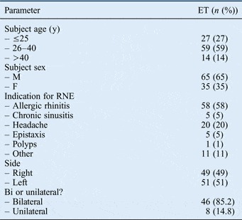
In total, 100 eustachian tubes of 54 subjects were included in the study (Table I). Due to selection criteria, for 8 individuals only one eustachian tube was included in the study, while both eustachian tubes were included for the remaining 46 individuals. The clinical profile of the eustachian tubes assessed is shown in Table I.
Clinical profile of eustachian tubes

ET = eustachian tubes; y = years; M = male; F = female; RNE = rigid nasal endoscopy
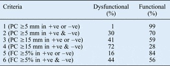
The percentages of eustachian tubes classified as functional and dysfunctional (according to the criteria proposed by Schuchman and Joachims) on the basis of the eustachian tube swallow test findings are shown in Table II.Reference Schuchman and Joachims10 The percentages of eustachian tubes classified according to the various criteria steadily decreased as the criteria became more stringent, ranging from 99 per cent in criterion 1, to 28 per cent in criterion 4.
Functional and dysfunctional eustachian tubes*

* Based on eustachian tube swallow test tympanometric criteria. PC = pressure change; +ve = positive external auditory canal pressure situation; –ve = negative external auditory canal pressure situation; FC = functional change
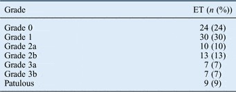
The profile of eustachian tube grading based on dynamic slow motion video endoscopic assessment showed that 24 per cent of tubes were structurally and functionally normal (Table III). Thirty per cent of the tubes opened fully with swallowing, but were oedematous. Twenty-three per cent of the tubes opened partially. Fourteen per cent of the tubes failed to open with swallowing. Nine per cent of the tubes were patulous. Among the eustachian tubes that opened partially or failed to open, five tubes were found to have a floppy medial cartilaginous lamina which seemed to contribute to the partial opening or failure to open with swallowing. Three of these five tubes opened partially with swallowing and were graded as 2a and two failed to open with swallowing and were graded as 3a.
Dynamic slow motion video endoscopy grading

ET = eustachian tubes
Analysis of the results revealed a statistically significant correlation between the eustachian tube swallow test and the dynamic slow motion video endoscopy findings when swallow test criteria 3 and 4 were used to assess the eustachian tube. The p values were 0.002 and 0.048 respectively. There was no significant correlation between the two tests when other tympanometric criteria were used.
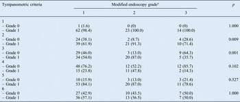
The dynamic slow motion video endoscopy findings were then re-graded on the basis of three functional grades as follows. Modified grade 1 included dynamic slow motion video endoscopy grades 0, 1 and P. Modified grade 2 comprised endoscopy grades 2a and 2b. Finally, grade 3 consisted of endoscopy grades 3a and 3b. The modified grade 1 therefore included all eustachian tubes that opened fully, grade 2 included all partially opened eustachian tubes and grade 3 included all eustachian tubes that failed to open. The results of video endoscopy using this modified grading were correlated with the results on tympanometry using the various tympanometric criteria (Table IV). The chi-square or Fisher's exact test were performed, which revealed a statistically significant correlation between the modified video endoscopy grading and the eustachian tube swallow test results when criteria 2 and 3 were used. The p values were 0.009 and 0.001 respectively. (The p values when other criteria were used were greater than 0.05.)
Association between tympanometric criteria and modified endoscopy grades

Tympanometric grades: 0 = dysfunctional, 1 = functional. *Data represent numbers (percentages) of eustachian tubes.
Discussion
Eustachian tube dysfunction is known to play a key role in the aetiopathogenesis of various middle-ear diseases including: acute suppurative otitis media; chronic suppurative otitis media; atticoantral disease and tubotympanic disease; otitis media with effusion; and middle-ear atelectasis.Reference Slattery, Glasscock and Gulya11 Furthermore, eustachian tube status is also an important prognostic indicator in the management of chronic suppurative otitis media and middle-ear atelectasis.Reference Slattery, Glasscock and Gulya11 Although difficult, the assessment of eustachian tube function is vital in otological practice.
Various diagnostic tests have been devised to directly or indirectly evaluate the anatomical and functional patency of the eustachian tube. Tests that examine functional patency assess pressure regulation, mucociliary clearance or the protective function of the eustachian tube. Of these, pressure regulatory function is considered to be of prime importance. Each of the eustachian tube function tests has its own merits and demerits; however, for various reasons such as the unavailability of equipment and expense of the tests, most of them are not used in routine clinical practice. Moreover, a gold standard test for the assessment of eustachian tube function is yet to be devised.
Dynamic slow motion video endoscopy of the eustachian tube is a recently described method of assessing dynamic eustachian tube function. It is simple to perform and can be carried out alongside diagnostic nasal endoscopy in the out-patient setting. The current study attempted to compare the efficacy of this test with an assessment of dynamic eustachian tube function based on tympanometry.
The eustachian tubes were classified as functional or dysfunctional based on the criteria described by Schuchman and Joachims.Reference Schuchman and Joachims10 The results of the current study were comparable to those obtained by these authors.Reference Schuchman and Joachims10 In their study, the respective percentages of functional eustachian tubes identified using criteria 1 to 6 were: 82.26, 66.13, 55.65, 21.77, 87.90 and 57.25 per cent. In the current study, the respective percentages of functional eustachian tubes obtained for criteria labelled 1 to 6 were: 99, 70, 59, 28, 84 and 56 per cent (Table II).
On the eustachian tube swallow test, when the tympanometric criterion of ≥5 dPa pressure change in the positive or negative external auditory canal pressure situation (criterion 1) was used to classify the eustachian tube as functional or dysfunctional, 99 per cent of eustachian tubes passed the test (Table II). As all the middle ears included in this study were normal both otoscopically and on the basis of the audiogram and tympanometry, it may be assumed that a pressure change of ≥5 dPa on the eustachian tube swallow test is adequate to consider the eustachian tube as functional for hearing and middle-ear aeration.
On video endoscopy, 24 per cent of eustachian tubes were found to be completely normal, with no evidence of any structural or functional abnormality; these were therefore graded as 0 (Table III). In the grading system proposed by Mathew et al., grades 0, 1 and P comprised tubes where the tubal lumen opened fully during swallowing.Reference Mathew, Kuruvilla and Job7 Grade 1 consisted of oedematous tubes, while grade P referred to patulous tubes. When considering only the functional status and ignoring structural abnormalities such as oedema or patulous tubes (i.e. considering grades 0, 1 and P together), 63 per cent of the tubes were found to open fully on video endoscopic assessment.
According to Mathew et al., grade 2 refers to eustachian tubes that open partially on swallowing, wherein grade 2a refers to partial opening due to oedema and grade 2b refers to partial opening due to muscle dysfunction. Twenty-three per cent of the eustachian tubes in the current study opened partially (grade 2a and 2b) on dynamic slow motion video endoscopy grading (Table III). When fully and partially opening eustachian tubes were considered together, 86 per cent of the tubes were functional and were maintaining the homeostasis of the middle ear.
Grade 3 of the classification system by Mathew et al. refers to eustachian tubes that fail to open on swallowing, in which 3a refers to failure to open secondary to oedema and 3b refers to failure to open due to muscle dysfunction. Despite a normal audiogram and tympanometry, 14 per cent of the eustachian tubes in the current study failed to open on swallowing (grade 3a and 3b) (Table III). These apparently dysfunctional eustachian tubes probably equalised middle-ear pressure by spontaneous opening that was secondary to the development of a pressure differential across the two ends of the eustachian tube, thereby maintaining the normalcy of the middle ear.Reference Thomsen12, Reference Ingelstedt, Ivarsson and Jonson13
An incidental observation was the presence of a floppy medial cartilaginous lamina in five eustachian tubes, three of which opened partially on swallowing and two failed to open on swallowing. This floppy cartilaginous lamina appeared to contribute to the dysfunctional status of the eustachian tube. The grading system proposed by Mathew et al. does not include this factor in the criteria for eustachian tube assessment.Reference Mathew, Kuruvilla and Job7 Further studies on the clinical significance and relevance of this entity are warranted in order to establish the role of a floppy medial cartilaginous lamina in the pathophysiology of eustachian tube dysfunction.
• Dynamic slow motion video endoscopy of the eustachian tube is a simple eustachian tube function assessment
• Endoscopy findings correlated with eustachian tube swallow test results for some lenient tube dysfunction (swallow test) criteria
• When used with other tests, dynamic slow motion video endoscopy can provide structural information of the pharyngeal end of the eustachian tube
The study by Mathew et al. on dynamic slow motion video endoscopy showed a significantly higher incidence of eustachian tube dysfunction in those with middle-ear disease compared with normal individuals. Although even a partially functional eustachian tube can maintain middle-ear pressure, dynamic slow motion video endoscopy has the advantage of revealing structural abnormalities such as oedema of the eustachian tube and tubal muscle dysfunction. The use of tympanometry alone does not pick up on such structural abnormalities. Thus, dynamic slow motion video endoscopy performed in addition to dynamic tympanometry can provide valuable information regarding the structural aspects of the eustachian tube. Knowledge of these structural and functional details can aid in early intervention for the prevention of eustachian tube dysfunction and middle-ear pathology. However, further studies of patients with eustachian tube dysfunction and middle-ear pathologies that use dynamic slow motion video endoscopy would be required to establish the value of this test in routine clinical practice.
Conclusion
Dynamic slow motion video endoscopy of the eustachian tube is a simple, easy to perform procedure for the assessment of eustachian tube function. The test appears to over-diagnose eustachian tube dysfunction when used alone. However, when used in combination with other eustachian tube function tests, it can provide additional information regarding the structural and functional status of the pharyngeal end of the eustachian tube. This would enable early intervention directed at the prevention and management of eustachian tube dysfunction related to middle-ear pathologies.
Acknowledgements
The authors would like to acknowledge with gratitude the contributions of Ms Tunny Sebastian and Mrs Nithya Joseph in the statistical analysis of the study results. We are also grateful for the financial support provided for the execution of this research by Christian Medical College, Vellore, India.