INTRODUCTION
Barnacles (Crustacea: Cirripedia) are composed of three well-distinguished superorders: Thoracica (filter-feeding barnacles); Rhizocephala (parasitic barnacles); and Acrothoracica (burrowing barnacles) (Glenner & Hebsgaard, Reference Glenner and Hebsgaard2006; Pérez-Losada et al., Reference Pérez-Losada, Harp, Høeg, Archituv, Jones, Watanabe and Crandall2008, Reference Pérez-Losada, Høeg and Crandall2009, Reference Pérez-Losada, Høeg and Crandall2012). The Acrothoracica contains approximately 63 species of burrowing barnacles that are divided into two orders: the Cryptophialida (with the family Cryptophialidae); and the Lithoglyptida (with the families Lithoglyptidae and Trypetesidae) (Tomlinson, Reference Tomlinson and Southward1987; Kolbasov, Reference Kolbasov2009; Chan et al., Reference Chan, Kolbasov and Cheang2012; WoRMS, 2012). The Trypetesidae, representing the largest sized acrothoracicans, contains highly derived species with uniramous cirri, one ventral ganglion, and lacking an anus (Tomlinson, Reference Tomlinson1969, Reference Tomlinson and Southward1987). All trypetesid species are obligate symbionts of hermit crabs and found only in gastropod shells inhabited by paguroid hosts. The co-evolution between hermit crabs and trypetesid barnacles dates back to at least the Miocene era (Baluk & Radwánski, Reference Baluk and Radwánski1991). It is likely that the trypetesids were once more widely distributed geographically and Tethyan in origin (Newman & Foster, Reference Newman and Foster1987).
Trypetesid barnacles are a completely sessile group of organisms, found in the littoral and sublittoral oceanic regions, and are widely distributed, being found in the North Sea, the Mediterranean, western Atlantic, California, the French coast of the English Channel, Japan, Madagascar, and the Philippines (Lambers & Boekschoten, Reference Lambers and Boekschoten1986; Williams & McDermott, Reference Williams and McDermott2004; Glenner & Hebsgaard, Reference Glenner and Hebsgaard2006; Williams & Boyko, Reference Williams and Boyko2006). The family Trypetesidae contains seven extant species in two genera, Trypetesa Norman, 1903 (five species) and Tomlinsonia Turquier, 1985 (two species). The seven extant species of trypetesids are: Tomlinsonia asymetrica (Turquier & Carton, Reference Turquier and Carton1976), T. mclaughlinae Williams & Boyko, Reference Williams and Boyko2006, Trypetesa habei Utinomi, Reference Utinomi1962, T. lampas (Hancock, 1849), T. lateralis Tomlinson, Reference Tomlinson1953, T. nassarioides Turquier, Reference Turquier1967, and T. spinulosa Turquier, Reference Turquier1976 (see Table 1; Figure 1). In addition to these extant species, two ichnofossil species of Trypetesa are known, T. caveata Tomlinson, 1963 and T. polonica Baluk & Radwánski, Reference Baluk and Radwánski1991, with fossil burrows dating back to the mid-Miocene when hermit crabs first inhabited shallow marine areas (Baluk & Radwánski, Reference Baluk and Radwánski1991). The specimens of T. caveata were found in myalinid bivalves (Tomlinson, Reference Tomlinson1969), causing Seilacher (Reference Seilacher1969) to question if they belong to this genus. Although similar in gross morphology, the genera Trypetesa and Tomlinsonia are distinguished in that members of Tomlinsonia have cushions on all three pairs of terminal cirri (4th, 5th and 6th), whereas members of Trypetesa have cushions on only the first two pairs of terminal cirri (4th and 5th) (Williams & Boyko, Reference Williams and Boyko2006). The cushions (also called ‘prominent excrescences': see Spivey, Reference Spivey1979, figure 2d; or ‘pillow-like hillocks': see Kolbasov, Reference Kolbasov2009, figure 27) are rounded protuberances on the distal end of the second segment of the terminal cirri that are covered in rows of small triangular denticules. Although they are easily observed on the cirri and have long been suspected to be involved in food processing (Darwin, Reference Darwin1854), their exact role in food capture or feeding remains unknown (Williams et al., Reference Williams, Gallardo and Murphy2011).
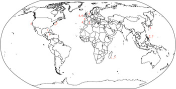
Geographical distribution of the seven extant species of trypetesid barnacles: (1) Tomlinsonia asymetrica (Turquier & Carton, Reference Turquier and Carton1976); (2) T. mclaughlinae Williams & Boyko, Reference Williams and Boyko2006; (3) Trypetesa habei Utinomi, Reference Utinomi1962; (4) T. lampas (Hancock, 1849); (5) T. lateralis Tomlinson, Reference Tomlinson1953; (6) T. nassarioides Turquier, Reference Turquier1967; (7) T. spinulosa Turquier, Reference Turquier1976.

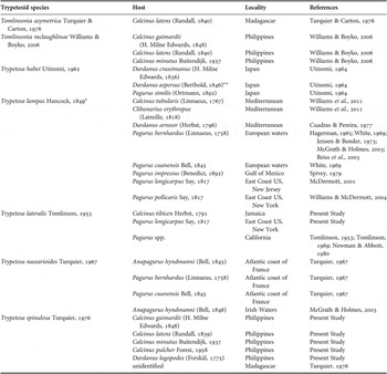
Geographical distribution of trypetesid species and their hosts.

*, the record of T. lampas from the Philippines by Rosell (1981) is erroneous (see discussion in text);
**, originally as Dardanus diogenes (De Haan).
The purpose of this research was to explore the taxonomy and natural history of two species of the genus Trypetesa, based on specimens inhabiting gastropod shells collected from the east coast of the United States, Jamaica and the Philippines. Based on a combination of light and scanning electron microscopy (SEM) techniques, we sought to: (1) identify the species of Trypetesa found associated with hermit crabs from these three regions and record host specificity and prevalence; (2) investigate the mouthparts and other taxonomically important morphological structures used in species delineation; and (3) analyse the gut contents of the trypetesids, investigating the occurrence of egg predation through histology. The geographical ranges of the two species of Trypetesa were evaluated, with discussion of their possible introductions. The symbiotic relationship between trypetesids and hermit crabs was re-evaluated based on the finding of their activity as egg predators.
MATERIALS AND METHODS
Specimen collection
Hermit crabs were collected from Long Island, New York, Jamaica and the Philippines. The Long Island collections occurred from 1 July 2005 to 15 July 2008, with data based on 2494 hermit crab hosts (McGuire, Dualan, and Murphy, coll.; see McGuire & Williams (Reference McGuire and Williams2010) for ecological data on hermit crab hosts). Collections from St Anne's Bay, Jamaica occurred from 14 May 2005 to 13 August 2006, with data based on 301 hermit crab hosts from multiple collection sites (Williams and Orensky, coll.; see Orensky & Williams (Reference Orensky and Williams2009) for map). Collections from the northern Philippines were made from 9 July 1997 to 17 June 2000, with data based on 2347 hermit crab hosts from multiple collection sites (Williams, coll.: see Williams (Reference Williams2002) for map). Specimens from all locations were collected haphazardly by hand in the intertidal or shallow subtidal. Hermit crabs from Long Island, New York were brought back to the laboratory and maintained in unfiltered, aerated seawater until shell cracking. Philippine and Jamaican specimens were collected as above and fixed in 70% ethyl alcohol (EtOH) or 4% formalin/seawater solution until cracking.
Measurements of specimens
Upon removal of the hermit crab from its shell, gastropod shells were identified, measured (total and aperture length) and examined for symbionts. Hermit crabs were identified, sexed, measured (SL, shield length) and examined for eggs and parasites. After hermit crab and external symbiont removal, the gastropod shells were cracked using bone cutters, starting at the aperture, working towards the apex, cracking off small pieces while evaluating for the presence of trypetesid and other endolithic symbionts. All means are reported with standard deviation for length measurements and ecological data (prevalence and intensity).
Microscopy and histology
Line drawings of the symbionts were made with a drawing tube attachment; original sketches were scanned and traced with Adobe Illustrator. Preserved specimens were prepared for SEM by dehydrating in an ascending ethanol series (70 to 100% EtOH), ending with four changes of 100% EtOH. A Samdri 795 Critical Point Dryer was used to dry specimens that were then mounted on aluminium stubs, coated with gold using an EMS-550 Sputter coater, and viewed with a Hitachi S-2460N SEM.
Histological techniques were used to evaluate the presence of yolk in the gut of trypetesids from all three locations. Preserved specimens were brought up to 100% EtOH, using an ascending dehydration series. After dehydration, specimens were placed in two Citra-Solv washes (one hour each) followed by two liquid paraplast washes (one hour each) at approximately 75°C. Specimens were transferred to tissue embedding moulds filled with liquid paraplast, allowed to harden, trimmed, and cut into ribbons using a Spencer 820 microtome. Ribbons were adhered to the slides with egg albumin and stained with Mallory I and II, which stains yolk a yellow/orange colour (Presnell & Schreibman, Reference Presnell and Schreibman1997). Slides were dried and permanently preserved using Permount. Stained slides were viewed using a light microscope and digital pictures were taken using an Olympus DP11 digital camera.
Voucher specimens were deposited in the National Museum of Natural History, Smithsonian Institution, Washington, DC, USA (USNM) and the Division of Invertebrate Zoology, California Academy of Natural Sciences, San Francisco, USA (CASIZ). In the ‘Material Examined' sections, the number of barnacles deposited from each host was one female trypetesid, unless otherwise indicated; specimens without catalogue numbers are deposited in the laboratory of J. Williams at Hofstra University.
RESULTS
SYSTEMATICS
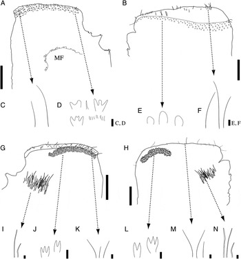
Trypetesa lateralis Tomlinson, Reference Tomlinson1953, internal and external mantle features (A–N, voucher specimen (USNM 1184481)): (A) left opercular bar, lateral outer view (MF, mantle flap); (B) right opercular bar, lateral outer view; (C) setae on top of left opercular bar, lateral outer view; (D) multifid denticules and setae on outer left opercular bar, lateral view; (E) blunt, ovate denticules on outer right opercular bar, lateral view; (F) setae on top of right opercular bar, lateral outer view; (G) left opercular bar and inner mantle, lateral inner view; (H) right opercular bar and inner mantle, lateral view; (I) pattern of inner mantle setae; (J) multifid denticules in an inner, curved ridge; (K) setae on top of left opercular bar; (L) multifid denticules on inner, curved ridge; (M) setae on top of left opercular bar; (N) pattern of inner mantle setae. Scale bars: A, B, G, H, 250 µm; C–F, I–N, 5 µm.

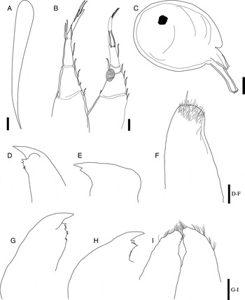
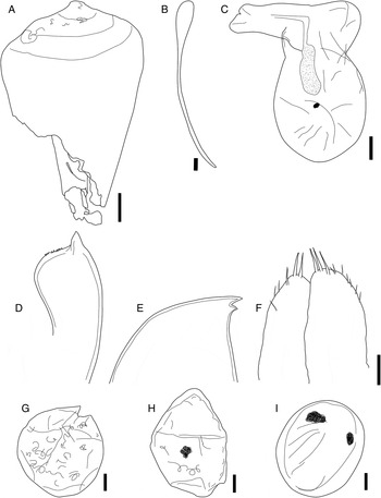
Trypetesa lateralis Tomlinson, Reference Tomlinson1953, aperture, terminal cirri, male, and mouthparts (A, voucher specimen (CASIZ 190072), B, D, E, G–I, voucher specimen (personal collection), C, voucher specimen (USNM 1184482), F, voucher specimen (USNM 1184486)): (A) aperture on columella of the gastropod shell Leucozonia nassa leucozonalis (Lamarck, 1822); (B) terminal cirri, lateral view; (C) male, lateral view; (D) mandible (Jamaica), lateral view; (E) maxilla I (Jamaica), lateral view; (F) maxilla II (Jamaica), lateral view; (G) mandible (New York), lateral view; (H) maxilla I (New York), lateral view; (I) maxilla II (New York), lateral view. Scale bars: A, 250 µm; B–I, 25 µm.

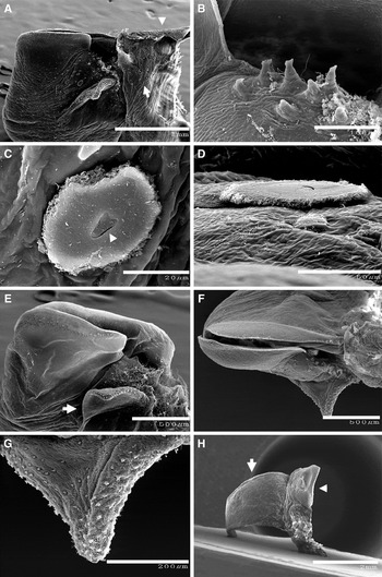
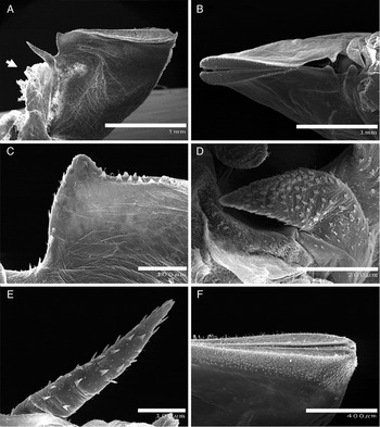
Trypetesa lateralis Tomlinson, Reference Tomlinson1953, external mantle features and opercular bars (A–D, F, G, voucher specimen (personal collection), E, H, voucher specimen (USNM 1184490)) scanning electron micrographs: (A) external mantle, left side, lateral view (arrowhead indicates attachment area, arrow indicates depressions surrounding denticules); (B) fleshy, digitiform extensions; (C) denticule surrounded by disc (arrowhead shows denticule), apical view; (D) denticule surrounded by disc, lateral view; (E) left side of mantle, lateral external view (arrow indicates mantle flap); (F) mantle aperture, apical view; (G) external mantle flap, apical view; (H) external mantle with flap, apical view (arrow indicates mantle flap, arrowhead indicates opercular bars). Scale bars: A, 1 mm; B, 100 µm; C, 20 µm; D, 50 µm; E, F, 500 µm; G, 200 µm; H, 2 mm.

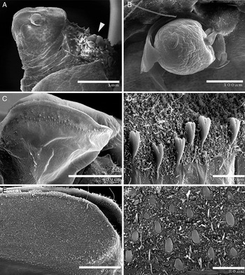
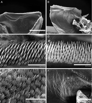
Trypetesa lateralis Tomlinson, Reference Tomlinson1953, external opercular bar features and males (A, B, voucher specimen (USNM 1184486), C, D, voucher specimen (USNM 1184490), E, F, voucher specimen (personal collection)), scanning electron micrographs: (A) female with attached males (shown by arrowhead), left lateral view; (B) single male; (C) left opercular bar, lateral external view; (D) multifid denticules and setae on outside of left opercular bar, lateral view; (E) right opercular bar, lateral view; (F) ovate denticules and setae on outside of right opercular bar, apical view. Scale bars: A, 1 mm; B, 100 µm; C, 400 µm; D, F, 20 µm; E, 200 µm.

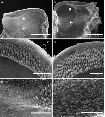
Trypetesa lateralis Tomlinson, Reference Tomlinson1953, internal mantle features (A–E, voucher specimen (USNM 1184494), F, voucher specimen (USNM 1184487)), scanning electron micrographs: (A) left side of mantle cavity, lateral internal view (arrow indicates denticule ridge, arrowhead indicates inner mantle setae); (B) right side of mantle cavity, lateral internal view (arrow indicates denticule ridge, arrowhead indicates inner mantle setae); (C) multifid denticules on inside of left opercular area, oblique view; (D) multifid denticules on inside of right opercular area, apical view; (E) internal mantle scales and denticules, apical view; (F) scales on internal mantle surface, apical view. Scale bars: A, 400 µm; B, 500 µm; C, D, 50 µm; E, 40 µm; F, 20 µm.

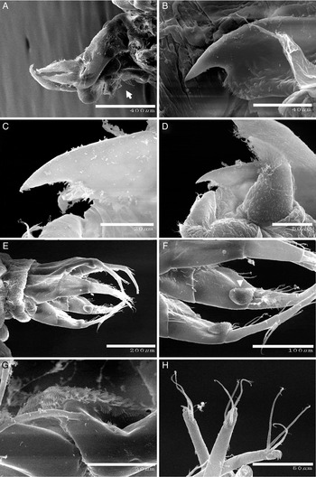
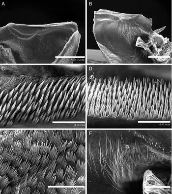
Trypetesa lateralis Tomlinson, Reference Tomlinson1953, mouth cirri, mouthparts, and terminal cirri (A–F, voucher specimen (personal collection), G, H, (USNM 1184487)), scanning electron micrographs: (A) mouth cirri and labrum, lateral view (arrow indicates labrum); (B) mandible, lateral view; (C) maxilla I, lateral view; (D) maxilla II, lateral view (outermost mouthpart shown); (E) terminal cirri, lateral view; (F) setae and cirral cushions (shown by arrowhead), lateral view; (G) cushion showing denticules, lateral view; (H) distal ends of terminal cirri with minute hooks and setae, lateral view. Scale bars: A, 400 µm; B, 40 µm; C, G, 20 µm; D, H, 50 µm; E, 200 µm; F, 100 µm.

Trypetesa lateralis Tomlinson, Reference Tomlinson1953: 373–380, figures 1–7, table 1; Tomlinson, Reference Tomlinson1955: 97–113, pls. I–IV; Utinomi, Reference Utinomi1964: 118, 125, 128; Seilacher, Reference Seilacher1969: 709, 715, figures 1, 6, pl. IV; Tomlinson, Reference Tomlinson1969: 7, 11, 14–15, 17, 19–20, 22, 24–26, 28, 126, 131–132, 147, 150, 153, figure 36, table 2; White, Reference White1969: 338; White, Reference White1970: 29, 33, figure 2; Turquier & Carton Reference Turquier and Carton1976: 392; Newman & Abbott, Reference Newman, Abbott, Morris, Abbott and Haderlie1980: 505–506, 509, 510, pl. 147; Grygier & Newman, Reference Grygier and Newman1985: 2, 18–19; Klepal, Reference Klepal1987: 251, 259–260, 294, 297; Barnes, Reference Barnes1989: 73–76, 78, table IV; Klepal, Reference Klepal1990: 362; Baluk & Radwánski, Reference Baluk and Radwánski1991: 1, 13, 22, 24, 30, 31, 32, 33, figure 7; Walker, Reference Walker1992: 545, table 5 (in part); Anderson, Reference Anderson1994: 162, 178; Kozloff, Reference Kozloff1996: 319; Williams & McDermott, Reference Williams and McDermott2004: 73–75, 90, table 1; Williams & Boyko, Reference Williams and Boyko2006: 294; Newman, Reference Newman and Carlton2007: 476, 481; Kolbasov & Høeg, Reference Kolbasov and Høeg2007: 129–130, figures 3, 5, 11; Kolbasov, Reference Kolbasov2009: 16, 39, 41, 43, 46, 49, 57, 69, 81, 89, 97, 144, 147, 163, 168, 177, 197, 260, 338, 339, 394, 395, figures 8–10, 12, 15, 20, 23, 27, 29, 41, 49, 53, 60, 118, 150; Murphy, Reference Murphy2009: 5, 6, 8–10, 15, 19, 21, 24–26, 33, 34, 40–47, 55, 56, 58–60, 62–64, figures 1–9; Williams et al., Reference Williams, Gallardo and Murphy2011: 14. not Walker, Reference Walker1992; table 5 (T. lateralis from Puerto Rico is an error; it should be listed as T. lampas, as originally recorded by Seilacher, Reference Seilacher1969).
MATERIAL EXAMINED
Jamaica
St Anne's Bay (18°27′N 77°13′25″W): Back Reef, from Cantharus tinctus Conrad, 1846 inhabited by male Calcinus tibicen Herbst, 1791 (6.15 mm SL), 14 May 2005 (USNM 1184476).—Christopher Cove, from Bursa sp. inhabited by female C. tibicen (4.70 mm SL), 15 May 2005 (USNM 1184477); from Bursa sp. inhabited by unidentified ovigerous female hermit crab (3.40 mm SL), 15 May 2005; from Leucozonia nassa leucozonalis (Lamarck, 1822) inhabited by unidentified female hermit crab, 15 May 2005 (USNM 1184478); from Cerithium sp. inhabited by ovigerous female C. tibicen (3.20 mm SL), 15 May 2005 (USNM 1184479).—Back Reef, from Tegula fasciata Born, 1778 inhabited by female C. tibicen (3.25 mm SL), 23 November 2005 (USNM 1184480); from Columbella mercatoria Linnaeus, 1758 inhabited by female C. tibicen, 23 November 2005 (USNM 1184481); from Cymatium nicobaricum Röding, 1798 inhabited by male C. tibicen (4.20 mm SL, three females), 23 November 2005 (USNM 1184482).—Drax Hall, from L. n. leucozonalis inhabited by ovigerous female C. tibicen (4.80 mm SL), 11 August 2006 (USNM 1184483); from L. n. leucozonalis inhabited by male C. tibicen (4.70 mm SL, three females, two males), 11 August 2006 (USNM 1184484); from L. n. leucozonalis inhabited by ovigerous female C. tibicen (4.15 mm SL, two females), 11 August 2006 (USNM 1184485); from L. n. leucozonalis inhabited by female C. tibicen (4.25 mm SL, two females), 11 August 2006 (USNM 1184486, on 2 SEM stubs); from L. n. leucozonalis inhabited by female C. tibicen (3.80 mm SL, two females), 11 August 2006 (USNM 1184487, on 4 SEM stubs); from L. n. leucozonalis inhabited by female C. tibicen (3.60 mm SL), 11 August 2006 (USNM 1184488); from T. fasciata inhabited by female C. tibicen (4.10 mm SL), 11 August 2006 (USNM 1184489); from T. fasciata inhabited by ovigerous female C. tibicen (4.20 mm SL, two females), 11 August 2006 (USNM 1184490, on 3 SEM stubs); from L. n. leucozonalis inhabited by ovigerous female C. tibicen (3.95 mm SL), 11 August 2006 (USNM 1184491).—Urchin Cove, from L. n. leucozonalis inhabited by male C. tibicen (3.60 mm SL, two females), 12 August 2006 (CASIZ 190070); from Engina sp. inhabited by female C. tibicen (4.10 mm SL, two females), 12 August 2006 (CASIZ 190071); from L. n. leucozonalis inhabited by ovigerous female C. tibicen (3.40 mm SL, one male), 12 August 2006 (CASIZ 190072); from L. n. leucozonalis inhabited by ovigerous female C. tibicen (4.50 mm SL), 12 August 2006 (CASIZ 190073); from L. n. leucozonalis inhabited by female C. tibicen (3.85 mm SL, two females), 12 August 2006 (CASIZ 190074); from Cerithium sp. inhabited by ovigerous female C. tibicen (3.10 mm SL), 12 August 2006 (CASIZ 190075).—Tide Pool Island, HUML, from unidentified shell inhabited by female C. tibicen (4.40 mm SL), 13 August 2006 (USNM 1184492, on 1 SEM stub).
New York
Oak Beach (40°38′20″N 73°17′10″W): from Ilyanassa trivittata (Say, 1822) inhabited by female Pagurus longicarpus (Say, 1817) (2.50 mm SL), 3 May 2005 (USNM 1184493); from Nassarius obsoletus (Say, 1822) inhabited by female P. longicarpus (3.0 mm SL), 3 May 2005 (USNM 1184494, on 2 SEM stubs); from I. trivittata inhabited by male P. longicarpus (2.90 mm SL), 3 May 2005 (USNM 1184495); from I. trivittata inhabited by female P. longicarpus (2.50 mm SL), 14 June 2005 (USNM 1184496); from I. trivittata inhabited by male P. longicarpus (3.80 mm SL), 14 June 2005 (USNM 1184497, on 2 SEM stubs); from I. trivittata inhabited by male P. longicarpus (3.30 mm SL), 14 June 2005 (USNM 1184498); from I. trivittata inhabited by female P. longicarpus (2.60 mm SL), 14 June 2005; from I. trivittata inhabited by ovigerous female P. longicarpus (2.90 mm SL), 14 June 2005; from I. trivittata inhabited by female P. longicarpus (2.30 mm SL), 1 August 2005; from I. trivittata inhabited by male P. longicarpus (2.70 mm SL), 1 August 2005 (USNM 1184499); from I. trivittata inhabited by male P. longicarpus (3.0 mm SL), 1 August 2005 (USNM 1184500); from I. trivittata inhabited by male P. longicarpus (2.50 mm SL), 1 August 2005; from I. trivittata inhabited by male P. longicarpus (2.60 mm SL), 1 August 2005 (USNM 1184501); from I. trivittata inhabited by male P. longicarpus (3.30 mm SL), 1 August 2005 (USNM 1184502, on 3 SEM stubs); from Urosalpinx cinerea (Say, 1822) inhabited by male P. longicarpus (3.75 mm SL), 1 August 2005; from I. trivittata inhabited by female P. longicarpus (3.10 mm SL; one male), 21 June 2007; from I. trivittata inhabited by female P. longicarpus (2.65 mm SL), 21 June 2007; from I. trivittata inhabited by male P. longicarpus (3.10 mm SL), 21 June 2007; from I. trivittata inhabited by female P. longicarpus (3.40 mm SL), 21 June 2007; from I. trivittata inhabited by male P. longicarpus (2.90 mm SL), 21 June 2007; from I. trivittata inhabited by male P. longicarpus (3.70 mm SL), 5 November 2007 (CASIZ 190076); from Lunatia heros (Say, 1822) inhabited by female P. longicarpus (4.15 mm SL), 5 November 2007 (CASIZ 190077); from I. trivittata inhabited by male P. longicarpus (2.85 mm SL), 5 November 2007 (CASIZ 190078); from I. trivittata inhabited by male P. longicarpus (3.40 mm SL), 5 November 2007; from I. trivittata inhabited by male P. longicarpus (3.30 mm SL), 5 November 2007; from I. trivittata inhabited by male P. longicarpus (3.0 mm SL), 30 May 2008; from I. trivittata inhabited by male P. longicarpus (2.80 mm SL), 30 May 2008 (CASIZ 190080); from I. trivittata inhabited by male P. longicarpus (3.20 mm SL), 1 July 2008; from I. trivittata inhabited by female P. longicarpus (3.10 mm SL), 15 July 2008; from I. trivittata inhabited by female P. longicarpus (3.40 mm SL), 15 July 2008; from I. trivittata inhabited by male P. longicarpus (4.35 mm SL), 15 July 2008; from I. trivittata inhabited by female P. longicarpus (3.15 mm SL), 15 July 2008; from I. trivittata inhabited by female P. longicarpus (3.0 mm SL), 15 July 2008 (CASIZ 190085).—West End Coast Guard Station, Jones Beach (40°35′27″N 73°33′5″W), from N. obsoletus inhabited by male P. longicarpus (3.60 mm SL), 16 June 2005 (CASIZ 190086); from I. trivittata inhabited by male P. longicarpus (3.2 mm SL), 21 June 2005 (CASIZ 190087); from I. trivittata inhabited by male P. longicarpus (3.90 mm SL), 21 June 2005 (CASIZ 190088); from N. obsoletus inhabited by male P. longicarpus (2.90 mm SL), 21 June 2005 (CASIZ 190089); from U. cinerea inhabited by male P. longicarpus (3.50 mm SL), 5 September 2005.
ADDITIONAL MATERIAL EXAMINED
California, Ford Point, Santa Rosa Island: from Olivella biplicata (Sowerby) inhabited by unidentified hermit crab, 3 February 1954 (USNM 122629).
DESCRIPTION
Female: largest complete specimen up to 4.52 mm in length, 5.11 mm in width (Jamaica), largest specimen from New York 3.58 mm in length, 2.69 mm in width. Body laterally compressed due to lateral orientation in respect to the aperture. Attachment disc, flattened area of mantle below rostral end of opercular bars (Figure 2), attaching upper part of animal to wall of burrow along columella of gastropod shell (Figure 4A). Aperture on columella of shell approximately straight, slit-like opening 2.85 mm in length and 0.39 mm in width at widest point on carinal end, tapering to rostral end, oriented toward apex of shell (Figure 3A).
Mantle muscular, oriented perpendicular to columella of shell, body rounded at distal end, with rounded lobes in larger individuals, burrow position in gastropod shell shown by striae in thin layer of shell overlaying barnacle. Orifice of mantle with small, rounded opening on carinal end of opercular bars, extending as narrow slit to rostral end.
Asymmetrical opercular bars (mantle lips). Left opercular bar approximately 3/4 length of right opercular bar, left opercular bar with notch on rostral end, short conical extensions below notch in left opercular bar (Figures 2A, B & 4A, B, E–H), conical extensions with simple rounded tip or bifid; outer surface of left opercular bar with one or two horizontal rows of blunt uni- or bifid denticules, simple setae of varying lengths above (Figures 2A & 5C, D). Right opercular bar thick with flattened distal margin and more setose; outer surface of right opercular bar with multiple horizontal rows of blunt, ovate denticules, surrounded by dense setae (Figures 2B & 5E, F). Left opercular bar inner surface (Figures 2G & 6A) with dense rows of mostly bi- and trifid denticules on ridge, ridge approximately 3/4 length of opercular bar, denticules replaced by clusters of approximately 40 acute spines in scale-like pattern immediately above denticule ridge (Figure 6C, F), patch of long, plumose setae inside mantle cavity, posterior to opercular bars and denticule rows (Figures 2G, H & 6A). Inner mantle with denticules randomly distributed, denticules change orientation, tapering towards aperture (Figure 6E). Outer mantle with denticules sparsely distributed, most abundant around mantle flap and at rostral end (Figure 4A), some with circular depressions around base (Figure 4A, arrow), some with flat discs (presumably calcium carbonate breakdown product from shell during growth, see ‘Remarks') surrounding denticules (Figure 4C, D). External mantle flap, triangular, from approximately 1/4 to two times length of opercular bars, extending from left side of mantle, with simple to trifid denticules and short setae on outer edge (Figures 2A & 4E–H).
Labrum edge with long setae along whole length (Figure 7A). Mandible broad and recurved with large spine above rounded plate-like projection with accessory spine and scales tapering toward mouth opening (Figures 3D, G & 7B). Maxilla I recurved with two large spines, upper spine about three times length of smaller, lower spine; lower spine with multiple, smaller tapered spines (Figures 3E, H & 7C). Maxilla II distally setose, with scales along inner edge where pairs meet, lacking accessory spines (Figures 3F, I & 7D).
Four pairs of cirri. Cirrus one (mouth cirri) biramous; protopod elongate, naked; endopod and exopod approximately half as long as protopod; simple exopod and endopod, both very setose, exopod slightly shorter than endopod (Figure 7A). Three pairs of uniramous terminal cirri with four segments each (Figures 3B & 7E); first segment elongate, flattened, with sparse row of setae along inner surface; second segment approximately half as long as first, tapered distally with protuberant cushion on distal inner surface (Figures 3B & 7F), cushion with approximately 20 rows of small, tapered blade-like denticules (Figure 7G); third segment half as wide as second segment, approximately as long as first segment, with long setae along inner surface; fourth segment narrower than third, tapering distally, terminating in pair of bifid hooks; two or three long setae positioned subterminally (Figure 7H).
Male (based on Jamaican specimens): up to 0.14 mm in length, 0.12 mm in width. One to multiple (N = 4) males attached near flap or near attachment disc (Figures 3C & 5A, B). Rounded, sac-like body with two antennules (Figure 5A, B). Irregular eyespot; other internal features not visible.
REMARKS
This is the first report of Trypetesa lateralis from the east coast of the United States and the Caribbean; the species was previously only known from California (Figure 1; Table 1). Females of T. lateralis have two diagnostic characters that are used to help distinguish them from other members of the genus: an external mantle flap and asymmetrical opercular bars. The mantle flap is present on the left side of the mantle and varies in length depending on maturity and shell thickness, similar to that found by Kolbasov (Reference Kolbasov2009, figure 8). Mantle flaps were found of varying lengths on specimens from New York and Jamaica, regardless of the presence of males (Figure 4F, H). In addition, males of T. lateralis lack a penis (Tomlinson, Reference Tomlinson1969) as found in the present study (Figure 3C).
Tomlinson (Reference Tomlinson1955) stated that the opercular bars of T. lateralis are asymmetrical, with the left opercular bar (the side of the mantle with the flap) being 20% longer than the right bar. However, re-examination of Tomlinson's specimens of T. lateralis from California (USNM 122629) and newly collected specimens showed the left opercular bar was shorter, indicating that Tomlinson (Reference Tomlinson1955) made a typographical error; the left opercular bar is at least 20% shorter than the right bar (Figure 4E, F). Kolbasov (Reference Kolbasov2009) showed the shorter left apertural lip with SEM (see his figures 8, 9 & 118). Tomlinson (Reference Tomlinson1955) also noted that T. lateralis lacked a labrum but current literature refers to the area denoted by Tomlinson (Reference Tomlinson1955) as the ‘head' as the labrum. The labrum is the dorsal region located posterior to the mouth cirri and has a characteristic linear row of long, simple setae that runs along the edge. The description of the mouth cirri by Tomlinson (Reference Tomlinson1955) agrees with our observations of the mouth cirri of specimens from Jamaica and New York.
Although mentioned in the original description as a minute hook, the distal ends of the terminal cirri terminate in bifid hooks (Figure 7H), as found in T. spinulosa (see below) and Tomlinsonia mclaughlinae (Williams & Boyko, Reference Williams and Boyko2006). The exact function of these hooks remains unknown; however, they may serve in feeding. The original description of the cushion states it is ‘wrinkled with fine, rounded ridges' (Tomlinson, Reference Tomlinson1955); when viewed using SEM, the cushion is actually composed of rows of sharp, blade-like denticules, possibly used to aid in food handling. These denticules are similar to those found in T. lampas (Williams et al., Reference Williams, Gallardo and Murphy2011) and the genus Tomlinsonia (Williams & Boyko, Reference Williams and Boyko2006).
Distinct discs were found surrounding denticules on the outer mantle and are herein interpreted as material resulting from the burrowing process, composed of splinters of calcium carbonate and an unidentified organic substance (as found by Kamens (Reference Kamens1981) in T. lampas). The discs are only found surrounding outer mantle denticules, and even denticules without discs are surrounded by circular indentations, indicating that a disc was previously present (Figure 4A). The SEM pictures show the splintered, fibrous nature of the disc (Figure 4C, D), providing further evidence that the discs are composed of shell fragments.
The only trypetesid previously documented along the east coast of the United States and Gulf of Mexico was T. lampas and, prior to this study, there were no reports of living specimens of Trypetesa from Jamaica or anywhere in the Caribbean. However, Seilacher (Reference Seilacher1969) showed recent borings made by T. lampas in ‘pagurized’ gastropod shells from Puerto Rico (not T. lateralis as cited by Walker (Reference Walker1992; table 5)). The present finding of T. lateralis on the east coast of the United States and in the Caribbean significantly extends the distribution of the species. Grygier & Newman (Reference Grygier and Newman1985) discussed an undescribed species from North Carolina reportedly discovered by J.D. Standing & J.T. Tomlinson; unfortunately, no description or work on this species has been published. Although T. lateralis and T. lampas are easily distinguished based on the external mantle flap and asymmetrical opercular bars, previous reports of T. lampas from the east coast of the United States should be re-evaluated because these morphological features can be overlooked. Finding T. lateralis on the east coast of the United States and Jamaica raises questions about the possible introduction of the species and its possible status as part of a sibling species complex (see ‘Discussion').
ECOLOGY
The prevalence of Trypetesa lateralis was 8.3% in the samples from Jamaica (25 of 301 hermit crabs). Trypetesa lateralis was associated with the most common species of hermit crab, Calcinus tibicen, from intertidal areas sampled in Jamaica. Of the 23 specimens of T. lateralis examined, 20 were found associated with female hermit crabs (80%); nine of these female hosts were ovigerous (45%) (data not recorded for two damaged host specimens). Although the sex-ratio of hermit crabs from Jamaica did not significantly differ from 50:50 (goodness of fit test χ2 (N = 291) = 0.168, P = 0.682, df = 1), the distribution of the barnacles among the hermit crabs was significantly different from random predicted values (test of independence χ2 (N = 25) = 12.565, P < 0.001, df = 1). Trypetesa lateralis was found most commonly in Leucozonia nassa leucozonalis (48%) but also in seven other species of gastropod shells: Bursa sp., Cantharus tinctus, Cerithium sp., Columbella mercatoria, Cymatium nicobaricum, Engina sp. and Tegula fasciata. Some shells were found to harbour more than one female T. lateralis, three being the maximum number of trypetesids found to occupy a single shell. The average intensity of trypetesids in Jamaica was 1.52 ± 0.07 (N = 25) trypetesids per shell.
The prevalence of T. lateralis in the samples from the east coast of the United States was 1.6% (41 of 2614 hermit crabs). Trypetesa lateralis was only sampled from the longwrist hermit crab, Pagurus longicarpus, from the intertidal area in Oak Beach, New York. Of the 39 specimens of T. lateralis examined, 14 were found associated with female hermit crabs (36%); only one of these females were ovigerous (7%) (data not recorded for three shells where hosts were absent); the rest (64%) were associated with male hermit crabs. The sex-ratio of hermit crabs from New York was biased significantly toward males (80%) (goodness of fit test χ2 (N = 839) = 294.409, P < 0.001, df = 1) and the distribution of the barnacles among the hermit crabs was not significantly different than predicted values (test of independence χ2 (N = 41) = 3.103, P = 0.078, df = 1). Trypetesa lateralis was found in four species of gastropod shells from New York: Lunatia heros, Nassarius obsoletus, Urosalpinx cinerea and most commonly Ilyanassa trivittata (78%). Shells were found to harbour more than one female T. lateralis, three being the most trypetesids found to occupy a single shell. In New York, the average intensity was 1.06 ± 0.23 (N = 41) trypetesids per shell.
FEEDING BIOLOGY AND EGG PREDATION
Mouth and terminal cirri of T. lateralis from New York were observed to pump rhythmically and constantly, at a rate of approximately 40 to 50 beats per minute, with a short pause between each repetition. Both sets of cirri (mouth and terminal) pump at the same time, and a beat consists of the two sets of cirri coming closer together and down into the mantle with each contraction, and then further apart and up near the aperture lips on the expansion. Occasionally, the barnacle appears to ‘stretch' (as termed by Tomlinson, Reference Tomlinson1955), which consists of a full expansion and non-rhythmic movement of the cirri, possibly to clean off the cirri and/or aperture, or dislodge food particles (Tomlinson, Reference Tomlinson1955). Our observations support the conclusions of Tomlinson (Reference Tomlinson1955) that the function of the pumping action is two-fold: to produce water currents that bring in fresh oxygenated water as well as a nutrient supply. In addition to the pumping action of the cirri, mantle contractions could create water currents.
The overall shape of the mouthparts of T. lateralis described by Tomlinson (Reference Tomlinson1955; see Figure 8) were consistent with our findings; however, SEM allowed more detailed analysis of these mouthparts of T. lateralis. The mandible has a pronounced, accessory spine on top and smaller, multifid scales and spines on the rounded, ventral area (Figures 3D, G & 7B). Maxilla I, like the mandible, has a pronounced spine at the top and a secondary spine (Figures 3E, H & 7C). Maxilla II is more rounded in shape and very setose at the distal tip, having no spines and a few multifid scales on the inner edges (Figures 3F, I & 7D). Kolbasov (Reference Kolbasov2009) provided SEM images of T. lateralis (based on specimens from California; Kolbasov, personal communication) showing a putative mandibular palp (Figure 20; Kolbasov, Reference Kolbasov2009) and asymmetrical mouthparts (both pairs of maxilla II on one side). No mandibular palp was noted in the present study and all mouthparts examined were symmetrical. The specimen examined by Kolbasov (Reference Kolbasov2009) may have been abnormal or the putative mandibular palp is not interpreted correctly.
Tomlinson (Reference Tomlinson1969) never observed cirri of trypetesids extending beyond the aperture; because of the reduced cirri in Trypetesa species it was assumed that food was captured through rhythmic pumping of the mantle, creating water current that brings in nutrients for the barnacle. In the present study, we observed the terminal cirri to occasionally protrude through the opercular bars of the mantle (upon muscle contraction) to approximately the second articulation. Although not a frequent occurrence, this action was observed numerous times, usually following a mantle stretch. Mouth cirri were never observed extending beyond the aperture.
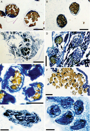
Of the specimens of T. lateralis from Jamaica associated with ovigerous host hermit crabs, 78% (seven of nine) had distended guts which were yellow/pale orange, the same coloration of preserved host eggs. Egg predation by T. lateralis from Jamaica was confirmed by histology of hermit crab host eggs, and comparison with barnacle gut content. When stained with Mallory I and II, yolk is represented by a yellow/orange colour, while the deeply blue stained areas are connective tissue (Figure 14C, D). Eggs of the hermit crab host Calcinus tibicen from Jamaica were ovoid and contained yellow staining yolk droplets; the eggs averaged 0.32 ± 0.03 mm in length (N = 19) and 0.28 ± 0.03 mm in width (N = 19) (Figure 14A). Specimens of T. lateralis from Jamaica contained yellow staining material in the gut (Figure 14C), and some ovoid structures with distinct edges were observed and averaged 0.28 ± 0.01 mm long (N = 4) and 0.25 ± 0.01 mm wide (N = 4) (Figure 14C), corresponding to the size of host eggs. In addition, large areas of yellow staining material had an irregular pattern (possibly representing host eggs in a more advanced stage of digestion). In the Jamaica collection, 36% of trypetesids (nine of 25) were found inhabiting shells with ovigerous hermit crabs.
Eggs of the hermit crab host Pagurus longicarpus from New York were ovoid and also contained yellow staining yolk droplets; the eggs averaged 0.25 ± 0.05 mm in length (N = 9) and 0.21 ± 0.04 mm in width (N = 9). Trypetesa lateralis from New York produced ovoid eggs that stained yellow and were 0.15 ± 0.01 mm in length (N = 11) and 0.12 ± 0.02 mm in width (N = 11) (Figure 14E). In New York, only 2% of trypetesids (one of 41) were found inhabiting shells with ovigerous hermit crabs, with no evidence of egg predation (Figure 14G).
Eggs of T. lateralis from Jamaica were 0.20 ± 0.01 mm in length (N = 20) and 0.23 ± 0.02 mm in width (N = 20). Cyprid larvae of T. lateralis were found inside the brood pouch of some specimens from New York (Figure 14H).
Trypetesa spinulosa Turquier, Reference Turquier1976, external mantle features (A, C, E, voucher specimen (USNM 1184475), B, D, F, voucher specimen (USNM 1184474)), scanning electron micrographs: (A) external mantle, right side, lateral view (arrow indicates attachment area); (B) mantle aperture, apical view; (C) small, blunt projection, external lateral view; (D) digitiform extension (orificial palp), external apical view; (E) digitiform extension, internal lateral view; (F) opercular bars, external lateral view. Scale bars: A, B, 1 mm; C, E, 100 µm; D, 200 µm; F, 400 µm.

Trypetesa spinulosa Turquier, Reference Turquier1976, aperture, male, and mouthparts, including egg and egg corions of host Dardanus lagopodes (A, B, G–I, voucher specimen (personal collection), C, F, voucher specimen (USNM 1184450), D, E, voucher specimen (USNM 1184471)): (A) shell of Conus sp. cut away to show barnacle burrow, view of columella with aperture; (B) aperture on columella of Conus sp. gastropod shell; (C) male, lateral view; (D) mandible, lateral view; (E) maxilla I, lateral view; (F) maxilla II, lateral view; (G) egg corion of D. lagopodes (Forskål, 1775) found in gut of T. spinulosa, lateral view; (H) egg of D. lagopodes found in gut of T. spinulosa, lateral view; (I) egg from pleopod of host hermit crab D. lagopodes, lateral view. Scale bars: A, 5.0 mm; B, 250 µm; C, 100 µm; D–F, 25 µm; G–I, 50 µm.

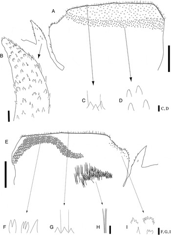
Trypetesa spinulosa Turquier, Reference Turquier1976, internal and external mantle features (A, C–I, voucher specimen (USNM 1184445), B, voucher specimen (USNM 1184450)): (A) right opercular bar, lateral outer view; (B) orificial palp, lateral outer view; (C) setae and scales lining outer right opercular bar; (D) blunt, ovate denticules on outer opercular bar; (E) right opercular bar and mantle, lateral inner view; (F) uni- to multifid denticules on inner ridge; (G) setae and scales lining outer right opercular bar; (H) inner mantle setae; (I) setae and scales from inner depression and low projection. Scale bars: A, E, H, 250 µm; B, 25 µm; C, D, F, G, I, 5 µm.

Trypetesa spinulosa Turquier, Reference Turquier1976, internal mantle features (A–F, voucher specimen (USNM 1184475)), scanning electron micrographs: (A) left side of mantle cavity, lateral internal view; (B) right side of mantle cavity, lateral internal view; (C) uni- to multifid denticules on inside of left opercular area, apical view; (D) uni- to multifid denticules on inside of right opercular area, apical view; (E) internal mantle scales, apical view; (F) internal setae and linear row of denticules, apical view. Scale bars: A, B, 500 µm; C, D, 40 µm; E, 20 µm; F, 200 µm.

Trypetesa spinulosa Turquier, Reference Turquier1976, mouth cirri, terminal cirri and mouthparts (A, D, E–H, voucher specimen (USNM 1184445), B, C, voucher specimen (USNM 1184450)), scanning electron micrographs: (A) mouth cirri and labrum, lateral view (arrow indicates labrum); (B) mandible, lateral view; (C) mandible showing rounded region below major spine, lateral view; (D) maxilla I (partially hidden) and maxilla II, lateral view; (E) terminal cirri, lateral view; (F) cirral cushion, apical view; (G) terminal cirri showing inner row of setae, lateral view; (H) distal ends of terminal cirri with bifid hooks and setae, lateral view. Scale bars: A, 1 mm; B, D, 50 µm; C, F, H, 20 µm; E, 200 µm; G, 100 µm.

Trypetesa spinulosa Turquier, Reference Turquier1976, internal mantle features (A–F, voucher specimen (USNM 1184475)), scanning electron micrographs: (A) left side of mantle cavity, lateral internal view; (B) right side of mantle cavity, lateral internal view; (C) uni- to multifid denticules on inside of left opercular area, apical view; (D) uni- to multifid denticules on inside of right opercular area, apical view; (E) internal mantle scales, apical view; (F) internal setae and linear row of denticules, apical view. Scale bars: A, B, 500 µm; C, D, 40 µm; E, 20 µm; F, 200 µm.

Histology of host eggs, barnacle guts, and barnacle eggs (A, C, voucher specimen (USNM 1184491), B, F, voucher specimen (USNM 190091), D, voucher specimen (USNM 1184449), E, G, H, voucher specimen (personal collection)), digital pictures from stained slides: (A) eggs of Calcinus tibicen Herbst, 1791 (Jamaica); (B) eggs of Calcinus gaimardii (H. Milne Edwards, 1848) (Philippines); (C) gut of Trypetesa lateralis Tomlinson, Reference Tomlinson1953 (Jamaica), with arrow showing a host egg; (D) gut of T. spinulosa Turquier, Reference Turquier1976 (Philippines), with arrow showing a host egg; (E) eggs of T. lateralis (New York); (F) eggs of T. spinulosa (Philippines); (G) gut of T. lateralis (New York) (negative for yolk staining); (H) cyprid larvae of T. lateralis (New York), inside brood pouch of mantle. Scale bars: A–E, G, 250 µm; F, 200 µm; H, 100 µm.

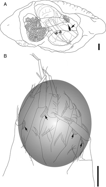
Egg predation by Trypetesa spinulosa Turquier, Reference Turquier1976: (A) semi-diagrammatic view of T. spinulosa in shell of Drupella cornus (Röding, 1798) with ovigerous female hermit crab Calcinus gaimardii (H. Milne Edwards, 1848). The semi-transparent host and eggs are positioned in a shell that has been cut away to show the position of the aperture (indicated by arrow) of T. spinulosa on the columella. Note that the host hermit crab is withdrawn in the shell; during movement by the crab the eggs would be positioned over the aperture of the barnacle, when egg predation is suspected to occur; (B) terminal cirri of T. spinulosa, showing size relative to a semi-transparent spheroid the size of host embryos. The cirral cushions (arrows) would be able to contact the circumference of the embryo when the 3 pairs of cirri are expanded. Scale bars: A, B, 1 mm.

Trypetesa spinulosa Turquier, Reference Turquier1976: 559–571, figures 1–9; Klepal, Reference Klepal1987: 258–259, 295, 297, 557; Klepal, Reference Klepal1990: 363, figure 4; Baluk & Radwánski Reference Baluk and Radwánski1991: 23, 24, 31, figure 8; Kolbasov, Reference Kolbasov, Schram and von Vaupel Klein1999: 142; Anderson, Reference Anderson1994: 162; Williams & McDermott, Reference Williams and McDermott2004: table 1; Williams & Boyko, Reference Williams and Boyko2006: 294, 296; Kolbasov, Reference Kolbasov2009: 18, 260, 338, 343, 394, figures 120, 150; Murphy, Reference Murphy2009: 5, 9, 19, 24, 47, 54–58, 61, 62, 66, figures 10–16; Williams et al., Reference Williams, Gallardo and Murphy2011: 14.
MATERIAL EXAMINED
Philippines
Bataan, Morong (14°39′53″N 120°16′28″E): from Latirus polygonus (Gmelin, 1791) inhabited by female Calcinus gaimardii (H. Milne Edwards, 1848) (4.65 mm SL), 25 April 1999 (USNM 1184437); from Cantharus undosus (Linnaeus, 1758) inhabited by female C. gaimardii (4.65 mm SL), 25 April 1999 (USNM 1184438); from C. undosus inhabited by female C. gaimardii (5.20 mm SL), 25 April 1999 (USNM 1184439); from C. undosus inhabited by female C. gaimardii (4.90 mm SL), 25 April 1999 (USNM 1184440); from Cypraea sp. inhabited by female C. gaimardii (3.60 mm SL), 25 April 1999 (USNM 1184441); from C. undosus inhabited by ovigerous female C. gaimardii (4.40 mm SL), 25 April 1999 (USNM 1184442); from Drupa ricinus ricinus (Linnaeus, 1758) inhabited by female C. gaimardii (3.75 mm SL), 25 April 1999 (USNM 1184443); from C. undosus inhabited by female C. gaimardii (5.0 mm SL), 25 April 1999 (USNM 1184444): from unidentified shell inhabited by female C. gaimardii (3.67 mm SL), 28 February 1999; from unidentified shell inhabited by female C. gaimardii (4.11 mm SL, one male), 28 February 1999 (USNM 1184445, on 3 SEM stubs).—Bataan, Mabayo (14°44′N 120°16′32″): from C. undosus inhabited by male C. gaimardii (5.55 mm SL), 21 February 1999 (USNM 1184446); from C. undosus inhabited by female C. gaimardii (4.15 mm SL), 21 February 1999 (USNM 1184447); from Latirolagena smaragdula (Linnaeus, 1758) inhabited by male C. gaimardii (6.45 mm SL), 21 February 1999 (USNM 1184448); from unidentified shell inhabited by ovigerous female C. gaimardii (5.30 mm SL), 21 February 1999 (USNM 1184449); from unidentified shell inhabited by male Calcinus minutus Buitendijk, 1937 (3.31 mm SL), 21 February 1999 (USNM 1184450, on 1 SEM stub); from unidentified shell inhabited by ovigerous female C. minutus (2.50 mm SL), 21 February 1999; from unidentified shell inhabited by ovigerous female C. minutus (3.10 mm SL), 21 February 1999; from unidentified shell inhabited by female C. gaimardii (2.98 mm SL, three males), 21 February 1999 (USNM 1184451); from unidentified shell inhabited by male C. gaimardii (6.29 mm SL), 21 February 1999 (USNM 1184452, on 2 SEM stubs); from L. smaragdula inhabited by female C. gaimardii (5.75 mm SL, two females), 28 February 1999 (USNM 1184453); from L. smaragdula inhabited by male C. gaimardii (4.20 mm SL), 28 February 1999 (USNM 1184454); from C. undosus inhabited by female C. gaimardii (4.35 mm SL), 28 February 1999 (USNM 1184455); from Gyrineum gyrinum (Linnaeus, 1758) inhabited by female C. gaimardii (4.80 mm SL), 28 February 1999 (USNM 1184456); from L. smaragdula inhabited by female C. gaimardii (6.60 mm SL, two females), 28 February 1999 (USNM 1184457); from C. undosus inhabited by female C. gaimardii (4.50 mm SL, two females), 28 February 1999 (USNM 1184458); from unidentified shell inhabited by ovigerous female C. minutus (3.0 mm SL), 28 February 1999 (USNM 1184459).—Batangas, Sombrero Island (13°41′51.4″N 120°49′47.5″E): from D. cornus inhabited by ovigerous female C. gaimardii (4.03 mm SL), 10 June 2000 (USNM 1184460).—Puerto Galera, Lalaguna Beach (13°31′32″N 120°58′8″E): from Drupella cornus (Röding, 1798) inhabited by ovigerous female C. gaimardii (3.50 mm SL), 19 July 1997 (USNM 1184461); from D. cornus inhabited by ovigerous female C. gaimardii (3.95 mm SL), 21 July 1997 (USNM 1184462); from D. cornus inhabited by ovigerous female C. minutus (3.50 mm SL), 21 July 1997 (USNM 1184463); from D. cornus inhabited by ovigerous female C. gaimardii, 31 July 1997; from D. cornus inhabited by ovigerous female C. gaimardii (two females), 31 July 1997 (USNM 1184464); from D. cornus inhabited by ovigerous female C. gaimardii, 31 July 1997 (USNM 1184465); from D. cornus inhabited by female C. gaimardii, 31 July 1997 (USNM 1184466); from D. cornus inhabited by female Calcinus latens (Randall, 1839), 31 July 1997 (USNM 1184467); from D. cornus inhabited by ovigerous female C. gaimardii, 31 July 1997 (USNM 1184468); from D. cornus inhabited by ovigerous female C. gaimardii, 31 Jul 1997 (USNM 1184469); from D. cornus inhabited by female C. gaimardii (two females), 31 July 1997; from D. cornus inhabited by ovigerous female C. gaimardii, 31 July 1997 (USNM 1184470); from unidentified, uninhabited gastropod shell, 31 July 1997; from unidentified gastropod shell inhabited by female C. latens, 31 July 1997 (USNM 1184471); from D. cornus inhabited by ovigerous female C. gaimardii, 31 July 1997; from Thais mancinella (Linnaeus, 1758) inhabited by male C. gaimardii, 31 July 1997; from Latirus turritus (Gmelin, 1791) inhabited by ovigerous female C. gaimardii, 31 July 1997; from Coralliophila neritoidea (Lamarck, 1816) inhabited by ovigerous female C. gaimardii (4.30 mm SL), 31 July 1997 (USNM 1184472); from unidentified shell inhabited by female C. gaimardii (3.39 mm SL), 27 March 1999 (CASIZ 190090); from unidentified shell inhabited by female C. gaimardii (5.60 mm SL), 27 March 1999 (USNM 1184473, on 1 SEM stub); from unidentified shell inhabited by ovigerous female C. gaimardii (3.31 mm SL), 27 March 1999 (USNM 1184474, on 1 SEM stub); from D. cornus inhabited by ovigerous female C. minutus (4.25 mm SL), 17 June 2000 (CASIZ 190091); from D. cornus inhabited by female C. gaimardii (2.45 mm SL), 17 June 2000 (CASIZ 190092).—Puerto Galera, Coco Beach (13°31′32″N 120°57′44″E): from Tectus fenestratus (Gmelin, 1791) inhabited by ovigerous female C. gaimardii, 12 January 1999 (CASIZ 190093); from D. r. ricinus inhabited by female C. gaimardii, 12 January 1999 (CASIZ 190094); from Morula granalata (Duclos, 1832) inhabited by ovigerous female C. latens (2.55 SL), 12 January 1999 (CASIZ 190095); from D. cornus inhabited by female C. minutus, 12 January 1999 (CASIZ 190096); from D. cornus inhabited by female C. gaimardii, 12 January 1999 (CASIZ 190097); from Drupella rugosa (Born, 1778) inhabited by ovigerous female C. minutus (unknown SL), 12 January 1999 (CASIZ 190098); from D. cornus inhabited by female C. gaimardii, 14 January 1999 (CASIZ 190099); from D. cornus inhabited by female C. minutus, 14 January 1999 (CASIZ 190100); from D. cornus inhabited by ovigerous female C. minutus, 14 January 1999 (CASIZ 190101); from D. cornus inhabited by female C. gaimardii, 14 January 1999 (CASIZ 190102); from D. cornus inhabited by male C. minutus, 14 January 1999 (CASIZ 190103); from Rhinoclavis fasciata (Bruguière, 1792) inhabited by female C. gaimardii, 14 January 1999 (CASIZ 190104).—Puerto Galera, Bayanan Beach (13°29′59″N 120°52′40″E): from Astralium sp. inhabited by female C. gaimardii, 13 January 1999 (CASIZ 190105).—Sulpa Island (10°14′15″N 124°0′29″E): from Astraea sp. inhabited by male Dardanus lagopodes (Forskål, 1775) (two females), 9 July 1997 (CASIZ 190106); from unidentified muricid shell inhabited by male Calcinus latens (Randall, 1839) (two females), 9 July 1997 (CASIZ 190107); from Astraea sp. inhabited by female C. gaimardii, 9 July 1997 (USNM 1184475, on 1 SEM stub); from Astraea sp. inhabited by male C. latens, 9 July 1997 (CASIZ 190108); from L. turritus inhabited by ovigerous female Calcinus pulcher Forest, 1958, 9 July 1997 (CASIZ 190109); from unidentified shell inhabited by female C. pulcher, 9 July 1997 (CASIZ 190110).—Boracay, Diniwid Beach (11°58′34″N 121°54′38″E): from unidentified shell inhabited by ovigerous female C. gaimardii (5.60 mm SL), 13 April 1999; from C. neritoidea inhabited by ovigerous female C. gaimardii (4.60 mm SL), 13 April 1999 (CASIZ 190111); from C. undosus inhabited by ovigerous female C. gaimardii (5.45 mm SL), 13 April 1999 (CASIZ 190112); from C. undosus inhabited by ovigerous female C. gaimardii (5.20 mm SL), 13 April 1999 (CASIZ 190113).—Boracay, Rocky Beach (11°57′42″N 121°55′25″E): from unidentified shell inhabited by female C. gaimardii (5.0 mm SL), 15 April 1999 (CASIZ 190114); from Strombus sp. inhabited by ovigerous female D. lagopodes (6.65 mm SL), 15 April 1999; from C. neritoidea inhabited by ovigerous female C. gaimardii (5.30 mm SL), 15 April 1999 (CASIZ 190115); from C. neritoidea inhabited by ovigerous female C. gaimardii (4.75 mm SL), 15 April 1999 (CASIZ 190116); from D. cornus inhabited by female C. gaimardii (3.30 mm SL), 15 April 1999 (CASIZ 190117); from D. cornus inhabited by ovigerous female C. gaimardii (4.15 mm SL), 15 April 1999 (CASIZ 190118); from D. cornus inhabited by female C. gaimardii (2.80 mm SL), 15 April 1999 (CASIZ 190119); from D. cornus inhabited by female C. latens (3.90 mm SL), 15 April 1999 (CASIZ 190120); from Peristernia nassatula Lamarck, 1822 inhabited by ovigerous female C. gaimardii (4.0 mm SL), 15 April 1999 (CASIZ 190121); from D. cornus inhabited by ovigerous female C. minutus (3.70 mm SL), 15 April 1999 (CASIZ 190122); from D. rugosa inhabited by ovigerous female C. latens (3.40 mm SL, two females), 15 April 1999; from C. undosus inhabited by ovigerous female C. gaimardii (4.60 mm SL), 12 April 1999 (CASIZ 190123); from D. rugosa inhabited by ovigerous female C. gaimardii (4.5 mm SL), 12 April 1999 (CASIZ 190124).
ADDITIONAL MATERIAL EXAMINED
Trypetesa habei Utinomi, Reference Utinomi1962: Japan: Honshu Island, Seto Marine Biological Laboratory, 27 September 1963 (USNM 122627).
DESCRIPTION
Female: largest complete specimen 6.22 mm, width 3.45 mm. Mantle laterally compressed, parallel to surface of gastropod shell. Attachment disc, flattened area of mantle below rostral end of opercular bars, attaching upper part of animal to wall of burrow along columella of gastropod shell (Figure 8A, arrow). Aperture on columella of shell approximately straight, slit-like opening 4.44 mm in length and 0.50 mm in width at widest point on carinal end, tapered toward rostral end, oriented toward apex of shell (Figure 9A, B).
Mantle muscular, oriented parallel to surface of host shell, extending towards apex of shell, body smoothly rounded at distal end toward apex, with one to multiple rounded lobes in larger individuals, burrow position shown by striae in thin layer of shell overlaying barnacle. Orifice of mantle with thin opening on carinal end of opercular bars, extending as narrow slit to rostral end (Figure 8B).
Symmetrical left and right opercular bars, top of opercular bars flattened and thick, rounded at carinal end, tapering to rostral end, opercular bars approximately 1 mm in length (Figure 8B). Rostral end of each opercular bar with shallow depression followed by low, rounded projection (Figures 8A–C & 10A, E), above large finger-like projection (orificial palp) covered with simple to multifid denticules, interspersed with minute setae (Figures 8A, B, D, E & 10B). Top surface of opercular bars very setose, with simple, ovate denticules, outer surface of left and right opercular bars with dense rows of simple denticules, row of long, simple setae on upper margin, sparser area toward rostral end (Figures 8F & 10A, C, D). Inner surfaces of left and right opercular bars with dense rows of simple to multifid sharply pointed denticules on ridge, ridge approximately 1/2 to 3/4 length of opercular bar (Figures 10E–I & 13A, B), denticules nearest mantle aperture mostly simple, becoming bi- and then multifid progressing downward into mantle cavity (Figure 13C, D). Denticules replaced by clusters of 10–20 acute spines in scale-like pattern (Figure 13E), beginning on rostral side (near shallow depression and low projection) sweeping down to denticule ridge, then up and over denticules, near mantle aperture (Figure 13C), acute spines orientated downward into mantle at rostral end, pointing toward aperture near denticule ridge, at ridge pointing towards carinal end. Patch of long, plumose setae inside mantle cavity (Figures 10H & 13F), linear row of simple denticules directly above setae.
Labrum large, edge with long setae along length (Figure 12A). Mandible strongly recurved with large upper spine (Figures 9D & 12B), smaller simple spines along edge of flattened area below large spine (Figure 12C). Maxilla I strongly recurved with two distal spines (Figures 9E & 12D). Maxilla II oblong, with few, large spines with dense patch of long setae on distal ends (Figures 9F & 12D).
Four pairs of cirri. Cirrus one (mouth cirri) biramous; protopod elongate, naked; endopod and exopod approximately half as long as protopod; simple exopod and endopod, very setose, exopod slightly shorter than endopod (Figure 12A). Three pairs of terminal cirri uniramous with four segments (Figure 12E); first segment elongate, tubular, with sparse row of long setae along inner surface, reaching second segment; second segment half as long as first, with protuberant cushion on distal inner surface, cushion with approximately 20 rows of small, tapered blade-like denticules (Figure 12F); third segment narrower than both preceding segments, approximately as long as first segment, with long setae along inner surface (Figure 12G); fourth segment narrower than third, tapering distally, terminating in pair of bifid hooks; two or three long setae positioned subterminally (Figure 12H).
Male: maximal length 0.78 mm, width 0.45 mm. One to multiple males, positioned near attachment disc. Rounded, sac-like body with protruding, elongate penis sheath (Figure 9C). Eyespot present. Testis with developing spermatozoa visible.
REMARKS
This is the first report of Trypetesa spinulosa from the Philippines; the species was known previously only from Madagascar (Turquier, Reference Turquier1976). The only other reports of trypetesids from the Philippines were T. lampas and Tomlinsonia mclaughlinae (Figure 1; Table 1). However, the description of T. lampas from the Philippines by Rosell (Reference Rosell1976) likely does not represent this species (see Williams & Boyko, Reference Williams and Boyko2006; Williams et al., Reference Williams, Gallardo and Murphy2011) and is distinct from T. spinulosa as evidenced by the lack of the large finger-like orificial palps and potentially the morphology of the mandibles (Rosell showed the mandible of his specimen with larger spines below the main spine). However, Rosell (Reference Rosell1976) did not provide a detailed description and did not record the host hermit crab species (or note if it was missing). Therefore, further studies are needed to definitively determine if the deep-water (≈200 m) trypetesid examined by Rosell (Reference Rosell1976) represents a known or undescribed species.
Trypetesa spinulosa resembles T. habei from Japan, particularly in the presence of orificial palps on both left and right sides of the rostral end of the symmetrical opercular bars (Utinomi, Reference Utinomi1964). However, the Philippine specimens lack the orificial knob (‘thorny dome’ of Turquier, Reference Turquier1976) occurring anterior to the orificial palp in T. habei; the primary characteristic that distinguishes T. habei and T. spinulosa. The opercular bars of the Philippines specimens are symmetrical, with a thick carinal end and a thinner rostral end. Following the tapered section, there is a region of mantle lip that slopes down, ending in a blunt projection covered with spines and setae. The orificial palps are positioned below the depression. Utinomi (Reference Utinomi1964) proposed that the function of the orificial palps was to aid in closing the rostral section of the aperture not closed by the opercular bars. Additionally, the orificial knob may help to maintain flexibility and mobility of the aperture (Tomlinson, Reference Tomlinson1969).
The original description of T. habei lacked any reference to the orificial knob; however, examination of museum specimens of this species from Japan (USNM 122627) confirmed their presence, as also found by Tomlinson (Reference Tomlinson1969). Observations of live specimens are required to determine the potential function of the orificial palps and orificial knob. Re-examination of T. habei in the course of this study led to some additional corrections regarding its description by Utinomi (Reference Utinomi1962, Reference Utinomi1964). Utinomi (Reference Utinomi1964) described the presence of a comb collar in T. habei, a structure found in other burrowing barnacles such as those in the genus Weltneria. The comb collar of Weltneria is located inside the mantle in the opercular area (sometimes extending externally) and is suspected to function in clearing the cirri of particulate matter gathered during cirral feeding (Tomlinson, Reference Tomlinson1969). Utinomi's (1964) description of the comb collar of T. habei appears to be of the long setae present on the labrum (see Utinomi, Reference Utinomi1964; Figure 11). Utinomi's (1964) use of the term ‘mantle flap’ refers to a protruding mantle lobe filled with eggs, not to be confused with the external mantle flap present on T. lateralis.
Turquier's (Reference Turquier and Carton1976) drawings of the mouthparts in the original description of T. spinulosa match the mouthparts of the Philippine specimens. Trypetesa spinulosa from Madagascar and the Philippines possess a mandible with a large, distal spine, maxilla I with two pronounced teeth and maxilla II with tufts of setae and spines at the distal end. The mandible of T. spinulosa also closely resembles that of T. habei, further supporting their taxonomic affinity; unfortunately, the maxillae of T. habei have not been described.
Males found attached to female Philippine specimens resembled the males of T. spinulosa from Madagascar; all observed males had a definite penis sheath and defined lobes, with complete regression of the antennules (Figure 9C), although the extension of the penis sheath was shorter in the specimens examined from the Philippines. The males of T. habei (Tomlinson, Reference Tomlinson1969; Kolbasov, Reference Kolbasov2009) are similar in morphology.
ECOLOGY
The prevalence of Trypetesa spinulosa was 3.7% in the samples from the Philippines (87 of 2347 hermit crabs). Trypetesa spinulosa was associated with the most common species of hermit crab Calcinus gaimardii from the intertidal areas sampled in the Philippines. Of the 83 specimens of T. spinulosa examined, 72 were found associated with female hermit crabs (87%); 38 of these females were ovigerous (53%) (data not recorded for four shells where hosts were absent). The sex-ratio of hermit crabs from the Philippines was significantly different from 50:50 (goodness of fit test χ2 (N = 916) = 17.886, P < 0.001, df = 1), with the majority of hermit crabs being female. The distribution of the barnacles among the hermit crabs was also significantly different than predicted values (test of independence χ2 (N = 87) = 44.831, P < 0.001, df = 1). Trypetesa spinulosa was found most commonly in Drupella cornus but also in 18 other species of gastropod shells: Astraea sp., Astralium sp., Cantharus undosus, Coralliophila neritoidea, Cypraea sp., Drupa ricinus ricinus, Drupella rugosa, Gyrineum gyrinum, Latirolagena smaragdula, Latirus polygonus, Latirus turritus, Morula granalata, Muricidae sp., Peristernia nassatula, Rhinoclavis fasciata, Strombus sp., Thais mancinella and Tectus fenestratus. Shells were found to harbour more than one female of T. spinulosa, three being the maximum of trypetesids found in a single shell; the average intensity was 1.11 ± 0.32 (N = 87) trypetesids per shell.
EGG PREDATION
Of the T. spinulosa specimens associated with ovigerous host hermit crabs, 66% (25 of 38) had distended guts filled with yellow/pale orange material similar to the coloration of preserved host eggs. Egg predation by T. spinulosa from the Philippines was confirmed by histology of barnacle gut contents and comparison to host hermit crab eggs. The hermit crab C. gaimardii produced eggs 0.40 ± 0.03 mm in length (N = 5) and 0.34 ± 0.05 mm in width (N = 5); C. minutus produced eggs 0.32 ± 0.01 mm in length (N = 3) and 0.23 ± 0.01 mm in width (N = 3) (Figure 14B). Specimens of T. spinulosa (from host C. gaimardii) contained yellow staining material in the gut (Figure 14D). Large areas of this material had an irregular, lace-like pattern that stained yellow (presumably representing digested egg material); in addition, ovoid structures with distinct edges (egg corions) were observed and matched the size of host eggs: 0.31 ± 0.05 mm long (N = 7) and 0.26 ± 0.06 mm wide (N = 7). Trypetesa spinulosa produced ovoid eggs that were 0.12 ± 0.02 mm in length (N = 32) and 0.11 ± 0.12 mm in width (N = 32) (Figure 14F). Some degree of digestion appears to have taken place in the samples so the number of host eggs ingested by T. spinulosa is unclear; however, there was evidence of at least 12 egg corions in one specimen from the Philippines. All host eggs ingested by T. spinulosa appeared to be at similar, early stages of development (i.e. eyespots were not present). The position of the aperture of the barnacle relative to the host embryos and size of cirri in relation to individual host embryos are shown in Figure 15.
DISCUSSION AND REVIEW OF THE BIOLOGY OF TRYPETESID BARNACLES
Most acrothoracicans gather food in a similar fashion to thoracicans, using a sweeping motion of the cirral net or fan (Tomlinson, Reference Tomlinson1969; Anderson, Reference Anderson1994). Typically, the terminal cirri are extended through the opercular bars and used to remove food particles from the water column. Use of the cirral net to feed has been observed in several species of Lithoglyptidae and Cryptophialidae, but never seen within the family Trypetesidae, whose cirri are reduced (in length and in their uniramous nature) and too short to form a net (Tomlinson, Reference Tomlinson1969). Tomlinson (Reference Tomlinson1969) did not observe cirri of trypetesids, specifically T. lampas and T. lateralis, to extend beyond the opercular bars. Kamens (Reference Kamens1981) concluded that T. lampas fed using a novel filter-feeding system. A pumping action of the barnacle creates water currents that enter the mantle during an expansion caused by an upward thrust of the terminal cirri that fills the mantle with water. Food particles, possibly dropped by the host or free-floating in the water, are thought to be brought in with each mantle expansion, and removed from the water by the cirri. Kamens (Reference Kamens1981) proposed that the blind gut of trypetesids probably required ingestion of very small-sized food particles, achieved by pressing the long setae of the labrum and the internal mantle scales together, creating a sieve through which incurrent water flows.
In spite of the suggestion that the reduced cirri cannot be used in a macrophagous mode of feeding, Williams et al. (Reference Williams, Gallardo and Murphy2011) showed that at least one species (T. lampas) is a predator of hermit crabs eggs in certain regions. In addition, Williams & Boyko (Reference Williams and Boyko2006) described Tomlinsonia mclaughlinae from the Philippines and provided evidence of host eggs in the gut of the barnacle (see also Williams, in press). Other possible sources of food for trypetesids include: faeces of hermit crab hosts, food particles dropped by the host, or perhaps other symbionts or the offspring of symbionts (Kolbasov & Høeg, Reference Kolbasov and Høeg2000; Williams & McDermott, Reference Williams and McDermott2004). Seilacher (Reference Seilacher1969) suggested that the switch to hermit crab hosts could have influenced burrowing barnacle evolution towards more simplified feeding structures (i.e. reduced cirri). Hermit crab shells provided a favourable environment for the barnacle, including a constant source of both food particles and well oxygenated water, thus the barnacles may no longer have required long cirri for suspension feeding.
Female trypetesid barnacles burrow into gastropod shells using a combination of mechanical and chemical mechanisms (Tomlinson, Reference Tomlinson1969; Williams & McDermott, Reference Williams and McDermott2004). Tomlinson (Reference Tomlinson1969) explained that Trypetesa spp. use chitinous teeth located on the mantle to initiate the burrowing and for continued abrasion; mantle teeth are rejuvenated at each moult. Kolbasov (Reference Kolbasov, Schram and von Vaupel Klein1999) disagreed that mantle teeth play a central role in the burrowing process due to their random distribution and concentration near the attachment disc. He instead proposed that the teeth are used to anchor the barnacle in place during the movements necessary for burrowing, as well as cirral beating and other body movements. The degree of development of the mantle teeth, as well as their concentration and distribution, vary between species and is another reason why Kolbasov (Reference Kolbasov, Schram and von Vaupel Klein1999) did not support a primary role for them in burrow formation. Kolbasov (Reference Kolbasov, Schram and von Vaupel Klein1999) suggested the multifid scales present on the opercular area of lithoglyptid and cryptophialid barnacles are used to create the burrow, via an abrading action. Kamens (Reference Kamens1981) found no signs of wear on the multifid scales of the external mantle in T. lampas but did show denticules near the opercular bar that appeared worn and may be involved in burrow formation.
Turquier (Reference Turquier1967) showed that T. nassarioides uses carbonic anhydrase (originating from the mantle tissue near the ovary and digestive tract) to breakdown calcium carbonate. Based on observations of cyprid settlement, T. lampas (and possibly all trypetesids) is able to begin penetration of the shell prior to establishment of mantle teeth (Kamens, Reference Kamens1981; Kolbasov, Reference Kolbasov, Schram and von Vaupel Klein1999). Kolbasov (Reference Kolbasov, Schram and von Vaupel Klein1999) agreed that chemicals are secreted from the opercular bar area, probably originating from the opercular papillae, and used to soften the calcareous substrate prior to burrowing. Shell-weakening is needed for each moult cycle as well; Turquier (Reference Turquier1968) discovered that the burrow wall of T. nassarioides is initially weakened by a chemical process about two weeks prior to moulting. Following the moult, the barnacle uses mechanical means to scrape away shell, although Turquier (Reference Turquier1968) suggested that the mechanical process has a limited role in burrow formation. Other acrothoracican barnacles are known to create burrows using solely chemical dissolution (e.g. Weltneria hessleri Newman, 1971) and Tomlinson's (Reference Tomlinson1973) work with cryptophialids indicated mantle papillae are responsible for chemical secretions and showed a secondary role of mantle teeth in burrow formation.
Burrows are usually initiated in the columellar region; some species (T. nassarioides) create helical burrows that conform to the spiral of the shell, whereas others are more linear or sinuous (Lambers & Boekschoten, Reference Lambers and Boekschoten1986; Baluk & Radwánski, Reference Baluk and Radwánski1991). The aperture (opening) of the burrow is elliptical, indicative of the initial larval attachment site, followed by a narrow, bent slit, which usually measures a few millimetres long, less than a millimetre wide, and is tapered to a fine point (Tomlinson, Reference Tomlinson1969; Grygier & Newman, Reference Grygier and Newman1985).
Female barnacles are held in the burrow via the attachment disc. Moulting occurs everywhere except this area, which causes a build-up of tegument resulting in the rough appearance of the disc; consequently it is also referred to as the horny disc or knob. The attachment disc is thought to function as a fulcrum, allowing the abrasive movement needed for mechanical burrow excavation (Tomlinson, Reference Tomlinson1969). The attachment disc adheres to the inside of the burrow via cement produced by glands located in the mantle (Tomlinson, Reference Tomlinson1969).
Dwarf male trypetesids develop following settlement on a female, lending support to the hypothesis that females, at least to some extent, have influence over male development. Attached males are non-feeding (Tomlinson, Reference Tomlinson and Southward1987), and further male maturation essentially stops post-settlement with the exception of reproductive development (Tomlinson, Reference Tomlinson and Southward1987). In addition to the breakdown and resorption of most functional systems (cement glands, musculature, compound eye and parts of the nervous system), males also lose all thoracic limbs, oral cone, mouthparts and adductor muscle (Turquier, Reference Turquier1971; Anderson, Reference Anderson1994). Post-settlement reproductive development happens quickly, and the single testis grows rapidly, becoming fully differentiated in less than a day; thus, males have reproductive capabilities within days of settling (Tomlinson, Reference Tomlinson and Southward1987).
Male trypetesids are typically found near the margins of the area of attachment (Tomlinson, Reference Tomlinson1969). Following embedding, sexual maturation of the male occurs, possibly initiated by chemical cues from the female (Tomlinson, Reference Tomlinson1969). There is a significant amount of variation between male morphology in the genus Trypetesa, some have a very well-developed penis (T. lampas, T. habei and T. spinulosa), others have a reduced penis (T. nassarioides), or lack a penis altogether (T. lateralis). Externally, the male's cuticle is layered and appears grooved; internally, developing spermatozoa are centrally contained (Tomlinson, Reference Tomlinson1969; Turquier & Pochon-Masson, Reference Turquier and Pochon-Masson1969).
The discovery of Trypetesa lateralis in Jamaica and on the east coast of the United States was unexpected due to the fact that T. lateralis has only been documented previously in central California, USA (Tomlinson, Reference Tomlinson1955) (Figure 1; Table 1). There are some marine invertebrate species that exhibit natural amphi-American distributions, spreading into the western Atlantic and eastern Pacific prior to the rise of the Isthmus of Panama (e.g. Jones & Hasson, Reference Jones, Hasson, Stehli and Webb1985; Laguna, Reference Laguna1990; Lessios, Reference Lessios2008). For example, the symbiotic polychaete worm Dipolydora commensalis (Andrews) is found associated with hermit crabs from temperate regions on both coasts of North America (Williams & McDermott, Reference Williams and McDermott2004; Dualan & Williams, Reference Dualan and Williams2011). However, the remarkably limited range of the species in California suggests the distribution of T. lateralis on both coasts of the United States and in the Caribbean could be due to human mediated introduction. Most estuarine and marine introductions (including examples of barnacles) are strongly linked with ‘human transport mechanisms', usually related to shipping (Carlton & Cohen, Reference Carlton and Cohen2003). Although dispersal of acrothoracicans by larval stages is considered limited compared to other barnacles (Kolbasov & Høeg, Reference Kolbasov and Høeg2007), the length of time spent in the water column by trypetesid larvae remains unknown. The best studied species, T. nassarioides (Turquier, Reference Turquier1967, Reference Turquier1970), has four lecithotrophic naupliar stages followed by a non-feeding, pelagic cyprid stage that was predicted to settle after approximately six days if suitable substrates are available. Trypetesa lateralis also releases pelagic cyprid larvae but has naupliar development within the body of the female (Tomlinson, Reference Tomlinson1969) whereas T. lampas releases free-swimming naupliar larvae that are potentially responsible for its broad distribution (Europe, east coast of the United States, Gulf of Mexico, Puerto Rico, and the Mediterranean, with an overall prevalence of 4.7% along the New Jersey coast: see McDermott, Reference McDermott2001). Typical thoracican larvae can survive for two to four weeks in the water column before settling (Lucas et al., Reference Lucas, Walker, Holland and Crisp1979), and have the potential to be transported over long distances in ballast water. Travelling from San Francisco to New York via the Panama Canal (or vice versa) takes approximately 11 days (Cohen, Reference Cohen, Gollasch, Galil and Cohen2006). It is thus possible that trypetesid larvae could survive the trip in ballast tanks and delay metamorphosis until finding a suitable calcareous substrate (shell inhabited by a hermit crab) on which to settle (see Cohen, Reference Cohen, Gollasch, Galil and Cohen2006 for review of the impacts of the Panama Canal on marine invasions). However, studies on the length of time that cyprids can delay metamorphosis are limited in the literature and cyprids may only retain capacity for metamorphosis for a few days (Høeg & Ritchie, Reference Høeg and Ritchie1987).
Whereas introductions of thoracican barnacles (e.g. Carlton & Zullo, Reference Carlton and Zullo1969; Carlton et al., Reference Carlton, Newman, Pitombo, Galil, Clark and Carlton2011) could have been mediated by transport of adults attached to the hulls of ships, this mode of introduction is not possible for Trypetesa because the barnacles are restricted to settling on and burrowing into gastropod shells inhabited by hermit crabs. However, it is possible that adult barnacles were introduced with hermit crab shells via their transport in materials such as those associated with bivalve aquaculture (Carlton & Zullo, Reference Carlton and Zullo1969). The abundance of gastropod shells occupied by pagurid hosts and the lack of hermit crab host and shell species specificity in Trypetesa may have aided in the successful establishment of the barnacles.
The most probable explanation for the disjunct distribution of T. lateralis on the east and west coasts of the United States and in Jamaica is human-mediated introduction. Hermit crabs along the east coast of the United States have been surveyed several times with the goal of examining their symbionts, and it is possible that T. lateralis was previously mistaken for T. lampas in some of these reports (see Williams & McDermott, Reference Williams and McDermott2004) especially considering that the external mantle flap can be overlooked when examining immature specimens. The most recent record of T. lampas (accompanied by a description correctly identifying this species) showed it occurred in the Gulf of Mexico (Spivey, Reference Spivey1979). Other more recent records of T. lampas from the east coast of the United States were not accompanied by any descriptive information (McDermott, Reference McDermott2001) and thus require confirmation of species identity with new samples.
If introduced, the historical range of T. lateralis is unknown; the species could have originated along the east coast and Caribbean and have been transported west prior to its discovery in 1953. Tomlinson (Reference Tomlinson1953) sampled extensively for the species along the coast of California and Oregon but did not find it beyond central California and it is still not present along the southern coast of Oregon (Williams, personal data: 500+ hermit crabs examined from Coos Bay, Oregon). Prevalence of T. lateralis in central California ranged from 7%–47% (Tomlinson, Reference Tomlinson1953), compared to 1.6% along the east coast of the United States and 8.3% in the Caribbean (herein). It is possible that T. lateralis was overlooked along the east coast prior to the 1950s when it was discovered in California. Alternatively, the species could have been introduced from California to the east coast of the United States, potentially representing a more recent introduction, but this is not the typical pattern seen in other invasive marine species (see Ruiz et al., Reference Ruiz, Fofonoff, Steves, Dahlstrom, Galil, Clark and Carlton2011 for review of marine crustacean invasions in North America). Reports of introduced marine hermit crabs are rare despite their prevalence in intertidal habitats (Zvyagintsev & Kornienko, Reference Zvyagintsev and Kornienko2008; Zvyagintsev et al., Reference Zvyagintsev, Radashevsky, Ivin, Kashin and Gorodkov2011) and none have been reported in the Atlantic. However, it is possible that hermit crabs and their shells were imported with aquacultural products and the hermit crabs did not survive whereas the trypetesids were successful after the shells were occupied by native hermit crabs. If true, this would represent the first instance of an obligate symbiotic acrothoracican barnacle becoming established with a non-native host (see Carlton et al., Reference Carlton, Newman, Pitombo, Galil, Clark and Carlton2011 for review of barnacle invasions), although examples of introduced parasitic rhizocephalan barnacles exist (Kruse & Hare, Reference Kruse and Hare2007; Innocenti & Galil, Reference Innocenti, Galil, Galil, Clark and Carlton2011).
Alternatively, T. lateralis could represent a cryptic species complex, including two or more morphologically indistinguishable yet reproductively isolated species, one in the western Atlantic and one in the eastern Pacific. Molecular data could be used to test these hypotheses. For example, Kruse & Hare (Reference Kruse and Hare2007) used molecular markers to determine the range of the introduced rhizocephalan barnacle Loxothylacus panopaei (Gissler, 1884) discovered in the Chesapeake Bay and documented that it originated from the Gulf of Mexico. Zardus & Hadfield (Reference Zardus and Hadfield2005) used variation in mitochondrial DNA sequences of thoracican barnacles of the family Chthamalidae to determine geographical origin, native ranges, and introduction points; this type of analysis would be useful in determining the same variables for Trypetesa.
Because the Indo-West Pacific (IWP) has been poorly sampled for trypetesids, the geographical range of T. spinulosa is largely unknown. Very few studies have focused on burrowing barnacles of the family Trypetesidae from the IWP region, and the presence of T. spinulosa may remain overlooked outside of its type locality (Madagascar) and the present records from the Philippines. Some thoracican barnacles are widely distributed throughout the IWP (Newman & Foster, Reference Newman and Foster1987), partly explained by the role of relatively long-lived planktonic larvae. However, compared to the thoracican barnacles, many acrothoracicans have relatively short larval dispersal phases and may have more limited distributions. Within the acrothoracican barnacles, Chan et al. (Reference Chan, Kolbasov and Cheang2012) used molecular data to show that Armatoglyptes taiwanus (Utinomi, 1950) was actually a complex of species, with a new species in the Mozambique Channel distinct from A. taiwanus sampled in the Philippines, Phuket and Taiwan. Thus, additional research is needed to determine if T. spinulosa is a widely distributed but poorly sampled species (which would explain its current, disjunct, distribution) or whether it may represent a sibling species complex.
Mouthpart morphology may aid in species distinction; however, more accurate representations are needed, similar to the SEM analysis of our paper, for T. lampas, T. habei and T. nassarioides, before mouthparts can be used as diagnostic characters for trypetesids. Utinomi (Reference Utinomi1964) described the mandible of T. habei as an oval structure with a ‘strong tooth', similar to the mandible of T. spinulosa except that the mandible of T. spinulosa appears more curved and has numerous teeth on the rounded area under a more pronounced, stout spine. The original descriptions of the maxillae I and II of T. habei are vague and lack any illustrations useful for a detailed comparison (Utinomi, Reference Utinomi1964); however, maxillae II were described as simple lobes with no mention of setae, whereas the distal ends of maxillae II of T. spinulosa were densely covered in setae and possess a stout spine on each distal tip. The mandible of T. lateralis was described by Tomlinson (Reference Tomlinson1955) as a ‘bent plate’ with sharp edges at the distal end; the mandible of T. lampas is broad and plate-like as well but has a prominent spine at the distal end (Genthe, Reference Genthe1905). Maxillae I of both T. lampas and T. lateralis are very similar in shape, both having two distinct teeth at the distal tip; maxillae II of T. lampas and T. lateralis are both lobe-shaped but Tomlinson (Reference Tomlinson1955) described ‘short bristles’ on the edges of maxillae II of T. lampas, whereas the distal ends of maxillae II of T. lateralis are thickly covered with setae.
Kolbasov (Reference Kolbasov2002) investigated acrothoracican males using SEM and examined cuticular structures and body form, which can be useful in grouping males among Acrothoracica. A similar SEM investigation encompassing all trypetesid males may identify additional taxonomically important characters but this work will require the collection of males of T. habei. SEM of other morphological features and stages (e.g. cyprid larval structures) has also been taxonomically informative (Kolbasov & Høeg, Reference Kolbasov and Høeg2007).
Regardless of the taxonomic issues, this is the second confirmation of egg predation by trypetesid barnacles, indicating that this behaviour is widespread in the family and perhaps found in all trypetesids (Williams et al., Reference Williams, Gallardo and Murphy2011). There are several examples of symbionts of hermit crabs that are known or suspected to consume their hosts' eggs, including: flatworms, polychaetes, cnidarians, and other crustaceans (Williams & McDermott, Reference Williams and McDermott2004). Ingestion of eggs could affect the reproductive potential of hermit crabs, especially those with small brood sizes. Although the negative impacts on the broods of individual hermit crabs could be high, this is unlikely to have an effect on most hermit crab populations because females produce many more offspring than can be supported by the limited number of empty gastropod shells available. However, the shell weakening caused by burrowing symbionts also increases the risk of death during a predation event by a shell crushing predator (e.g. crab) and thus fitness of hermit crabs can be negatively impacted (Buckley & Ebersole, Reference Buckley and Ebersole1994). Further studies are needed to explore if decreased shell strength caused by trypetesid burrows increases predation on hermit crab hosts and how burrowing symbionts affect host reproductive success and survivorship.
The mechanics of egg capture and ingestion by trypetesids is unknown. The cushions on the terminal cirri are thought to aid in the handling or maceration of food (Darwin, Reference Darwin1854; Tomlinson, Reference Tomlinson1969) but no direct observations have been made on exactly how the cirri are used in feeding or the role of the cushions in the process. The cushions were initially theorized by Darwin (Reference Darwin1854) to macerate food but this is unlikely based on their morphology and the fact that intact egg corions were found within the gut of some of the barnacles. The terminal cirri may be used to grasp a host egg, possibly gripped by the cushions, and then bring it to the mouth for ingestion (see Figure 15). Although never observed by Tomlinson (Reference Tomlinson1969) in T. lampas, we have seen terminal cirri of some specimens of T. lateralis extended beyond the aperture lips, as noted by Utinomi (Reference Utinomi1964) for T. habei. Whether this behaviour is a consequence of stress after shell cracking is unknown but cirral extension beyond the opercular bars should be considered when discussing egg predation mechanisms. The minute hooks present at the ends of the terminal cirri (Figures 7H & 12H) may also be involved in capturing or manoeuvering of host eggs.
The diet of trypetesids inhabiting shells with non-ovigerous females and male hermit crabs needs to be investigated. Trypetesids may ingest host faeces, as has previously been proposed (Kolbasov & Høeg, Reference Kolbasov and Høeg2000; Williams & Boyko, Reference Williams and Boyko2006) but no one has tested this possibility. Kamens (Reference Kamens1981) described blue-green algae-like particles in the gut of T. lampas specimens but did not clarify the sex of the hermit crab inhabiting the shell. It is possible that trypetesids filter feed and ingest tiny particles from the water column (e.g. algae, faeces and pieces of food dropped by hermit crab) when inhabiting a shell with a male hermit crab, and engage in egg predation when in a shell with an ovigerous hermit crab. The spherical particles in the gut found by Kamens (Reference Kamens1981) resemble the yolk material found in the gut of the egg predators in the present study. He concluded that the diet of trypetesids consists of tiny particles (gut particles measured 1.5 µm in diameter, thought to be algae). However, the present studies show trypetesids can ingest eggs which range from 0.20 mm to 0.35 mm in diameter, probably representing the maximum size of particles that can be ingested, based on the barnacles' aperture size.
The barnacles could capture host eggs while they are being deposited by the hermit crab, prior to attachment to pleopod setae, using the pumping action of the mantle. Based on the studies of Kamens (Reference Kamens1981), the gut residence time of trypetesids is at least 18 hours and the barnacles could be non-feeding for these periods. Trypetesids lack an anus, a feature that may also increase gut residence time and may be explained, in part, due to the behaviour of egg predation. It is possible that eggs provide ample nutrition for the trypetesid, perhaps enough to allow for their survival while associated with non-ovigerous females and males.
Regardless of the mechanics of egg capture, the burrows of trypetesids are typically very close to the location of a female hermit crab's egg clutch, making it feasible that barnacles could remove host eggs from the pleopods when the host body moves over the columella during walking and is withdrawn into the shell (Figure 15A). Our findings, combined with those of Boyko & Williams (2006) and Williams et al. (Reference Williams, Gallardo and Murphy2011), indicate that most, if not all, members of Trypetesidae are egg predators. Although presently labelled as an obligate commensal relationship (one partner benefits while the other is neither harmed nor helped: see Bush et al., Reference Bush, Fernández, Esch and Seed2001), trypetesids may more accurately be categorized as transient parasites of hermit crabs (i.e. the barnacles harm female hosts by feeding on their eggs but can live for periods with no harmful impacts on the hosts). Further studies of these symbionts are needed to determine the compounding effects of multiple egg predators inhabiting the same shell, in terms of both egg predation and shell weakening abilities (Williams, Reference Williams2002). In addition, it is possible that the barnacles could have some presently unknown positive impact on hermit crabs, such as reducing rhizocephalan barnacle parasites by feeding on the settling cyprids.
Trypetesid barnacles from Jamaica and the Philippines (including T. spinulosa and T. mclaughlinae) are associated more often with female hermit crabs. The confirmation of egg predation raises questions about the barnacle's ability to detect the sex of the inhabiting hermit crab prior to settlement. It is unclear what cues (physical or chemical) are used in larval settlement, but the high incidence of these trypetesids from Jamaica and the Philippines in shells inhabited by female hermit crabs indicates that trypetesids may have a mechanism for determining host sex prior to settlement on the gastropod shell. Chemosensory organs (lattice organs) located in the head region of acrothoracican cyprid larvae (including Trypetesa spp.) transmit sensory stimuli and may play a role in detection of female hermit crabs or their eggs (Kolbasov & Høeg, Reference Kolbasov and Høeg2007). Rittschof (Reference Rittschof1990) documented the role of peptides in crustacean behaviours such as shell attraction and larval release and settlement; peptides are commonly used for communication in the marine environment and may also be used for detection of female hermit crabs by cyprid larvae. However, it seems likely that the cyprids settle on gastropod shells regardless of the sex of hermit crabs originally inhabiting the shells. What remains unexplained is why female hermit crabs do not switch shells (as is done when shells are inhabited by other negatively-impacting symbionts: see review in Williams & McDermott, Reference Williams and McDermott2004). It may be that empty shells are limited (as found in many hermit crab populations: Kellogg, Reference Kellogg1976; Scully, Reference Scully1979) and that costs from switching shells such as exposure to predation outweigh potential brood reduction. Alternatively, but less likely, the barnacles could induce female hermit crabs to stay in shells via a currently unknown mechanism such as a chemical cue.
The impact of trypetesids as egg predators on their host hermit crabs requires additional investigation to more fully understand the ecological interactions between these symbionts. Additional behavioural and ecological studies should address the following: does egg predation occur in all members of the family Trypetesidae?; how many eggs can be ingested and in what time period?; what are the overall effects on the host and host fecundity?; and what do trypetesids eat when inhabiting a shell with a non-ovigerous female or male hermit crab? It is noteworthy that thoracican barnacles of the genus Pagurolepas are found in the lumen of gastropod shells inhabited by hermit crabs (Zevina & Kolbasov, Reference Zevina and Kolbasov1997) and show analogous features with trypetesids. For example, species of Pagurolepas have reduced cirri and cannot form a cirral net (Keeley & Newman, Reference Keeley and Newman1974). Members of Pagurolepas are presently considered commensals and are presumed to feed on host faeces based on their position in hermitted shells. However, the findings of the present study indicate that researchers should investigate whether members of Pagurolepas are also egg predators, possibly representing an interesting example of convergence between these derived groups of thoracican and acrothoracican barnacles.
KEY TO THE FAMILY TRYPETESIDAE*
1. Cushions on the second segment of all three pairs of terminal cirri (4–6) ……………… 2 ……………… (Tomlinsonia Turquier, 1985)
Cushions on the second segment of only two pairs of terminal cirri (4 & 5) ……………… 3 ……………… (Trypetesa Norman, 1903)
2. All segments of the terminal cirri approximately of equal length and width ……… Tomlinsonia asymetrica ……………… (Turquier & Carton, Reference Turquier and Carton1976)
Second segment of the terminal cirri shorter than 1st and 3rd segments, 3rd terminal segment much narrower than 1st and 2nd segments ……… Tomlinsonia mclaughlinae ……………… Williams & Boyko, Reference Williams and Boyko2006
3. Female with external mantle-flap on left side of mantle ……………… Trypetesa lateralis Tomlinson, Reference Tomlinson1953
External mantle-flap lacking in females ………… 4
4. Attachment area of female oval, not twisted ……… 5
Attachment area of female elongated, twisted spirally ……………… Trypetesa nassarioides Turquier, Reference Turquier1967
5. With finger-like projection (orificial palp) on opercular bars; brooded nauplius larvae ……………… 6
Lacking finger-like projection (orificial palp) on opercular bars; free-swimming nauplius larvae ………Trypetesa ……………… lampas Hancock, 1849
6. Orificial knob positioned anterior (rostral) to the orificial palp ……………… Trypetesa habei Utinomi, Reference Utinomi1962
Lacks orificial knob positioned anterior (rostral) to the orificial palp ……… Trypetesa spinulosa Turquier, 1976
* Based on a modified translation of Kolbasov (Reference Kolbasov2009).
ACKNOWLEDGEMENTS
We thank Drs Russell Burke (Hofstra University), Peter Daniel (Hofstra University), and Christopher Boyko (Dowling College) for comments on a previous draft of this work. The comments of Dr Benny Chan (Biodiversity Research Center, Academia Sinica, Taipei) and an anonymous referee improved the quality of this work. The support of Hofstra University is greatly appreciated.