Implications
Muscle hypertrophy has been extensively studied in meat-producing animals. In double-muscled (DM) cattle, a loss-of-function mutation in the myostatin gene (MSTN) is responsible for muscle hypertrophy. In order to get a thorough knowledge of the molecular mechanisms of MSTN action, we have examined the molecular phenotype of DM muscle based on our previous data in MSTN-null mice. The data bring new elements to the understanding of the DM muscle phenotype and of the regulation of muscle mass in farm animals.
Introduction
In cattle double muscling (DM), a recessive trait, results in a generalised hypertrophy of muscles. DM cattle display heavier muscled carcasses that have less fat and bone than the conventional ones. They are known to have a high yield of lean meat and very tender meat. Their meat is pale, mainly due to an increased proportion of white fast-twitch glycolytic fibres, with reduced flavour due to low marbling. It is tender mainly due to the lower collagen content and higher percentage of fast-twitch glycolytic fibres which favours beef ageing and hence tenderness (Ashmore et al., Reference Ashmore, Parker, Stokes and Doerr1974; Uytterhaegen et al., Reference Uytterhaegen, Claeys and Demeyer1994). The DM phenotype is controlled by the mh (muscle hypertrophy) gene, mapped to the centromeric end of B. taurus chromosome 2. Grobet et al. (Reference Grobet, Martin, Poncelet, Pirottin, Brouwers, Riquet, Schoeberlein, Dunner, Menissier, Massabanda, Fries, Hanset and Georges1997) showed that the myostatin (MSTN) gene maps to the mh locus. Mutations disrupting MSTN lead to the DM phenotype in cattle (Kambadur et al., Reference Kambadur, Sharma, Smith and Bass1997; McPherron and Lee, Reference McPherron and Lee1997). DM in the European cattle breeds is characterised by allelic heterogeneity and a number of independent natural mutations have been observed (Georges et al., Reference Georges, Grobet, Poncelet, Royo, Pirottin and Brouwers1998). MSTN-null mice generated by gene targeting show a dramatic and widespread increase in skeletal muscle mass with approximately twice as much muscle as normal mice (McPherron et al., Reference McPherron, Lawler and Lee1997a). The MSTN gene comprises three exons and two introns and encodes a member of the TGF-β family specifically expressed in skeletal muscle (McPherron et al., Reference McPherron, Lawler and Lee1997a) that binds to activin IIB receptors (Lee, Reference Lee2004) and recruits Smads 2 and 3. This growth factor controls myogenesis at crucial stages of development and is responsible for the maintenance of muscle mass by regulating myogenic cell proliferation and differentiation: for a review, see Joulia-Ekaza and Cabello (Reference Joulia-Ekaza and Cabello2006). It also regulates atrophy and hypertrophy pathways in muscles.
Muscular hypertrophy is due to an increase in the number of muscle fibres (hyperplasia; Gerrard and Judge, Reference Gerrard and Judge1993) and to fibre enlargement (hypertrophy) as shown in mice (McPherron et al., Reference McPherron, Lawler and Lee1997b). The properties of DM muscles appear during foetal development (Picard et al., Reference Picard, Gagnière, Robelin and Geay1995; Deveaux et al., Reference Deveaux, Cassar-Malek and Picard2001). MSTN expression is detected from 16 days in bovine embryos (Kambadur et al., Reference Kambadur, Sharma, Smith and Bass1997) and is regulated throughout gestation (Deveaux et al., Reference Deveaux, Picard, Bouley and Cassar-Malek2003). An increase in the total number of fibres has been observed in DM muscles during the foetal period (Swatland and Kieffer, Reference Swatland and Kieffer1974; Gagnière et al., Reference Gagnière, Ménissier, Geay and Picard2000). This is likely to be the consequence of disinhibition of myoblast proliferation at crucial stages of myogenesis (Picard et al., Reference Picard, Depreux and Geay1998; Thomas et al., Reference Thomas, Langley, Berry, Sharma, Kirk, Bass and Kambadur2000; Deveaux et al., Reference Deveaux, Cassar-Malek and Picard2001). Muscle contractile and metabolic differentiation of DM animals is delayed compared to that of conventional animals (Gagnière et al., Reference Gagnière, Picard, Jurie and Geay1997) as their muscles express fewer mature myosin heavy chains at the same gestational age during the first two-thirds of foetal life (Picard et al., Reference Picard, Gagnière, Robelin and Geay1995; Martyn et al., Reference Martyn, Bass and Oldham2004). However, whether the delay of muscle differentiation is strictly related to the initial difference in proliferation or whether it is related to some other process has not been elucidated yet.
Recently, we have examined the molecular profiles in the muscles of DM cattle (Bouley et al., Reference Bouley, Meunier, Chambon, De Smet, Hocquette and Picard2005; Cassar-Malek et al., Reference Cassar-Malek, Passelaigue, Bernard, Leger and Hocquette2007) and of MSTN-null mice (Chelh et al., Reference Chelh, Meunier, Picard, Reecy, Chevalier, Hocquette and Cassar-Malek2009b) v. their controls. In the absence of functional MSTN, we showed a differential expression of genes and proteins related to the muscle energy metabolism (Bouley et al., Reference Bouley, Meunier, Chambon, De Smet, Hocquette and Picard2005; Cassar-Malek et al., Reference Cassar-Malek, Passelaigue, Bernard, Leger and Hocquette2007), extracellular-matrix (Cassar-Malek et al., Reference Cassar-Malek, Passelaigue, Bernard, Leger and Hocquette2007), and cell survival/anti-apoptotic pathway (Cassar-Malek et al., Reference Cassar-Malek, Passelaigue, Bernard, Leger and Hocquette2007; Chelh et al., Reference Chelh, Meunier, Picard, Reecy, Chevalier, Hocquette and Cassar-Malek2009b). Activation of the PI3K/Akt pathway (Chelh et al., Reference Chelh, Meunier, Picard, Reecy, Chevalier, Hocquette and Cassar-Malek2009b) and of anti-apoptotic processes (Chelh et al., Reference Chelh, Meunier, Picard, Reecy, Hocquette and Cassar-Malek2009a) represents new potential mechanisms to account for muscle hypertrophy as these processes are crucial determinants for muscle protein biosynthesis.
In this study, we have examined the molecular signatures previously identified in mice in the muscles of foetuses and cows harbouring MSTN loss-of-function (DM animals). We show that similar mechanisms participate in the development of muscle hypertrophy in cattle as in mice.
Material and methods
Muscle samples
Three 260-day-old DM foetuses (homozygotes for the Q204X mutation) were obtained by artificial insemination of Charolais heifers by transplantation of frozen INRA95 strain embryos. This strain comprised a mixture of breeds and the transplanted embryos contained around 75% Charolais. Three non-DM Charolais foetuses (controls) were obtained by artificial insemination of Charolais heifers using Charolais sperm from non-DM sires. After slaughter at the INRA (Theix) Experimental slaughterhouse, the foetuses were collected and the Semitendinosus (ST) muscle was excised, snap frozen and stored at −80°C before analysis.
Four DM cows from INRA95 strain (homozygotes for the Q204X mutations) and four controls were bred at the Carmeaux INRA cattle facility (INRA Domaine de la Verrerie 81400 Blaye-les-Mines). They were compared to four cows of the same strain that did not harbour the Q204X mutation. Samples of ST muscle were collected by biopsy, snap frozen and stored at −80°C until analysis.
Western blot analysis
Western blotting was used to compare the abundance of candidate proteins that were potential MSTN targets in the ST of DM v. control animals. Thirty micrograms of proteins were separated by gel electrophoresis using SDS-PAGE for 2 h according to Laemmli’s method (Laemmli, Reference Laemmli1970). After migration, the proteins were transferred onto PVDF transfer membrane Millipore (Dutscher Dominique SAS, Brumath, France). The membranes were then blocked with 5% non-fat milk in T-TBS 1X buffer (prepared from T-TBS 10X buffer: 113 mM Tris-HCl, 87.5 mM Tris-base, 1.37 M NaCl and 0.5% Tween 20) and incubated under gentle agitation overnight at room temperature in the presence of the following primary antibodies: DJ-1, 1 : 2000 dilution of purified rabbit polyclonal anti-DJ-1 antibody (sc 32874, Santa Cruz, USA); MyBP, 1 : 8000 dilution of purified mouse monoclonal anti-MyBP antibody (3B8H4-MO1, ABNOVA); PGM1, 1 : 250 dilution of purified mouse monoclonal anti-PGM1 antibody (H0000 4608-MO1, ABNOVA); translationally controlled tumour protein (TCTP), 1 : 2000 dilution of purified rabbit polyclonal anti-HRF antibody (sc 30124); 14-3-3ε, 1 : 1000 dilution of purified goat polyclonal anti-14-3-3ε protein antibody (sc 1615); α-1 syntrophin, 1 : 250 dilution of purified rabbit polyclonal anti-SNTA1 antibody (sc-50460); H-FABP, 1 : 2000 dilution of purified rabbit polyclonal anti-H-FABP (a gift from Dr Jacques Veerkamp and Dr Herman Van Moerkerk, Department of Biochemistry, Nijmegen, the Netherlands); mortalin, 1 : 100 dilution of purified mouse monoclonal anti-GRP75 antibody (MAB3584, Clone 419612, R&D systems Europe Limited, Lille, France); PINK1, 1 : 1000 dilution of purified rabbit polyclonal anti-PINK1 antibody (sc-33796); GSK-3β, 1 : 1000 dilution of purified rabbit polyclonal anti-GSK-3β antibody (sc-9166); GSK-3β (ser9), 1 : 2000 dilution of purified goat polyclonal anti-p-GSK-3β (Ser9) antibody (sc-11757); PTEN, 1 : 2000 dilution of purified rabbit polyclonal anti-PTEN antibody (sc-9145); CASP8, 1 : 1000 dilution of purified rabbit polyclonal anti-CASP8 antibody (a gift from Dr Ahmed Ouali, QuaPA unit, INRA Centre Clermont-Ferrand/Theix, France); CASP3, 1 : 500 dilution of purified rabbit polyclonal anti-CASP3 antibody (sc-7148); CASP9, 1 : 1000 dilution of purified rabbit polyclonal anti-CASP9 antibody (sc-7885); BID, 1 : 500 dilution of purified rabbit polyclonal anti-BID antibody (sc-11423); Id2, 1 : 250 dilution of purified rabbit polyclonal anti-Id2 antibody (sc-489); Daxx, 1 : 150 dilution of purified mouse monoclonal anti-Daxx antibody (sc-8043); Actin, 1 : 20000 dilution of purified mouse monoclonal anti-actin antibody (IgG2a, Clone AC40, Sigma Chemical Company, St Louis, MO, USA). The specificity of the antibodies was determined in preliminary experiments (data not shown).
The blots were extensively washed with Tris buffer and incubated under gentle agitation with the secondary antibodies (anti-mouse, anti-goat or anti-rabbit IgG conjugated with horseradish peroxidase 1 : 20 000 dilution) for 1 h. Enzyme activity was revealed by ECL Western Blotting Kit (Perbio Science France SAS, Brerbières, France). The Western blot photographic film was scanned with an Expression 10 000XL PRO scanner (Seiko Epson Corp., Nagano, Japan) calibrated in optical density. The band volumes were quantified in the images using ImageQuant TL v2003 software (Amersham) and expressed as arbitrary densitometry units.
For each protein, the fold change (FC) was calculated as the ratio of protein abundance in DM animals to protein abundance in control animals.
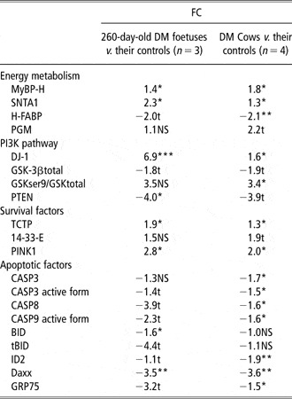
The power analysis (Lenth, Reference Lenth2007) allowed proteins to be retained which presented a minimum difference of 50% corresponding to a threshold FC of 1.5 in the foetus groups (n = 3 animals/group, individual samples were analysed in replicate) and cow groups (n = 4 animals/group). This is the case for most of the data shown in Table 2.
Real-time polymerase chain reaction analysis
Total RNA was extracted from muscle tissue samples with TRIZOL® reagent (Life Technologies) according to the manufacturer’s recommendation. RNA integrity was checked using Agilent RNA 6000 Nano Kit (AGiLENT technologies, Massy, France), as previously described (Cassar-Malek et al., Reference Cassar-Malek, Passelaigue, Bernard, Leger and Hocquette2007).
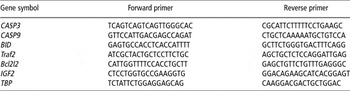
Six genes were selected from the data obtained in MSTN-null mice (Chelh et al., Reference Chelh, Meunier, Picard, Reecy, Chevalier, Hocquette and Cassar-Malek2009b). The abundance of transcripts was measured by quantitative RT-PCR (qPCR) using SYBR Green I dye. qPCRs were performed using gene-specific primers pairs (Table 1). The ΔΔCt (Cycle threshold) method (Wong and Medrano, Reference Wong and Medrano2005) was used to analyse the relative change in gene expression between DM foetuses and their controls and between DM cows and their controls (individual samples were analysed in replicate). The ΔΔCt value for each sample was calculated as follows: ΔΔCt = (CtTarget − CtReference) in DM animals – (CtTarget − CtReference) in control animals. The fold difference for each sample relative to the control sample: FC = 2−ΔΔCt. qPCR experiments were checked using the TBP as a reference gene. Values of 2−ΔΔCt >2 and 2−ΔΔCt <0.50 were considered, respectively, to declare that a transcript was upregulated and downregulated.
Primer sequences used in quantitative real-time PCR

DM = double muscled; CASP3 = caspase-3; CASP9 = caspase-9; BID = Bcl-2 interacting protein; Traf2 = Tnf receptor-associated factor 2; Bcl2l2 = Bcl2-like 2; IGF2 = insulin growth factor 2; TBP = Tata box binding protein.
All primer sequences were designed with the Primer3 software (http://primer3.sourceforge.net/).
Differential abundance of putative myostatin targets in DM foetuses and DM cows v. their controls

DM = double muscled; FC = fold change; MyBP-H = Myosin binding protein H; SNTA1 = α-1-syntrophin; H-FABP = heart-fatty acid binding protein; PGM1 = Phosphoglucomutase; DJ-1 = Cancer- and Parkinson’s disease-associated protein (park7); GSK-3β = glycogen synthase kinase 3β; GSK-3β (ser9) = glycogen synthase kinase 3β (phosphorylated at serine 9); PTEN = tumour-suppressor phosphatase with tensin homology; TCTP = translationally controlled tumour protein; 14-3-3E: 14-3-3ε protein; PINK1 = PTEN-induced putative kinase 1; CASP3 = caspase-3; CASP9 = caspase-9; BID = Bcl-2 interacting protein; ID2 = inhibitor of differentiation-2; Daxx = death domain-associated protein 6; GRP75 = Mortalin.
Results are expressed as FCs and the differences were declared significant by Student’s t-test: tP < 0.10; *P < 0.05; **P < 0.01; ***P < 0.001 except for no significant differences (NS: P > 0.10).
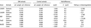
The power analysis (Lenth, Reference Lenth2007) allowed genes to be retained which presented a minimum difference of 100% corresponding to a threshold FC of 2.0. This is the case for most of the data shown in Table 3.
Differential expression of genes related to apoptosis confirmed by real time PCR analysis in DM foetuses and cows v. their controls

DM = double muscled.
Results are expressed as fold difference and for data interpretation (Fold up- or downregulation) the absolute ΔΔCt values were used. The mean of Ct values in each condition was calculated (data not shown) and the differences were declared significant by Student’s t-test: tP < 0.10; *P < 0.05; **P < 0.01; ***P < 0.001 except for no significant differences (NS: P > 0.10).
Statistical analyses
The differences in protein and transcript levels between DM animals and their controls were assessed by Student t-test. The results are expressed as the mean ± s.e.m. A difference between groups was considered significant when P < 0.05.
Results
Muscle energy metabolism
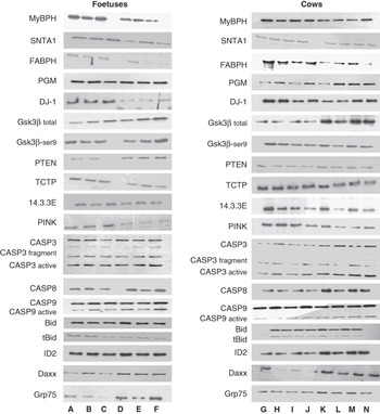
Western blot analysis revealed upregulation of the proteins related to glycolytic metabolism (Figure 1; Table 2) including myosin-binding protein (H-MyBP) and α1-syntrophin (SNTA1). H-MyBP was highly expressed in the muscles of DM foetuses and DM cows (FC = 1.4; P < 0.05 and FC = 1.8; P < 0.05, respectively). A high SNTA1 protein level was also detected in both DM foetuses and DM cows compared to controls (FC = 2.3; P < 0.05 and FC = 1.3; P < 0.05, respectively). No significant differences in phosphoglucomutase 1 (PGM1) abundance were revealed in the muscles of DM foetuses v. controls. There was a trend to a higher abundance in the muscles of DM cows v. controls. In contrast, the abundance of H-FABP was lower in the muscles of DM foetuses and DM cows v. controls (FC = −2.0; P < 0.1 and FC = −2.1; P < 0.05, respectively).
Abundance of putative myostatin targets in double-muscled (DM) foetuses and DM cows v. their controls as revealed by western blot analysis. A–C: 260-day-old DM foetuses; D–F: control foetuses; G–J: DM cows; K–N: control cows. MyBP-H: myosin-binding protein H; SNTA1: α-1-syntrophin; H-FABP: heart-fatty acid-binding protein H; PGM1: Phosphoglucomutase; DJ-1: cancer- and Parkinson’s disease associated protein (park7); GSK-3β: glycogen synthase kinase 3β; GSK-3β (ser9): glycogen synthase kinase 3β (phosphorylated at serine 9); TCTP: translationally controlled tumour protein; 14-3-3E: 14-3-3ε protein; PINK1: PTEN-induced putative kinase 1; PTEN: tumour-suppressor phosphatase with tensin homology; CASP3: procaspase-3 p32 kDa; CASP3 fragment: caspase-3 cleaved intermediate fragment p17&20 kDa; CASP3 active: caspase-3 p11 kDa; CASP8: procaspase-8 p57 kDa; CASP9: procaspase-9 p49 kDa; CASP9 active: caspase-9 p35 kDa; BID: Bcl-2 interacting protein; tBID: truncated 14 kDa form of BID; ID2: Inhibitor of differentiation-2; Daxx: death domain-associated protein 6; GRP75: mortalin.

PI3K/Akt signalling pathway
Based on our previous data in mice, we analysed the abundance of some components and regulators of the PI3K/Akt signalling pathway in the muscles of DM foetuses and DM cows, especially Gsk-3βser9/Gsk-3β total ratio, PTEN and DJ-1 (Figure 1; Table 2). The abundance of total GSK-3β tended to decrease in the muscles of both DM foetuses and DM cows. However, the ratio of serine-9 phosphorylated GSK-3β/total GSK-3β, an indicator of GSK-3β inactivation, was significantly increased in the muscles of DM cows only. This indicated that the inactive form of Gsk-3β was predominant in the absence of active MSTN. The PTEN protein, an antagonist of PI3K function, showed a decreased abundance in DM animals v. controls (FC = −4.0; P < 0.05 and FC = −3.9 P < 0.1, respectively, in foetuses and cows). The abundance of DJ-1, a regulator of the PI3K-PTEN pathway, was higher in DM animals v. controls (FC = 6.9; P < 0.01 and FC = 1.6; P < 0.05, respectively, in DM foetuses and cows).
All together, these data showed an activation of the PI3K/Akt signalling pathway in the muscles of DM foetuses and DM cows compared to their controls.
Cell survival pathway
Western blot analysis (Figure 1, Table 2) showed an upregulation of anti-apoptotic/survival factors such as TCTP, and 14-3-3E protein. The abundance of TCTP was higher in both DM foetuses and DM cows v. controls (FC = 1.9 and FC = 1.3, respectively; P < 0.01). There was a trend for higher 14-3-3E abundance in the muscles of DM cows v. controls (FC = 1.9, P < 0.1).
The PTEN-induced putative kinase 1 (PINK1), a crucial protein for cell survival, was significantly more abundant in the muscles of DM foetuses and DM cows compared to their controls (FC = 2.8 and FC = 2.0, respectively; P < 0.01).
Apoptotic pathway
The amount of proteins involved in apoptosis was examined by western blot including members of the caspase family and other apoptosis regulators (Figure 1; Table 2).
Caspases
Lower steady state levels of caspases-3, -8 and -9 playing a central role in the execution of apoptosis were detected in DM animals. A significant decrease was observed for pro-caspase-3 (FC = −1.7; P < 0.05) and active caspase-3 (FC = −1.5; P < 0.05), pro-caspase-8 (FC = 1.6; P < 0.05) and active caspase-9 (FC = −1.6; P < 0.05) especially in the muscles of cows harbouring MSTN loss-of-function. In addition, the levels of caspase-3 and -9 transcripts were checked by real time PCR analysis (Table 3). Caspase-9 mRNA was decreased significantly especially in DM foetuses (2−ΔΔCt = 0.20, P < 0.05), while caspase-3 mRNA was lower in DM cows (2−ΔΔCt = 0.20, P < 0.01).
Other regulators of apoptosis
Bid, a Bcl-2 interacting protein, was revealed by western blot as two distinct bands, a 22 kDa and its truncated 14 kDa active form tBID. A significant reduction of the p22 content (FC = −1.6, P < 0.05) and a tendency to be lower for the truncated form p14 (FC = −4.4, P < 0.1) were noted. The transcript of Bid was also downregulated in DM foetuses and DM cows compared to their controls as shown by RT-PCR (2−ΔΔCt = 0.30, respectively; P < 0.05). A decreased abundance of two positive regulators of apoptosis, Id2 (Inhibitor of differentiation-2) and Daxx (Death domain-associated protein 6) was also found. Id2 tended to decrease in DM foetuses (FC = −3.5 P < 0.1), whereas a significant decrease was found in DM cows (FC = −1.9; P < 0.01). Daxx level was significantly decreased in both DM foetuses muscle and DM cows muscle (FC = −3.5 and FC = −3.6, respectively; P < 0.01). There was only a trend for decreased abundance of GRP75 (mortalin) in DM foetuses.
Moreover, there was a significant increase in the abundance of two anti-apoptotic transcripts Bcl2l2 (Bcl2-like 2), a member of the Bcl2 family, and Traf2 (TNF receptor-associated factor 2), a member of the Traf family, in the muscles of DM animals (2−ΔΔCt = 12.1 for Bcl2l2 in DM cows and 2−ΔΔCt = 42.6 and 2−ΔΔCt = 3.8 for Traf2 in DM foetuses and DM cows, respectively, P < 0.05) except for Bcl2l2 in DM foetuses, which seemed to be increased (2−ΔΔCt = 2.3, P < 0.1).
All together, these data indicated that abundance of proteins and transcripts involved in the induction of the apoptotic pathway was decreased, whereas abundance of those involved in the anti-apoptotic pathway was increased.
Discussion
In an earlier study, comparing molecular profiles in the muscles of MSTN-null mice v. their controls, we found differential expression of many genes and proteins related to muscle energy metabolism, activation of the PI3K/Akt signalling pathway and cell survival/apoptosis pathway (Chelh et al., Reference Chelh, Meunier, Picard, Reecy, Chevalier, Hocquette and Cassar-Malek2009b). In this study, we examined some components of these pathways in the muscles of DM foetuses and DM cows harbouring MSTN loss-of-function.
Signatures related to muscle energy metabolism and growth
As expected (Bouley et al., Reference Bouley, Meunier, Chambon, De Smet, Hocquette and Picard2005; Cassar-Malek et al., Reference Cassar-Malek, Passelaigue, Bernard, Leger and Hocquette2007; Chelh et al., Reference Chelh, Meunier, Picard, Reecy, Chevalier, Hocquette and Cassar-Malek2009b), our results revealed additional evidences of glycolytic shift within skeletal muscle in the absence of functional MSTN with increased abundance of fast-twitch proteins (e.g. H-MyBP and PGM1) and decreased abundance of H-FABP in DM muscles. H-MyBP is involved in the contractile apparatus and is principally associated with fast twitch fibres (Bennett et al., Reference Bennett, Craig, Starr and Offer1986). PGM1 catalyses the interconversion of glucose-1-phosphate and glucose-6-phosphate. It plays a central role in the glycolytic pathway at the branch point leading to glycogen metabolism. H-FABP is a major fatty acid-binding protein in the heart and skeletal muscles which is involved in oxidative metabolism (Hocquette et al., Reference Hocquette, Ortigues-Marty, Pethick, Herpin and Fernandez1998). Our data are consistent with the observation that cattle with mutations in MSTN express more of fast, glycolytic isoforms (Wegner et al., Reference Wegner, Albrecht, Fiedler, Teuscher, Papstein and Ender2000). In addition, SNTA1, a predominant syntrophin isoform in skeletal and cardiac muscle, was significantly higher in both DM foetuses and DM cows v. controls. SNTA1 is necessary for muscle activity through the regulation of the calcium influx in skeletal muscle (Vandebrouck et al., Reference Vandebrouck, Sabourin, Rivet, Balghi, Sebille, Kitzis, Raymond, Cognard, Bourmeyster and Constantin2007). It is thought to link several signalling proteins to the actin cytoskeleton and to the extra-cellular matrix via dystrophin. Increased abundance of SNTA1 protein may participate in maintaining muscle metabolism and contraction in hypertrophied muscles of DM animals.
Activation of the PI3K/Akt signalling pathway
The PI3K/Akt pathway is a signalling network that promotes cell survival and proliferation, and plays a role in oncogenic transformation and in prevention of apoptosis (Lin et al., Reference Lin, Adam, Santiestevan and Freeman1999; Chang et al., Reference Chang, Lee, Navolanic, Steelman, Shelton, Blalock, Franklin and McCubrey2003). Our microarray experiments previously identify components of the PI3K/Akt/GSK-3β as upregulated in MSTN-null mice. In this study, differential expression of some components of this pathway was also found in the muscles of DM foetuses and DM cows v. their controls. The abundance of DJ-1 (also called park7), a regulator of the PI3K/Akt pathway, was increased in DM muscles. This protein protects cells from toxic stress, is implicated in mitochondria and oxidative stress-related survival pathways (Lev et al., Reference Lev, Roncevich, Ickowicz, Melamed and Offen2006), and is a novel regulator of the PI3K/Akt pathway (Kim et al., Reference Kim, Peters, Jang, Shi, Pintilie, Fletcher, DeLuca, Liepa, Zhou, Snow, Binari, Manoukian, Bray, Liu, Tsao and Mak2005). The abundance of PTEN, the most important negative regulator of the PI3K/Akt pathway, was found to be downregulated in DM muscles compared to control ones. The level of total GSK-3β was decreased and the serine-9 phosphorylated GSK-3β/total GSK-3β ratio was increased, especially in the muscles of DM cows. GSK-3β is a constitutively active component of the PI3K/Akt pathway that is inactivated upon serine-9 phosphorylation by Akt (Stambolic and Woodgett, Reference Stambolic and Woodgett1994), and the GSK-3β ser9/total GSK-3β ratio is an accurate indicator of GSK3b inactivation (Grimes and Jope, Reference Grimes and Jope2001). Therefore, GSK-3β activity was inhibited in DM muscles, which was a pre-requisite for the induction of the PI3K/Akt downstream survival signalling pathway.
These data demonstrated an activation of the PI3K/Akt signalling pathway in response to MSTN loss-of-function in cattle as previously shown in mice. They are in agreement with data indicating a crucial role of this pathway in muscle hypertrophy (Glass, Reference Glass2003) for example, increase in fibre size induced by constitutive activation of Akt (Pallafacchina et al., Reference Pallafacchina, Calabria, Serrano, Kalhovde and Schiaffino2002) and hypertrophy of type IIb muscle fibres induced by muscle-specific Akt1 transgene expression (Izumiya et al., Reference Izumiya, Hopkins, Morris, Sato, Zeng, Viereck, Hamilton, Ouchi, LeBrasseur and Walsh2008).
Increased abundance of cell survival factors
Interestingly, we also found that the abundance of cell survival/anti-apoptotic factors, namely TCTP, 14-3-3E and PINK1 was increased in DM muscles, while that of GRP75 a mitochondrial protein with predicted involvement in proteasomal degradation of proteins (Kaul et al., Reference Kaul, Deocaris and Wadhwa2007) seemed to decrease. TCTP is a crucial factor for cell growth, cell cycle progression, malignant transformation and is involved in the protection of cells against various stress conditions and apoptosis (Bommer and Thiele, Reference Bommer and Thiele2004). 14-3-3E is a member of the 14-3-3 family of phosphoserine-binding adapter molecules that mediate a general survival-promoting function through interaction with target proteins, by enhancing pro-survival signalling while suppressing activity of pro-apoptotic proteins (Porter et al., Reference Porter, Khuri and Fu2006). PINK1 encodes a Ser/Thr kinase with a mitochondrial-targeting signal, and acts with DJ-1 to protect cells against oxidative stress-induced cell death (Tang et al., Reference Tang, Xiong, Sun, Zhang, Wang, Hu, Zhu, Ma, Pan, Xia, Xia and Zhang2006).
Increased abundance of survival factors with anti-apoptotic function may participate in apoptosis resistance in DM muscle. This process could contribute to increase protein synthesis at the expense of protein degradation and therefore participate in muscle hypertrophy in DM cattle.
Genes and proteins related to the apoptotic pathway
Our previous genomic data in mice indicated that the apoptosis pathway is a downstream target of MSTN invalidation through altered expression or abundance of many pro- and anti-apoptotic factors (Chelh et al., Reference Chelh, Meunier, Picard, Reecy, Hocquette and Cassar-Malek2009a). In this study, we have demonstrated that MSTN loss-of-function is associated with changes in the expression and abundance of genes and proteins involved in the apoptotic pathway. The abundance of caspases and pro-apoptotic factors was decreased in DM muscles compared to control muscles. Caspases belong to a family of proteolytic enzymes, which play a key role in both the initiation and execution of apoptosis. Caspases-3 and -9 play, respectively, an effector and executor role in the apoptosis pathway. The levels of caspases-3 and -9 transcripts and the abundance of active caspases-3 and -9 were demonstrated to be lower in DM muscles than control muscles. The abundance of procaspase-8 protein was also lower in agreement with our previous observations in mice (Chelh et al., Reference Chelh, Meunier, Picard, Reecy, Hocquette and Cassar-Malek2009a). Thus, the caspase-dependent apoptotic pathway was attenuated in the muscles of DM foetuses and DM cows related to MSTN loss-of-function. The expression and abundance of Bid, a pro-apoptotic member of the Bcl-2 family and an important substrate of caspase-8 (Li et al., Reference Li, Zhu, Xu and Yuan1998), also differed between muscles of DM foetuses and their controls. Accordingly, the abundance of the truncated form of Bid (tBid), which results from a cleavage by caspase-8, tended to be lower in DM foetuses. Consistently two regulators of apoptosis (Daxx and Id-2) also tended to be less abundant in both DM foetuses and DM cows compared to their controls. The pro-apoptotic protein Daxx plays a role in the activation of ASK1, which is essential to JNK activation and then apoptosis induction (Yang et al., Reference Yang, Khosravi-Far, Chang and Baltimore1997; Chang et al., Reference Chang, Nishitoh, Yang, Ichijo and Baltimore1998). Daxx also interacts with the Fas receptor and potentiates its pro-apoptotic action (Yang et al., Reference Yang, Khosravi-Far, Chang and Baltimore1997). The Inhibitor of differentiation-2 (Id2) has been associated with apoptosis-associated atrophy of skeletal muscles (Alway et al., Reference Alway, Martyn, Ouyang, Chaudhrai and Murlasits2003).
Conversely, the abundance of anti-apoptotic factors was increased in DM muscles. The Bcl2l2 transcript was upregulated in the muscles of DM foetuses and DM cows v. their controls. The expression of this anti-apoptotic member of the Bcl2 family has been shown to reduce cell apoptosis and promote of cell survival (Ross et al., Reference Ross, Waymire, Moss, Parlow, Skinner, Russell and MacGregor1998; Denisov et al., Reference Denisov, Madiraju, Chen, Khadir, Beauparlant, Attardo, Shore and Gehring2003). In DM animals, there was also an increased expression of Traf2, a member of the Traf family of signal transducers of the Tumour Necrosis Factor (TNF) pathway (Arch et al., Reference Arch, Gedrich and Thompson1998) that inhibits TNFα-induced cell death by recruiting anti-apoptotic proteins (Zhang et al., Reference Zhang, Blackwell, Thomas, Sun, Yeh and Habelhah2009). Interestingly, Traf2−/−mice are born with severely decreased skeletal muscle mass indicating that this factor is necessary for normal muscle development (Yeh et al., Reference Yeh, Shahinian, Speiser, Kraunus, Billia, Wakeham, de la Pompa, Ferrick, Hum, Iscove, Ohashi, Rothe, Goeddel and Mak1997; Timothy and Antonio, Reference Timothy and Antonio1998). Traf2 might therefore be a new marker of muscle hypertrophy in the context of MSTN inactivation.
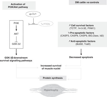
All together, the results of this study are in agreement with our previous study conducted in the context of MSTN inactivation in mice (Chelh et al., Reference Chelh, Meunier, Picard, Reecy, Hocquette and Cassar-Malek2009a and Reference Chelh, Meunier, Picard, Reecy, Chevalier, Hocquette and Cassar-Malek2009b). They confirm that MSTN loss-of-function in DM muscles results in the activation of a cell survival pathway through activation of PI3K pathway and alteration of the balance between survival/apoptotic factors in favour of mitigation of or resistance to apoptosis within the muscle (Figure 2). As previously hypothesised in mice, this may promote myonuclei or satellite cell survival. In addition, attenuated caspase-3 expression and activity could also lead to lower proteolysis in DM muscle (Gao et al., Reference Gao, Ordas, Klein and Price2008). This could account for muscle hypertrophy in both DM foetuses and DM cows. However, attenuated apoptosis is not likely to contribute to meat tenderness as to the hypothesis of Ouali et al. (Reference Ouali, Herrera-Mendez, Coulis, Becila, Boudjellal, Aubry and Sentandreu2006) in conventional meat-producing animals, especially as caspases have been identified as potential contributors to post-mortem tenderization. Indeed, tenderness of DM cattle would rather be linked to the collagen content of their muscles (Ashmore et al., Reference Ashmore, Parker, Stokes and Doerr1974; Uytterhaegen et al., Reference Uytterhaegen, Claeys and Demeyer1994). The existence of similar changes in some biological processes in the muscles of MSTN KO mice (gene inactivation) and DM cattle (loss-of-function mutation) reinforces the biological relevance of the proposed mechanisms of action of this growth factor.
In the muscles of double-muscled (DM) cattle v. their controls, regulators of the PI3K pathway and cell survival/apoptosis contribute to muscle hypertrophy. In DM cattle, a combination of increased potential for PI3K activation and of increased cell survival/anti-apoptotic factors leading to decreased apoptosis and increased cell survival likely participates in muscle hypertrophy. PI3K signalling pathway: PI3K, Phosphatidyl Inositol 3 Kinase; AKT, protein kinase B; GSK-3β, Glycogen synthase kinase 3β (a substrate for Akt inactivated upon phosphorylation); PTEN, tumour-suppressor phosphatase with tensin homology (the most important negative regulator of the cell survival signalling initiated by PI3K); DJ-1, a cancer- and Parkinson’s disease-associated protein (a regulator of the PI3K-PTEN pathway). Survival/anti-apoptotic factors: PINK1, PTEN-induced putative kinase; TCTP, Translationally controlled tumour protein; 14-3-3E, 14-3-3 protein epsilon. Apoptotic factors: CASP3: caspase-3; CASP9: caspase-9; BID: Bcl-2 interacting protein; BID: Bcl-2 interacting protein; Id2: inhibitor of differentiation-2; Daxx: death domain-associated protein 6; Traf2: Tnf receptor-associated factor 2; Bcl2l2: Bcl2-like 2.

Acknowledgements
The authors thank members of the Herbivore Research Unit for producing the foetuses, slaughtering the animals and collecting muscle samples, F. Ménissier for production of DM foetuses and Q204X genotyping, G. Gentes, for excellent technical assistance in molecular biology, A. Delavaud and S. Bes for helpful discussions. Dr A. Ouali (UR370 QuaPA, INRA, Clermont-Ferrand/Theix, FRANCE) for kindly providing the Caspase 8 antibody. The study received a grant from ANR (for the MYOTROPHY project: ANR BLAN08-2_309209).