Introduction
Otosclerosis is a primary disease of the bony labyrinth, which only occurs in humans and affects the temporal bone.Reference Wiatr, Składzień, Świeży and Wiatr1 It is associated with progressive conductive and/or sensorineural hearing loss due to abnormal bone resorption and deposition in the otic capsule. The aetiology of the disease has not yet been understood.Reference Carvalho, Hamerschmidt, Telles and Richter2 Otosclerosis manifests in three histopathological patterns: spongiotic (cellular), fibrotic, and sclerotic. Given the dynamic nature of the disease, these patterns can be observed concomitantly.Reference Djeric3
Otosclerosis histopathology is characterised by increased cellularity, areas of bone resorption and deposition (osteoblasts and osteoclasts), increased vascularity, basophilic/eosinophilic staining pattern, and narrow/wide pseudovascular–perivascular spaces.Reference Quesnel, Ishai and McKenna4, Reference Cureoglu, Schachern, Ferlito, Rinaldo, Tsuprun and Paparella5 The most commonly involved parts of the otic capsule in otosclerosis are the fissula ante fenestram anterior to the oval window, the round window niche, and the apical-medial wall of the cochlea.Reference Schuknecht and Barber6
There are many previous studies on histopathologic changes in the base of the stapes in otosclerosis, yet only a few studies that have histologically investigated the stapes superstructure (stapes head and crura) are known to us. Nevertheless, these few studies did not evaluate histopathologic findings in combination with audiometric data.
The classical methods in otosclerosis surgery are the removal of the stapes superstructure, stapedotomy, and piston prosthesis placement. This study aimed to investigate the stapes superstructure removed during surgical procedure under a light microscope to see whether it was directly or indirectly affected by otosclerosis and to assess the results in combination with audiometric data.
Materials and methods
This study was commenced upon approval No. 2023/178 of the Ethics Committee of our hospital. Intra-operatively removed stapes superstructures of patients operated witha preliminary diagnosis of otosclerosis in the Department of Otorhinolaryngology were sent to the Department of Pathology for pathologic examination. Here, the superstructures were stained using haematoxylin-eosin, and the preparations were examined by a pathologist. In the present study, preparations between 2016 and 2023 were retrospectively re-examined. All surgeries were performed by the same surgeon. The study had no control group.
After preliminary microscopic examination, preparations containing insufficient material and therefore not suitable for thorough microscopic examination were excluded from the study. All remaining preparations were included in the study. Patients who developed complications during surgery and whose pure tone audiometry (PTA) tests were unavailable were also excluded from the study.
The stapes superstructure preparations included in the study were examined and recorded by two pathologists to determine whether they included the stapes head and crura and contained otosclerotic foci, and if so, the stage of otosclerosis (active/inactive) and any other histopathologic alterations. Age, sex, and PTA data of the patients included in the study were accessed via the hospital information system, the patient files, and audiology archive records.
During PTA, the mean thresholds for air conduction and bone conduction at frequencies of 500, 1000, 2000, and 3000 Hz were calculated pre-operatively and post-operatively for each patient’s operated ear pursuant to the recommendations of the Hearing and Balance Committee of the American Academy of Otolaryngology and Head and Neck Surgery.Reference Monsell, Balkany, Gates, Goldenberg, Meyerhoff and House7 The Statistical Package for the Social Sciences (SPSS) Software, Windows version was used for statistical analyses. A paired t-test was used to compare the pre-operative and post-operative air conduction, bone conduction, and air–bone gap (ABG) PTA of the patients. Independent t and Mann–Whitney U tests were used to compare the audiometric data of the groups, and a p-value of less than 0.05 was considered statistically significant.
Results
The study commenced with a retrospective review of 53 patients. As a result of the preliminary microscopic examinations, all preparations with stapes superstructures suitable for histopathologic examination were included in the study (n = 30). Preparations with insufficient material and not suitable for the main examination were excluded from the study (n = 23).
Upon exclusion of the ineligible patients, stapes superstructures, age, sex, and PTA data of the remaining 30 patients were noted. The participants’ ages ranged between 10 and 54 years (mean = 36 years), and there were 17 male and 13 female participants (1.3:1).
Upon examination of the stapes superstructures included in the study under a light microscope, the stapes head was intact in 27 (90%) of the preparations, while it was defective in 3 (10%). Upon review of the preparations by stapes crura, both stapes crura were intact in 11 (37%), both were defective in 2 (7%), and one of the two crura was defective in 17 (57%). No distinction was made on which stapes crura (anterior/posterior) was defective. It was considered that surgical trauma during removal of the stapes superstructures and/or technical conditions during histopathologic preparation might account for the fact that there were defective stapes heads in some cases and crura in others.
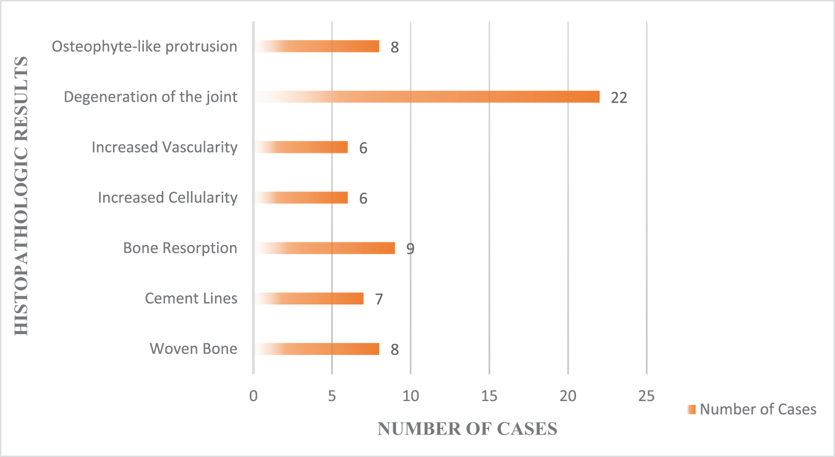
There were no obvious active/inactive otosclerotic foci in the investigated stapes superstructures. Nevertheless, stapes superstructures had certain histopathologic findings that were not normally expected (Figures 1 and 2). Among these, the remarkable findings included woven bone and cement lines, which were not specific to otosclerosis but indicative of bone regeneration and the construction/destruction cycle that could be seen in otosclerosis disease. About 8 (27%) out of 30 preparations had woven bone, and 7 (23%) had cement lines. Furthermore, there were resorptive alterations in the bone (stapes crura) in 9 (30%) cases. Bone resorption also can be seen in otosclerosis and was considered significant in patients with no middle-ear pathology (cholesteatoma, chronic otitis media, granulation tissue, tumour, etc.) other than otosclerosis.
Microscopic images of woven bone and cement line. (A) Woven bone (rectangle) in the stapes crura; (B) woven bone (ring) and cement line (arrow) in the stapes crura.

Microscopic images showing bone resorption areas indicated by a bracket (A) and arrows (B) in the stapes crura.

Some of the investigated preparations had signs of joint degeneration similar to osteoarthritis of long bones, which would not be expected in healthy people. About 22 (73%) out of 30 preparations had degenerative changes in the stapes head (incudostapedial joint), and 8 (27%) had osteophyte-like protrusions (Figures 3 and 4). The histopathological findings associated with the stapes superstructures in question are summarised in Figure 5.
Microscopic images of osteophyte-like protrusions (circles) on the stapes head from three different patients (A–C).

Microscopic images of degenerative findings in the stapes head. (A, B), degenerative findings in the stapes head, cartilage regeneration, and asymmetric and irregular cartilage thickness (arrow). (C) Significantly increased cartilage thickness in the stapes head, cartilage regeneration.

Summary of histopathological findings in the stapes superstructure.

Upon examination of preparations by histopathologic changes, 15 cases with significant findings with regard to otosclerosis (woven bone, cement lines, and resorption areas) were classified as Group 1, and 15 cases with only secondary degenerative findings (joint degeneration and osteophytes) were classified as Group 2. There was no statistically significant difference between the two groups by age and sex (p = 0.320, p = 0.232, respectively). Pure tone audiometry data of these groups are given in Table 1. There was no statistically significant difference between the groups with and without otosclerotic alterations by pre-operative and post-operative air conduction, bone conduction, and air–bone ABG PTA.
A comparison of the groups with and without otosclerotic alterations

* Mann–Whitney U test, **Independent t test (PTA: pure tone average; SD: standard deviation; AC: air conduction; BC: bone conduction, and ABG: air–bone gap)
Discussion
Otosclerosis is a localised disease of the otic capsule characterised by irregular bone resorption and deposition.Reference Chole and McKenna8 There is a wealth of previous studies on histopathologic changes in the base of the stapes in otosclerosis. Yet, we know of only a few studies that have histologically investigated the stapes superstructure (stapes head and crura). Given that the stapes superstructure does not belong to the otic capsule and has a different embryologic origin (second branchial arch), it may not be involved in the process of otosclerosis.Reference Carvalho, Hamerschmidt, Telles and Richter2 This study aimed to investigate the morphology and histopathology of intra-operatively removed stapes superstructures of patients with otosclerosis under light microscopy and evaluate the findings in combination with audiometric data. Otosclerosis histopathology is characterised by osteoclastic bone resorption, osteoblastic new bone formation, increased vascularity, cellularity, wide pseudovascular spaces in the spongiotic phase, and decreased cellularity and narrow pseudovascular spaces in the sclerotic phase.Reference Quesnel, Ishai and McKenna4
In the present study, no obvious active/inactive otosclerotic foci were similar to the characteristics described by previous studies in the investigated stapes superstructures. Nevertheless, stapes superstructures had certain histopathologic findings that were not normally expected. The noteworthy alterations included woven bone, cement lines, and bone resorption areas in the stapes crus, which might be considered significant for otosclerosis. Similar to the results of a study by Carvalho et al.,Reference Carvalho, Hamerschmidt, Telles and Richter2 we do not know whether the histopathologic changes in the present study were associated with the involvement of the stapes superstructure in otosclerosis or to the extension of otosclerosis at the base of the stapes to the stapes superstructure, or whether they were secondary to trauma during surgery or incidental findings. Nevertheless, it was concluded that the areas of reactive bone formation, woven bone, and cement lines indicative of new bone regeneration in certain cases were unexpected findings in normal stapes histology (since a cycle of repair and bone formation/destruction was not expected after birth). Therefore, the stapes superstructure might be directly or indirectly affected by otosclerosis.
Similar to the results of a study by Clark-Brodber et al.,Reference Clarke-Brodber and Taxy9 this study included cases with cartilage degeneration, suggesting osteoarthritis of the long bones and osteophyte-like protrusion in the stapes head. These findings were considered independent of age and related to possible persistent trauma in the stapes head induced by incus due to stapes ankylosis.
Unlike the limited studies in the literature, in this study there was no statistically significant difference between the groups with and without otosclerotic alterations by pre-operative and post-operative air conduction, bone conduction, and ABG PTA. We think that the presence of otosclerotic or degenerative changes in the stapes superstructure may negatively affect pre-operative hearing outcomes more than their absence. However, since all our cases exhibited such changes, we are unable to verify this assumption.
The present study was designed as retrospective research, and therefore, the main limitations of the study included the lack of information on whether the patients had diseases other than otosclerosis, such as osteoarthritis,Reference Rawool and Harrington10 osteoporosis,Reference Babich, Hoffmeister and Doughty11 and rheumatoid arthritis,Reference Milisavljevic, Stankovic, Zivic, Radovanović and Stankovic12 which could affect the middle-ear ossicles and especially the incudostapedial joint. Another limitation was the lack of access to post-operative PTA tests of certain patients, and the relatively small number of cases included in the study because almost half of the specimens became ineligible for further examination during the preparation phase.
• Otosclerosis histopathology is characterised by areas of bone resorption and deposition (osteoblasts and osteoclasts), basophilic/eosinophilic staining pattern, and narrow/wide pseudovascular–perivascular spaces
• The stapes superstructure does not belong to the otic capsule and has a different embryologic origin, it may not be involved in the process of otosclerosis
• There are many previous studies on histopathologic changes in the base of the stapes in otosclerosis, yet only a few studies have histologically investigated the stapes superstructure
• This is the first study that investigated the stapes superstructure from the otosclerosis perspective, and the cases were assessed in combination with audiometric data
• There were no obvious active/inactive otosclerotic foci in the investigated stapes superstructures but stapes superstructures had certain histopathologic findings (woven bone, cement lines, bone resorption areas) that were not normally expected
• There was no statistically significant difference between the groups with and without otosclerotic alterations by pre-operative and post-operative pure tone audiometry data
Conclusion
Although there was no statistically significant difference by pre-operative and post-operative air conduction, bone conduction, and ABG PTA between the groups with and without alterations that might be considered significant vis-à-vis otosclerosis, the present study is the first of its kind known to us that investigated the stapes superstructure from the otosclerosis perspective, and for which the findings were evaluated in combination with audiometric data.
Ethics approval and consent to participate
This study was performed in line with the principles of the Declaration of Helsinki. Approval was granted by the Ethics Committee of University of Health Sciences Gulhane Training and Research Hospital (No. 2023/178).
Availability of data and material
The data used in this study have not been used/published in any other publications.
Competing interests
The authors declare that they have no competing interests.
Funding
The authors have no relevant financial or non-financial interests to disclose.
Authors’ contributions
BS designed the study; BK collected the data, did the literature search, and FCAO helped in data analysis. OG, EA, and GS did the histopathological examination. BK wrote the manuscript with critical inputs from BS.