Introduction
Snapping shrimps of the family Alpheidae Rafinesque 1815 are ubiquitous components of tropical and subtropical benthic communities of marine habitats (Mathews & Anker, Reference Mathews and Anker2009). With more than 313 valid species (e.g. De Grave & Fransen, Reference De Grave and Fransen2011; Salgado-Barragán et al., Reference Salgado-Barragán, Ayón-Parente and Zamora-Tavares2017; Komai & Ohtomi, Reference Komai and Ohtomi2018; Dehghani et al., Reference Dehghani, Sari and Naderloo2019), of which 34 were registered in Brazilian waters (Soledade & Almeida, Reference Soledade and Almeida2013; Anker et al., Reference Anker, Tavares and Mendonça2016; Almeida et al., Reference Almeida, Terossi, Buranelli, Castilho, Costa, Zara and Mantelatto2018), the snapping shrimp genus Alpheus Fabricius, 1798 is the most representative of the family Alpheidae, and the second most speciose genus of infraorder Caridea Dana, 1852, after Caridina H. Milne Edwards, 1837 (in H. Milne Edwards, 1834–1840) (De Grave & Fransen, Reference De Grave and Fransen2011; Soledade & Almeida, Reference Soledade and Almeida2013). Their representatives occupy a wide variety of environments, from deep sea to shallow waters, inhabiting from coral reefs to the estuarine intertidal region (Bauer, Reference Bauer2004; Anker et al., Reference Anker, Ahyong, Noël and Palmer2006).
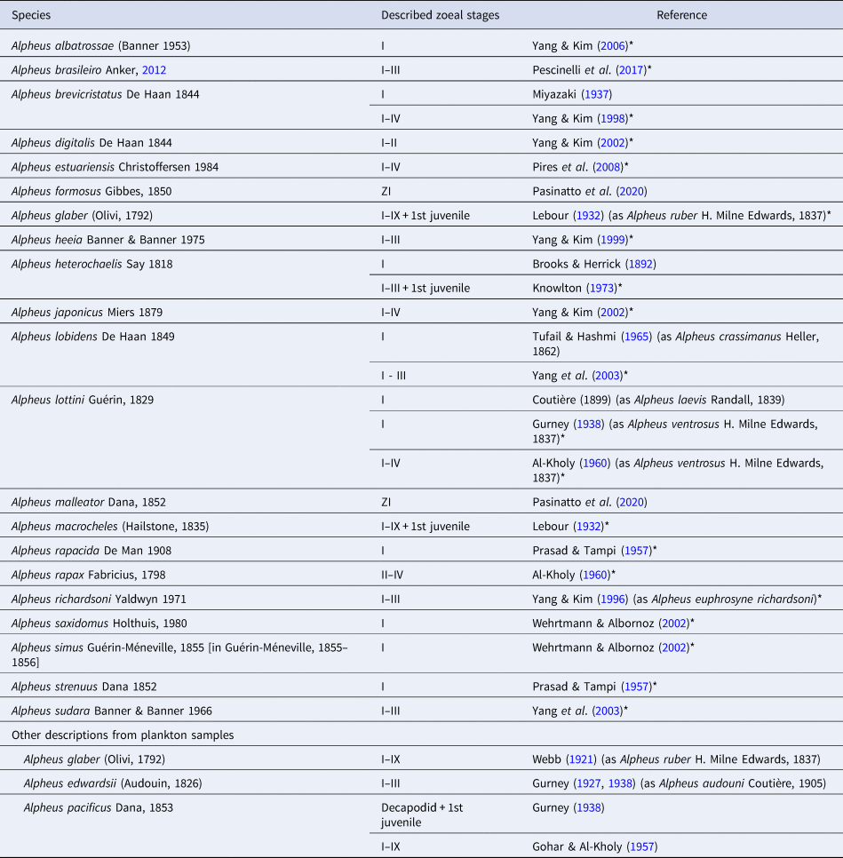
Despite the high representativeness of the genus and its larval abundance in meroplanktonic communities (Knowlton, Reference Knowlton1973), larval descriptions are very poorly reported when considered in terms of the total number of species described (Pescinelli et al., Reference Pescinelli, Pantaleão, Mantelatto and Costa2017). After a review of the larval descriptions of the genus we noted that ~7% of the species had at least the first larval stage described. This percentage does not include some morphological descriptions of various larval stages attributed to Alpheus species from plankton samples because there is no assurance of identification of species.
New techniques used on the systematics of genus Alpheus led to many taxonomic rearrangements in the last decade, as well as the number of described species (Pescinelli et al., Reference Pescinelli, Pantaleão, Mantelatto and Costa2017; Almeida et al., Reference Almeida, Terossi, Buranelli, Castilho, Costa, Zara and Mantelatto2018). That is the case of Alpheus armillatus H. Milne Edwards, 1837, which was considered a species complex (see Anker, Reference Anker2012). The description of larvae of more Alpheus species belonging or not to this complex is important to verify if larval morphology data support the relatively recent separation of species previously considered under the same taxonomic entity. Therefore, here we conducted unedited descriptions of the first zoea of two species, one that belongs to the complex and another that does not, aiming to generate a favourable scenario for comparisons within the genus.
Alpheus carlae Anker, Reference Anker2012 is one of the 11 species of the Alpheus armillatus species complex. Its preliminary distribution ranges appear to be continuous, from Florida (USA) to São Paulo (Brazil) and throughout the Caribbean Sea, where specimens are found in various intertidal and shallow subtidal habitats (Anker, Reference Anker2012). Biological information on this species was generated prior to taxonomic rearrangements proposed with the species complex (as Alpheus armillatus H. Milne Edwards, 1837). Mossolin et al. (Reference Mossolin, Shimizu and Bueno2006) detected the formation of male and female pairs under rocks, and Pavanelli et al. (Reference Pavanelli, Mossolin and Mantelatto2008) showed that the reproductive strategy of this species is marked by extrusion of small and numerous eggs, with an increase in reproductive effort during the rainy season. Larval information about species belonging to A. armillatus complex is available only for Alpheus brasileiro Anker, Reference Anker2012 (Pescinelli et al., Reference Pescinelli, Pantaleão, Mantelatto and Costa2017).
Alpheus intrinsecus Spence Bate 1888 has an amphi-Atlantic geographic distribution (Christoffersen, Reference Christoffersen1979; Soledade & Almeida, Reference Soledade and Almeida2013; Cunha et al., Reference Cunha, Terossi, Mantelatto and Almeida2017; Almeida et al., Reference Almeida, Terossi, Buranelli, Castilho, Costa, Zara and Mantelatto2018). Despite this wide distribution, it was confirmed by morphological and molecular data that it is a single species, with no cryptic species as was previously suspected (Cunha et al., Reference Cunha, Terossi, Mantelatto and Almeida2017). Information on its biology is practically non-existent; the single available biological information about the species is that it inhabits sandy environments, with shells and calcareous algae, from the inter-tides to 40 m depth (Christoffersen, Reference Christoffersen1979). Apheus intrinsecus is not a species of commercial interest, but it is recurrent in the trawling fishery, being one of the carcino-bycatch species of Xiphopenaeus kroyeri (Heller, 1826), the most commercially important penaeid shrimp in south-eastern Brazil (Mantelatto et al., Reference Mantelatto, Bernardo, Silva, Bernardes, Cobo and Fransozo2016; Almeida et al., Reference Almeida, Terossi, Buranelli, Castilho, Costa, Zara and Mantelatto2018).
Descriptions of larval morphology are important in taxonomic and phylogenetic contexts and allow the elaboration of identification keys (González-Gordillo & Rodriguez, Reference González-Gordillo and Rodríguez2000; Vela & González-Gordillo, Reference Vela and González-Gordillo2016), enabling studies about larval ecology (Iorio et al., Reference Iorio, Scelzo and Boschi1990). The aim of this study was to describe and illustrate the first larval stages of A. carlae and A. intrinsecus. Remarks from a semi-quantitative comparison of the morphology of zoea I of Alpheus species are also provided.
Materials and methods
Descriptions and illustrations were based on larvae from two parental females of each species. For Alpheus intrinsecus, larvae from two distinct locations in south-eastern Brazil were obtained. One female with embryos was collected in July 2011, at Macaé, Rio de Janeiro state (22°22′21.9″S 41°44′17.7″W), and another specimen at Ubatuba, São Paulo state, Brazil (23°26′35.6″S 45°03′33.2″W), in July 2013. The collections were made at depths of 5–10 m, using similar shrimp-fishing boats equipped with otter-trawl nets (mesh size 20 mm and 18 mm in the cod end). Both A. carlae females were captured manually, removing the rocks from the substrate during low tide, in an intertidal estuary in Araçá, São Sebastião, São Paulo state, Brazil (23°48′78.1″S 45°24′46.9″W) in February 2017.
Females with embryos were transported alive to the laboratory and maintained in 2-litre containers with water from the sampling site until the larvae hatched. After hatching, females were conserved (ethyl alcohol 98%) and their carapace length (CL in mm, excluding rostrum) was measured. In an attempt to obtain later stages of larval development, the larvae were mass-reared at constant 25°C and a salinity of 18 and 33 ppt (for A. carlae and A. intrinsecus, respectively), under continuously moderated aeration in small beakers (500 ml, about 100 larvae per beaker). Every day, newly hatched nauplii of Artemia sp. (source: Bio Artemia A. Ferreira De Melo ME; state of Rio Grande do Norte, Brazil) was offered ad libitum (Broad, Reference Broad1957; Chamberlain, Reference Chamberlain1957), the water partially renewed, and the larvae checked for moulting and mortality in order to reach other stages. Larvae were conserved in a mixture (1:1) of 70% ethyl alcohol and glycerin.
Zoeae appendages were dissected for examination under a Zeiss Stemi 2000C trinocular stereomicroscope and mounted on semipermanent slides. Drawings and measurements were made using a Leica DM750 microscope equipped with a camera lucida. All drawings and measurements were based on at least 10 larvae of each parental female. The following measurements were taken in the larvae: carapace length (CL) as the maximum length from the posterior margin of the ocular orbit to the posterior margin of the carapace; total length (TL) as the distance from the tip of the rostrum to the posterior margin of the telson, excluding setae. Larval description is arranged based on the standards proposed by Clark et al. (Reference Clark, Calazans and Pohle1998) and Clark & Cuesta (Reference Clark, Cuesta, Castro, Davie, Guinot, Schram and von Vaupel-Klein2015), and setal classification followed Garm (Reference Garm2004). Long terminal plumose natatory setae on exopods of the maxillipeds were drawn truncated.
The CL of hatches was compared (from two locations for A. intrinsecus and with A. carlae). The normality and homoscedasticity were tested by the Shapiro–Wilk and Levene tests, respectively. Since the data did not meet the requirements for parametric statistics, the non-parametric Kruskal–Wallis test followed by Dunn's test were used, adopting the significance level of α = 0.05 (Zar, Reference Zar1996). The statistical analysis was performed using the software GraphPad Prism 8 (GraphPad Software Inc., San Diego, CA, USA).
Voucher of the parental females and respective larvae were deposited at the Crustacean Collection of the Biology Department of FFCLRP, University of São Paulo, Brazil (CCDB/FFCLRP/USP) under numbers: CCDB 6152 and 6153 for A. carlae; CCDB 4492 and 6154, for A. intrinsecus from Macaé and Ubatuba, respectively.
Results
The parental females of Alpheus carlae have a CL of 11.71 mm and 12.37 mm; A. intrinsecus females have a CL of 13.20 mm (from Macaé) and 9.80 mm (from Ubatuba).
Newly hatched larvae of both species did not show interest for Artemia nauplii, nevertheless, the hatchlings of A. intrinsecus from Ubatuba reached the second zoeal stage (in ~24 h). Since we did not obtain zoea II from A. carlae and the discussion presented here focuses on the first larval stage of Alpheus species, illustrations of the zoea II of A. intrinsecus are presented in a supplementary figure (Supplementary Figure S1). The CL did not differ between hatches of A. intrinsecus from two distinct locations, but differed between species (Kruskal–Wallis, H = 18.329, P < 0.001). Since the external morphology of newly hatched larvae of A. intrinsecus did not differ among the hatches, only larvae from Ubatuba were used for the illustrations.
A semiquantitative comparison of selected morphological characters of the first zoeal stage of species of Alpheus was carried out using the original descriptions and illustrations of the species. Information on larval morphology of Alpheus is currently limited to 21 species, excluding descriptions from plankton samples. However, some older descriptions that do not show basic details were not used in this comparison (Tables 1 & 2).
Previous larval descriptions of the genus Alpheus Fabricius, 1798

(*) Descriptions used in Table 2.
Comparison of selected morphological characters of zoea I of the genus Alpheus

ae, aesthetascs; ds, denticulate seta; ps, plumose seta; se, serrate seta; seg., segmented; sp, spine; ss, simple seta; sps, sparsely plumose seta; st, setulate seta; pd, plumodenticulate seta; NA, not available in descriptions. Modified from Pescinelli et al. (Reference Pescinelli, Pantaleão, Mantelatto and Costa2017).
Bold characters indicate differences between species of A. armillatus complex.
Larval description
Order DECAPODA Latreille, 1802
Infraorder CARIDEA Dana, 1852
Family ALPHEIDAE Rafinesque, 1815
Genus Alpheus Fabricius, 1798
Alpheus carlae Anker, Reference Anker2012
(Figure 1)
ZOEA I (Figure 1A–N)
Alpheus carlae Anker, Reference Anker2012, Zoea I: (A) dorsal view; (B) lateral view; (C) magnification of the carapace, lateral view; (D) antennule; (E) antenna; (F) maxillule; (G) maxilla; (H) first maxilliped; (I) second maxilliped; (J) third maxilliped; (K) first pereiopod; (L) second pereiopod; (M) fifth pereiopod; (N) telson. Arrows indicate exclusive characteristics of the species. Scale bars: A–B, 0.5 mm; C, 0.5 mm; F–G, 0.05 mm; D–E, 0.25 mm; H–N, 0.25 mm.

Dimensions: CL: 0.52 ± 0.03 mm; TL: 2.22 ± 0.05 mm (10 larvae of each parental female = 20).
Carapace (Figure 1A–C): without rostrum; with one pair of acute spines in the pterygostomian region; eyes sessile.
Antennule (Figure 1A, B, D): peduncle unsegmented; primary flagellum with 4 terminal aesthetascs and 1 terminal plumose seta; accessory flagellum as a long plumose seta.
Antenna (Figure 1A, B, E): peduncle unsegmented, with a terminal spine near endopod; endopod with 1 long plumose seta and 1 terminal inner spine; exopod (antennal scale) 5-segmented, with 11 plumose setae, 9 on the inner side (2, 1, 1, 1, 4), 2 shorter on the outer side (1, 1, 0, 0, 0) plus 1 small simple seta on the outer side of fifth segment.
Mandibles (not illustrated): incisor and molar process developed; palp absent.
Maxillule (Figure 1F): coxal endite with 6 (5 simple and 1 sparsely plumose) setae; basial endite with 2 stout spines and 2 small (1 simple and 1 sparsely plumose) setae; endopod with 1 terminal denticulate seta.
Maxilla (Figure 1G): coxal endite not bilobed, with 3 (2 simple and 1 sparsely plumose) setae; basial endite bilobed, with 4 and 5 simple setae on proximal and distal lobe, respectively, proximal lobe with one terminal protuberance; endopod with 3 (1 small proximal and 1 distal simple, and 1 distal plumose) setae; exopod (scaphognathite) margin with 5 plumose setae; microtrichia on inner and outer margins of endopod as illustrated.
First maxilliped (Figure 1H): coxa with 1 simple seta; basis with 7 (4 strong and 3 slender) simple setae (arranged 2 + 2 + 2 + 1); endopod unsegmented, with 4 (1 proximal and 3 terminal) simple setae; exopod unsegmented, with 1 subterminal simple and 4 long plumose natatory setae terminally.
Second maxilliped (Figure 1I): coxa without seta; basis with 4 (3 strong and 1 slender) simple setae (arranged 1 + 2 + 1); endopod 4-segmented with 1 terminal simple, 0, 1 terminal serrate, 5 (1 proximal and 3 terminal simple, plus 1 terminal serrate) setae, respectively; exopod unsegmented, with 2 (1 small simple and 1 long plumose) subterminal, and 4 long terminal plumose natatory setae.
Third maxilliped (Figure 1J): coxa without setae; basis with 1 strong simple seta; endopod 4-segmented with 0, 0, 0, 5 (1 proximal, 2 subterminal and 2 terminal) simple setae, respectively; exopod unsegmented, with 3 (1 subterminal small simple and 2 subterminal long plumose), and 4 long terminal plumose natatory setae.
Pereiopods (Figure 1K–M): first as a biramous bud; second and fifth as a uniramous bud; third and fourth pereiopods absent.
Pleon (Figure 1A, B): with five rounded pleonites without setae, pleonite 6 fused with the telson; pleopods and uropods absent.
Telson (Figure 1A, N): broad at posterior margin, with 7 + 7 plumose setae (the outer 2 + 2 setae plumose only in the inner margin), outermost pair subterminal, inner pair shorter; one row of minute spinules on distal margin between and around bases of the 4 + 4 inner setae.
Alpheus intrinsecus Spence Bate, 1888
(Figure 2 and Supplementary Figure S1)
ZOEA I (Figure 2A–M)
Alpheus intrinsecus Spence Bate, 1888, Zoea I: (A) dorsal view; (B) lateral view; (C) magnification of the carapace, lateral view; (D) antennule; (E) antenna; (F) maxillule; (G) maxilla; (H) first maxilliped; (I) second maxilliped; (J) third maxilliped; (K) first pereiopod; (L) fifth pereiopod; (M) telson. Arrows indicate exclusive characteristics of the species. Scale bars: A–B, 0.5 mm; C, 0.4 mm; F–G, 0.05 mm; D–E, 0.2 mm; H–M, 0.2 mm.

Dimensions: CL: 0.34 ± 0.01 mm; TL: 1.85 ± 0.04 mm (N = 10 larvae of each parental female, total = 20 larvae).
Carapace (Figure 2A–C): without rostrum; with one pair of acute spines in the pterygostomian region; eyes sessile.
Antennule (Figure 2A, B, D): peduncle unsegmented; primary flagellum with 4 terminal aesthetascs and 1 terminal plumose seta; accessory flagellum as a long plumose seta.
Antenna (Figure 2A, B, E): peduncle unsegmented, with a terminal spine near endopod; endopod with 1 long plumose seta and 1 terminal inner spine; exopod (antennal scale) 5-segmented, with 11 plumose setae, 9 on the inner side (2, 1, 1, 1, 4), 2 shorter on the outer side (1, 1, 0, 0, 0), plus 1 small simple seta on the outer side of fifth segment.
Mandibles (not illustrated): incisor and molar process developed; palp absent.
Maxillule (Figure 2F): coxal endite with 5 (3 small simple and 2 sparsely plumose) setae; basial endite with 2 stout spines and 2 small simple setae; endopod with 1 terminal denticulate seta.
Maxilla (Figure 2G): coxal endite not bilobed, with 2 simple setae; basial endite bilobed, with 3 and 5 simple setae on proximal and distal lobe, respectively, proximal lobe with one terminal protuberance; endopod with 3 (1 proximal and 1 distal simple, and 1 distal sparsely plumose) setae; exopod (scaphognathite) margin with 5 plumose setae; microtrichia on inner and outer margins of endopod as illustrated.
First maxilliped (Figure 2H): coxa with 1 simple seta; basis with 5 (4 strong and 1 slender) simple setae (arranged 1 + 1 + 2 + 1); endopod unsegmented, with 3 (2 simple and 1 serrate) terminal setae; exopod unsegmented, with 1 subterminal simple and 4 long plumose natatory setae terminally.
Second maxilliped (Figure 2I): coxa without seta; basis with 4 (3 strong and 1 slender) simple setae (arranged 1 + 2 + 1); endopod 4-segmented with 1 terminal simple, 0, 1 terminal serrate, 4 (1 proximal plus 2 subterminal simple and 1 terminal serrate) setae, respectively; exopod unsegmented, with 2 (1 small simple and 1 long plumose) subterminal, and 4 long terminal plumose natatory setae.
Third maxilliped (Figure 2J): coxa without setae; basis with 1 strong simple seta; endopod 4-segmented with 0, 0, 0, 4 (3 subterminal and 1 terminal) simple setae, respectively; exopod unsegmented, with 3 (1 subterminal small simple and 2 subterminal long plumose), and 4 long terminal plumose natatory setae.
Pereiopods (Figure 2K, L): first as a biramous bud and fifth as a uniramous bud; second, third and fourth pereiopods absent.
Pleon (Figure 2A, B): with five rounded pleonites without setae, pleonite 6 fused with the telson; pleopods and uropods absent.
Telson (Figure 2A, M): broad at posterior margin, with 7 + 7 plumose setae (the outer 2 + 2 setae plumose only in the inner margin), outermost pair subterminal, inner pair shorter; one row of minute spinules on distal margin between and around bases of the 4 + 4 inner setae.
ZOEA II (Supplementary Figures S1A–M)
Dimensions: CL: 0.42 ± 0.03 mm; TL: 2.04 ± 0.06 mm (N = 10 larvae).
Carapace (Figure S1A–C): rostrum almost reaching the extremity of the eyes stalks; eyes compound and stalked; otherwise unchanged.
Antennule (Figure S1A, B, D): peduncle 2-segmented; proximal segment without setae and distal segment with two terminal plumose setae; primary flagellum with 5 terminal aesthetascs; accessory flagellum unchanged.
Antenna (Figure S1A, B, E): exopod (antennal scale) 4-segmented, with 11 plumose setae, 9 on the inner side (3, 1, 1, 4), 2 shorter on the outer side (2, 0, 0, 0), plus 1 small simple seta on the outer side of fourth segment; otherwise unchanged.
Mandibles (not illustrated): incisor and molar process developed; palp absent.
Maxillule (Figure S1F): coxal endite with 6 (4 simple and 2 sparsely plumose) setae; distal stout spine of basial endite with two rows of spinules; endopod unchanged.
Maxilla (Figure S1G): coxal endite not bilobed, with 1 simple and 1 plumose setae; basial endite bilobed, with 4 and 5 simple setae on proximal and distal lobe, respectively; endopod with 3 (1 proximal and 1 distal simple, and 1 distal plumose) setae; exopod (scaphognathite) unchanged.
First maxilliped (Figure S1H): coxa without seta; basis with 7 (4 strong and 3 slender) simple setae (arranged 2 + 2 + 2 + 1); endopod unchanged; exopod unsegmented, with 4 long plumose natatory setae terminally.
Second maxilliped (Figure S1I): endopod 5-segmented with 1 terminal plumodenticulate, 0, 0, 1 terminal serrate, 5 (2 proximal plus 2 subterminal simple and 1 terminal serrate) setae, respectively; otherwise unchanged.
Third maxilliped (Figure S1J): endopod 5-segmented with 0, 0, 0, 2 subterminal simple, 3 proximal simple setae, respectively; otherwise unchanged.
Pereiopods (Figure S1K, L): first and fifth as biramous buds; second, third and fourth pereiopods absent.
Pleon (Figure S1A, B): with five rounded pleonites without setae, pleonite 6 fused with the telson; pleopods and uropods absent.
Telson (Figure S1A, M): with 8 + 8 plumose setae (the outer 1 + 1 setae plumose only in the inner margin), one row of minute spinules on distal margin between and around bases of the 5 + 5 inner setae.
Discussion
General remarks
When we compare the morphology of the zoea I of Alpheus carlae and A. intrinsecus with other representatives of the genus, merging our results with the review performed by Pescinelli et al. (Reference Pescinelli, Pantaleão, Mantelatto and Costa2017) and including some species not considered previously, we confirm that the zoeae I of Alpheus species can be considered relatively similar, with the exceptions of species with abbreviated development, A. heterochaelis and A. simus; and two recently described species with extended development, A. formosus and A. malleator (Pasinatto et al., Reference Pasinatto, Mantelatto and Terossi2020). To compare the morphology of A. carlae and A. intrinsecus with the other representatives of the genus that already have the zoea I described, we used a table modified from Pescinelli et al. (Reference Pescinelli, Pantaleão, Mantelatto and Costa2017), including also A. lottini (Gurney, Reference Gurney1938; Al-Kholy, Reference Al-Kholy1960), A. saxidomus and A. simus (Wehrtmann & Albornoz, Reference Wehrtmann and Albornoz2002), A. formosus and A. malleator (Pasinatto et al., Reference Pasinatto, Mantelatto and Terossi2020) (Table 2).
To characterize the entire family at larval level, Saito et al. (Reference Saito, Nakajima and Konishi1998) proposed two simple characters to separate Alpheidae from all other carideans. According to these authors, alpheid zoeas are easily distinguished from those of all the other caridean families by means of the presence of spinules instead of setae on the basal part of maxillipeds, and by the presence of a stout spine on the distal segment of endopod of third maxilliped. In fact, both species described here show these characteristics, but together with these spinules (called here ‘strong simple setae’, see Figure 1H, for an example) there are very fine simple setae in the first and second maxillipeds of both species, which are very difficult to see under light microscope. The strong spine (or seta) on the distal segment of the endopod of third maxilliped was also observed in both species of the present study; however, there are many other species with very similar structures which can be confused with those from alpheids. A good example can be observed in Gurney (Reference Gurney1938, figure 7), in the description of the palaemonid Palaemon pacificus (Stimpson, 1860) (as Leander pacificus Stimpson, 1860). Thus, according to our observations, the proposal of Saito et al. (Reference Saito, Nakajima and Konishi1998) can probably be applied for the identification of alpheids, but only the presence of spinules (or strong simple setae) on the basis of the maxillipeds seems to be informative.
According to Pescinelli et al. (Reference Pescinelli, Pantaleão, Mantelatto and Costa2017), the majority of newly hatched larvae in the genus share the following characteristics (with exceptions of A. heterochaelis and A. simus which have abbreviated development, and A. glaber, A. macrocheles, that were not described in detail): rostrum absent; eyes compound and sessile; all pleonites without spines; antennule with unsegmented peduncle and accessory flagellum as a long plumose seta; antenna with unsegmented peduncle with a terminal spine near the endopod, and endopod with 1 long plumose seta and 1 small inner spine; pleon with 5 pleonites, sixth pleonite fused with telson; telson triangular, with shallow postero-median concavity, margin with 7 + 7 plumose setae.
Most of the features mentioned by Pescinelli et al. (Reference Pescinelli, Pantaleão, Mantelatto and Costa2017) are common not only for Alpheus, but even for the entire group of carideans. According to the revision performed by Guerao & Cuesta (Reference Guerao, Cuesta, Martin, Olesen and Hoeg2014) there are exceptions, but the majority of newly hatched carideans with extended larval development share the following characteristics: sessile eyes; antenna with segmented exopod (scaphocerite) and unsegmented endopod; the three maxillipeds natatory; pereiopods absent or present as buds; pleopods and uropods absent; sixth pleonite fused with telson, and telson with 14 (7 + 7) setae.
After the inclusion of seven more species in the comparative table and considering more specific characteristics, we could note that the majority of the described species with extended development also have: maxilla with basial endite bilobed, endopod with 3 setae (different types) and exopod with 5 plumose setae; first maxilliped coxa with 1 simple seta; and third maxilliped basis with 1 simple seta. The continuity of studies on the larval morphology of the genus, including redescriptions of some species that were not described in detail (e.g. A. glaber and A. macrocheles) will enable verification as to whether the mentioned characteristics are authentic patterns for the group. Therefore, we suggest attention to the above features during future descriptions.
Some remarkable exceptions to the aforementioned similarities were observed in the recently described first larval stage morphology of A. formosus and A. malleator (Table 2). Both have the peduncle of antenna, endopod of first maxilliped and exopods of all maxillipeds segmented, pleonites with simple setae, and presence of anal spine. Additionally, A. formosus have 2 setae on the basis of third maxilliped (Pasinatto et al., Reference Pasinatto, Mantelatto and Terossi2020). Unfortunately, there are not enough phylogenetic studies to date for us to interpret these conspicuous morphological differences, but we suggest special attention for these two species in future phylogenetic investigations. Additionally, the description of the complete larval development is necessary to clarify whether these two species have any type of abbreviation in larval development.
Even with the similarity among the zoeae I of Alpheus species with extended development, it is possible to distinguish the zoea I of both species described here by means of some characteristics, or a combination of them. Alpheus carlae is identifiable by means of two characteristics: basial endite of maxillule with 2 spines and 2 setae, 1 simple and 1 plumose (A. intrinsecus, A. japonicus and A. lobidens also have this number of spines and setae, but both setae are simple in A. intrinsecus and plumose in the two latter species); and basial endite of maxilla bilobed with 4 and 5 simple setae on proximal and distal lobes, respectively (rest of species with more or less than 9 setae in total) (Figure 1; Table 2).
Alpheus intrinsecus can also be identified by means of three morphological characteristics: 2 spines and 2 simple setae on the basial endite of maxillule (A. carlae, A. japonicus and A. lobidens also have this number of setae, but one of them is plumose in A. carlae and both are plumose in the other two species); basial endite of maxilla bilobed with 3 and 5 simple setae on proximal and distal lobes, respectively (A. lobidens also has this number of setae, but they are plumose); and 3 setae (irrespective of types) on the endopod of first maxilliped (rest of species more or less, but never 3 setae) (Figure 2; Table 2). These features will be useful to distinguish these species in future ecological studies with plankton samples.
In the present study we found no intraspecific variation in the morphology and setation pattern of the first zoeal stage of A. intrinsecus from two locations in south-eastern Brazil (Macaé, RJ and Ubatuba, SP) and nor between two broods from the same region, for A. carlae (São Sebastião, SP); even though the morphology of caridean zoeal stages shows intraspecific variability, the degree of morphological variability increases in the later larval stages (see Anger Reference Anger2001, for details).
Comparison between extended and abbreviated development
On the description of A. heterochaelis, Knowlton (Reference Knowlton1973) summarized and compared the morphological characteristics of distinct larval stages of this species (with abbreviated development) with those alpheids exhibiting extended (or normal) development, based on literature and some personal observations. According to the morphological characteristics, we can infer that A. carlae and A. intrinsecus larval development fits into the ‘extended development’ category, the common pattern of larval development of most alpheid species, with larvae hatching as typical zoeae and many moults required to complete the larval phase (Knowlton, Reference Knowlton1973).
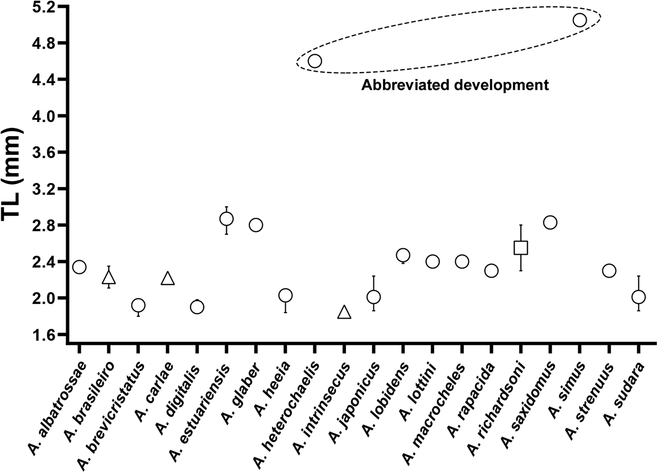
After the publication of other descriptions of Alpheus species with extended and also abbreviated development (A. simus clearly shows an advanced hatching stage), and focusing on the first post-embryonic stage of the genus, we could note that both species with abbreviated larval development described to date (A. heterochaelis and A. simus) hatch with some structures at the same level of development as in species with extended larval development (e.g. telson formula of 7 + 7, segmentation of antennal scale and endopods of maxillipeds). Nevertheless, subsequent morphogenesis is faster, as observed in A. heterochaelis (Knowlton, Reference Knowlton1973). Regarding more specific morphological patterns of species with abbreviated development, we can notice: exopod of maxilla (scaphognathite) margin with 8–11 plumose setae, while all the species with extended development have 4–5; and presence of rudimentary pleopods and all 5 pereiopods, including the presence of chelae on the first and second ones (Knowlton Reference Knowlton1973, tables 1 and 4, pp. 274 and 301, respectively). Some aforementioned characteristics (presence of pleopods and number of plumose setae on scaphognathite) are thus candidates to be used as indicative of the type of larval development of Alpheus species, based on the morphology of newly hatched larvae. In respect to size of larvae, species with abbreviated development were revealed to be the largest Alpheus larvae (4–6 mm for A. heterochaelis and 5.05 mm of TL for A. simus), approximately double the other species (Figure 3). Therefore, size at hatching is another robust indicator of development type, as suggested by Knowlton (Reference Knowlton1973).
Total length (TL mm) values of first zoeal stage (Zoea I) of already described species of Alpheus Fabricius, 1798. Data sources are referenced in Table 1. (○) Mean with minimum and maximum values, when applicable; (Δ) Mean and standard deviation values; (□) Values calculated from the data provided by the studies.

Some structures showed marked differences between the two species with abbreviated development. Alpheus heterochaelis has sessile eyes and rostrum absent (as most other Alpheus species) while A. simus descriptions mention the eyes as ‘semi-pedunculated’ and rostrum present. However, these characteristics are not visible in the illustrations of Wehrtmann & Albornoz (Reference Wehrtmann and Albornoz2002, figure 1, p. 606). The peduncle of antennule of A heterochaelis and A. simus also differed in both descriptions (unsegmented in the former species and 2-segmented in the latter). Intriguingly, this structure was also described as 2-segmented for A. saxidomus in the same study (Wehrtmann & Albornoz, Reference Wehrtmann and Albornoz2002), a species with typical characteristics of extended development. Some possible explanations for this fact (presence of the 2-segmented peduncle of antennule) in zoeae I of the Alpheus species are discussed below.
As pointed out by Pescinelli et al. (Reference Pescinelli, Pantaleão, Mantelatto and Costa2017), some Alpheus species were not described in detail, which makes comparisons difficult to perform. That is the case of A. rapacida and A. strennus (Prasad & Tampi, Reference Prasad and Tampi1957), and A. glaber and A. macrocheles (Lebour, Reference Lebour1932). In the description of A. rapacida there are some characteristics (broad rostrum, stalked eyes, telson with 8 + 8 marginal setae, and the endopod dactyl of the third maxilliped with long spine) that are common in the second larval stage of Alpheus with extended development. Similarly, Lebour (Reference Lebour1932) pointed out some advanced characteristics, such as pedunculated eyes and rostrum showing for A. glaber. It is plausible to consider that these species have some morphological exceptions in zoea I, as reported for A. formosus and A. malleator by Pasinatto et al. (Reference Pasinatto, Mantelatto and Terossi2020), and for another alpheoid shrimp, Athanas nitescens (Leach, 1813), described by Bartilotti et al. (Reference Bartilotti, Calado and Dos Santos2005). The larvae of A. nitescens hatch with some advanced features such as the presence of stalked eyes and the antennular peduncle segmented, but still with a telson formula 7 + 7.
On the other hand, in some alpheid species the first moult (zoea I to II) occurs relatively very fast (e.g. A. heterochaelis in 6 h, A. lobidens and A. sudara in 18 h) as already described by Brooks & Herrick (Reference Brooks and Herrick1892). Thus, it is possible that Prasad & Tampi (Reference Prasad and Tampi1957) and Lebour (Reference Lebour1932) have lost the first stage of A. rapacida and A. glaber, respectively, as reported by Al-Kholy (Reference Al-Kholy1960) for A. rapax. Interestingly, Lebour (Reference Lebour1932) mentioned that A. glaber ‘jumped’ the second stage, changing to a typical third stage with just one moult, in the laboratory. A possible loss of the first stage in the laboratory could explain the telson formula of 8 + 8 (A. rapacida), which is a characteristic of a second larval stage, more than a characteristic of a species with abbreviated development, according to the revision presented here and the generalization of Guerao & Cuesta (Reference Guerao, Cuesta, Martin, Olesen and Hoeg2014), and even considering the case of A. nitescens. In such cases, a redescription of the species seems to be necessary to elucidate these doubts, as was carried out with A. nitescens by Bartilotti et al. (Reference Bartilotti, Calado and Dos Santos2005). Additionally, special attention should be given to conserve zoea I samples immediately after hatching.
Taxonomic considerations
The description of the second species (A. carlae) of a recently described complex, A. armillatus (see Anker, Reference Anker2012), and the merger of the generated data to the comparative table initially performed by Pescinelli et al. (Reference Pescinelli, Pantaleão, Mantelatto and Costa2017), leads us to speculate whether the results obtained to date support the recent separation of the species based on morphology (Anker, Reference Anker2012) and molecular characteristics (Almeida et al., Reference Almeida, Terossi, Araújo-Silva and Mantelatto2013, 2014). In fact, there are differences between the zoeae of A. brasileiro and A. carlae.
The main differences to separate the larvae of two species of A. armillatus complex were found at basial endite of maxillule and maxilla, coxal endite of maxilla, types of setae on the endopods of first maxilliped, number of setae on the basis of first and second maxillipeds, and number of setae on the endopod of third maxilliped. Regarding similarities, the following structures showed the same patterns on both species, and also with two other species (A. formosus and A. malleator): coxal endite of maxillule (with 6 setae) and endopod of second maxilliped (4-segmented with 1, 0, 1, 5 setae) (Table 2).
After the inclusion of recent descriptions in the review of the genus, the single characteristic that now enables the distinction of A. brasileiro is the presence of 4 simple setae and 2 sparsely plumose setae on the coxal endite of maxillule (A. carlae, A. formosus and A. malleator also have 6 setae, but 5 of them are simple in these species). Considering the number of species of the genus in relation to the number of species with at least the zoea I described (N = 313/21), it would be premature to affirm if the larval morphology supports this complex.
Challenges for the advance of knowledge and conclusions
In the present study, some structures evidenced the difficulty in generalizing when we work with descriptions conducted at different times, equipment, accuracy and researchers. A good example is the antennule. Although the number of outgrowths (e.g. aesthetascs, setae and spines) found in the primary flagellum is practically constant, some species have notable exceptions, such as A. rapacida and A. strenuus (Prasad & Tampi, Reference Prasad and Tampi1957) (Table 2). Even for the other species with extended development, besides a plumose seta in the primary flagellum, there are 4 other outgrowths, for which the definition is very variable in the literature, being referred to sometimes as simple setae and other times as aesthetascs. This example illustrates the need to use caution to generalize morphological characteristics, since in many cases it is not possible to interpret if they are real variations or possible misidentifications during the descriptions. Nevertheless, it was possible to separate the two species described here from the others, based on the morphology of zoea I.
Even if we consider only Brazilian species of Alpheus, of the 34 species already registered and considering the present study, only six (17.6%) had at least the first larval stage described: Alpheus estuariensis by Pires et al. (Reference Pires, Abrunhosa and Maciel2008); A. brasileiro by Pescinelli et al. (Reference Pescinelli, Pantaleão, Mantelatto and Costa2017); A. formosus and A. malleator by Pasinatto et al. (Reference Pasinatto, Mantelatto and Terossi2020); A. carlae and A. intrinsecus from the present study. Therefore, we conclude that the quantity of larval descriptions available is still insufficient to draw conclusive assertions in relation to taxonomic proposals. We leave here an appeal for new descriptions, which will allow the advancement of other important areas, such as the clarification of the phylogenetic relationships of the group and the investigation of larval ecology.
Supplementary material
The supplementary material for this article can be found at https://doi.org/10.1017/S0025315420000855
Acknowledgements
The authors thank Dr Robert E. Knowlton who kindly sent us a copy of his manuscript and Dr Alexandre O. Almeida for help in confirming identification of parental females and with some updated literature. We thank anonymous reviewers for their suggestions that helped to improve the quality of this manuscript during the review process.
Financial support
Support for this study was provided by grants from the multidisciplinary research project by São Paulo Research Foundation – FAPESP (Thematics BIOTA 2010/50188-8 and INTERCRUSTA 2018/13685-5), which aims to produce a fine-scale assessment of the marine decapod biodiversity of the State of São Paulo. Complementary support came from the Coordination for the Improvement of Higher Education Personnel – CAPES – Financial Code 001 (Ciências do Mar II Proc. 2005/2014–23038.004308/2014-14). Thanks are also due to the Brazilian National Council for Scientific and Technological Development – CNPq to research scholarships, to J.A.F.P. (PDJ 151105/2019-7), to R.A.P. (140608/2015-0), to R.C.C. (PQ 306672/2018-9) and to F.L.M. (PQ 302253/2019-0). J.A.F.P. thanks the CAPES for the award of a doctoral and post-doctoral PNPD scholarships.