Highlights
-
Hyperbaric oxygen therapy (HBOT) improves best-corrected visual acuity in retinal artery occlusion (RAO) patients with a favorable safety profile.
-
Patients receiving HBOT show lower rates of glaucoma (7.89% vs. 15.79%) and stroke (4.3% vs. 16.6%) compared to controls.
-
Further large-scale prospective trials are needed to validate HBOT’s role in RAO treatment due to study variability.
Introduction
Retinal artery occlusion (RAO) is a rare but vision-threatening condition that demands prompt intervention. It is often referred to as the stroke of the eyes, since it leads to a rapid and profound reduction in retinal perfusion, resulting in irreversible retinal damage if not treated promptly.Reference Chen, Singh, Madike and Cugati1 Despite its critical nature, effective therapeutic options for RAO remain elusive, and current management strategies are largely supportive, focusing on mitigating further vascular insults rather than reversing ischemic damage.
Hyperbaric oxygen therapy (HBOT) has emerged as a potential treatment modality for RAO, leveraging its ability to enhance tissue oxygenation and potentially salvage ischemic retinal tissue. By delivering high concentrations of oxygen under increased atmospheric pressure, HBOT aims to restore oxygenation in ischemic areas, reduce hypoxic damage and improve visual outcomes.Reference Lee, Choi, Jeon, Chang, Wang and Hong2,Reference Chiabo, Kauert and Casolla3 However, the use of HBOT in RAO remains debatable, with limited evidence from small-sample studies and inconsistent reporting of clinical outcomes.
In this context, we conducted a systematic review and meta-analysis to evaluate the efficacy and safety of HBOT in RAO management. By synthesizing data from available studies, we aim to provide insights into HBOT’s potential role in improving visual outcomes and reducing complications associated with RAO.
Methods
We followed guidelines in the Preferred Reporting Items for Systematic reviews and Meta-Analyses (PRISMA) guidelines and the Cochrane Handbook for Systematic Reviews of Interventions while conducting this systematic review and meta-analysis.Reference Higgins, Thomas and Chandler4,Reference Moher, Liberati, Tetzlaff, Altman and Group5
Search and eligibility criteria
A search process was conducted from inception until November 9, 2024, across PubMed, Scopus and Cochrane CENTRAL databases using the following search strategy: ([“Hyperbaric oxygen therapies” OR “Hyperbaric oxygen therapy” OR “Hyperbaric oxygen treatment” OR “Hyperbaric oxygenation” OR “Hyperoxic therapy” OR “Hyperoxic treatment” OR “Oxygenation therapy” OR “Oxygenation treatment” OR “HBOT”] AND [“Central retinal artery occlusion” OR “Central retinal artery obstruction” OR “CRAO” OR “retinal artery obstruction” OR “retinal artery occlusion” OR “Retinal artery stenosis” OR “Ischemic retinal disease” OR “Retinal infarction”]).
We included all clinical trials and observational studies that assessed the efficacy or safety of HBOT specifically for RAO. Studies were required to have two groups: the HBOT group and a control group. We excluded review articles, editorials and abstracts. Additionally, single-arm studies that included only the HBOT group were also excluded.
Screening and data extraction
Screening and data extraction were performed by two independent authors, with disagreements resolved through consultation with the first author and, if necessary, a fourth co-author. Screening was conducted using Rayyan softwareReference Ouzzani, Hammady, Fedorowicz and Elmagarmid6 in two stages: title and abstract screening and full-text screening. Data extraction was carried out using structured Excel sheets, which included the following information:
-
i. Summary data: Study ID, study design, study duration, end follow-up time, population characteristics, details of the intervention, comparator details, outcomes measured, p-values for the comparison of intervention versus control and an overall summary of the study.
-
ii. Baseline data: Sample size, age, sex, baseline visual acuity (VA), time from symptom onset to treatment and comorbidities.
-
iii. Outcomes: VA, best-corrected visual acuity (BCVA), number of lines of improvement in VA and adverse events (AEs) including neovascular glaucoma, seizures, anxiety, asymptomatic bradycardia, stroke, ear barotrauma, epistaxis and others.
Risk of bias assessment
We conducted an evaluation of bias risk using the Newcastle-Ottawa Scale (NOS) tool.Reference Wells, Wells, Shea, Shea, OConnell and Peterson7 The overall quality of each included study was assessed using the NOS based on three domains: Selection (maximum 4 points), Comparability (maximum 2 points) and Outcome (maximum 3 points). Studies were rated as “Good Quality” if they scored 3–4 points in the Selection domain, 1–2 points in Comparability and 2–3 points in Outcome. Studies were considered to be of “Fair Quality” if they scored exactly 2 points in Selection, 1–2 in Comparability and 2–3 in Outcome. Studies were classified as “Poor Quality” if they scored 0–1 point in Selection, 0 in Comparability or 0–1 in Outcome.
Statistical analysis
The statistical evaluation was carried out using RevMan software. We evaluated either the pooled mean difference (MD) with a 95% confidence interval (CI) for continuous variables or the pooled odds ratio (OR) with a 95% CI for categorical variables. Across all studies, we used the Meta-Analysis Accelerator tool to calculate the change from the baseline.Reference Abbas, Hefnawy and Negida8 It was used to calculate the pooled mean and standard deviation (SD) for individual patients’ data in the study conducted by Aisenbery and colleagues.Reference Aisenbrey, Krott, Heller, Krauss, Rossler and Heimann9 Furthermore, it was used to convert median and range to mean and SD in the study conducted by Kim et al.,Reference Kim, Nam and Park10 and in the study conducted by Kalaw et al.,Reference Kalaw, Chartrand and Wedekind11 it was used to convert CI to SD. We utilized a fixed-effect model in the absence of significant heterogeneity or a random-effects model when significant heterogeneity was present.Reference Higgins, Thomas and Chandler4
We examined statistical heterogeneity among the studies employing the chi-square test and the I-squared (I 2) statistic. An I 2 value exceeding 50% or a P-value lower than 0.1 indicated substantial heterogeneity. In instances of marked heterogeneity, a sensitivity analysis (leave-one-out) was performed by excluding outlier studies to mitigate the heterogeneity to an insignificant level. Due to the small number of studies included in each analysis (fewer than 10), we were unable to reliably conduct Egger’s regression test for publication bias following Cochrane guidelines.Reference Higgins, Thomas and Chandler4
Results
Search and screening
Out of 269 total studies found in the searched database, 221 were retained after removing duplicates. Title and abstract screening further reduced this to 24 studies, and ultimately, 9 studiesReference Lee, Choi, Jeon, Chang, Wang and Hong2,Reference Aisenbrey, Krott, Heller, Krauss, Rossler and Heimann9–Reference Rozenberg, Hadad and Peled16 were included in this systematic review and meta-analysis (Figure 1).
PRISMA flow diagram of the search and screening process.

Summary of included studies and baseline characteristics
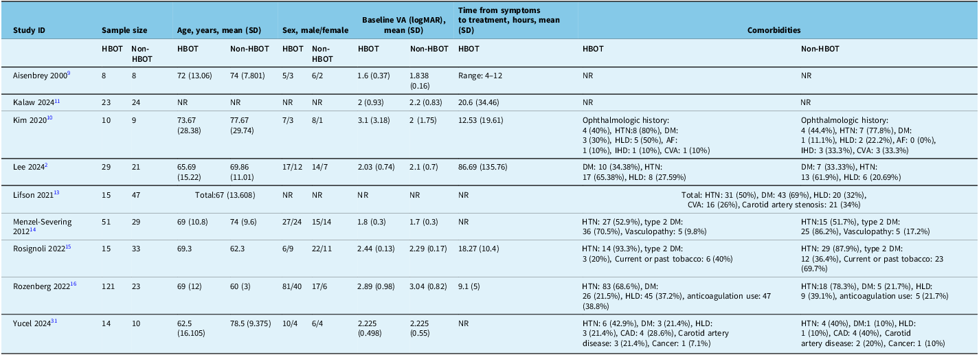
Across the nine studies, the HBOT group consisted of 286 patients, while the non-HBOT group included 213 patients. The mean age across studies ranged from 62.5 to 73.67 years in the HBOT group and 60 to 78.5 in the non-HBOT group, predominantly representing an older patient population. Gender distribution was relatively balanced, with a slight male predominance in the HBOT group. Baseline VA measurements showed considerable variation, with logMAR values ranging from 1.6 to 3.1. Common comorbidities included hypertension, diabetes mellitus and hyperlipidemia. Time from symptom onset to treatment varied between studies, with reported ranges spanning from 9.1 to 86.69 hours. Further details regarding patient characteristics, baseline measurements and study-specific information are provided in Tables 1 and 2.
Summary of included studies

Table 1 Long description
The table compares nine studies on hyperbaric oxygen therapy (HBOT) for central retinal artery occlusion (CRAO). It includes columns for study ID, study design, duration, end follow-up time, population, intervention, comparator, outcomes measured, P-value of intervention versus control, and summary. Each row provides detailed information for a specific study, including the study design, duration, population characteristics, intervention details, comparator details, outcomes measured, P-values, and a summary of the study's findings. The table highlights the variability in study designs, populations, interventions, and outcomes, providing a comprehensive overview of the research on HBOT for CRAO.
CRAO = central retinal artery occlusion; HBOT = hyperbaric oxygen therapy; VA = visual acuity; NVG: neovascular glaucoma; logMAR = logarithm of the minimum angle of resolution; SD = standard deviation; BCVA = best-corrected visual acuity; EDI-OCT = enhanced depth imaging optical coherence tomography; CFT = central foveal thickness; CCT = central choroid thickness; ORLT = outer retinal thickness; IRLT = inner retinal layer thickness; CVA = cerebrovascular accident; NR = not reported.
Baseline characteristics

Table 2 Long description
The table compares baseline characteristics of patients in studies involving HBOT and non-HBOT groups. It has 10 rows and 8 columns. The columns are Study ID, Sample size, Age, Sex, Baseline VA, Time from symptoms to treatment, and Comorbidities. The rows list individual studies with their respective data. Each study provides details on the number of patients in HBOT and non-HBOT groups, mean age with standard deviation, gender distribution, baseline visual acuity (VA) in logMAR with standard deviation, mean time from symptoms to treatment with standard deviation, and common comorbidities. Notable trends include variations in baseline VA and time from symptom onset to treatment across studies. Comorbidities frequently mentioned include hypertension, diabetes mellitus, and hyperlipidemia.
HBOT = hyperbaric oxygen therapy; VA = visual acuity; logMAR = logarithm of the minimum angle of resolution; NR = not reported; SD = standard deviation; DM = diabetes mellitus; HTN = hypertension; HLD = hyperlipidemia; AF = atrial fibrillation; IHD = ischemic heart disease; CVA = cerebrovascular accident; CAD = coronary artery disease.
Risk of bias assessment
The nine studies were assessed using the NOS tool. Five of these studies received an overall good quality score of 8 out of 9. Reference Lee, Choi, Jeon, Chang, Wang and Hong2,Reference Aisenbrey, Krott, Heller, Krauss, Rossler and Heimann9,Reference Kalaw, Chartrand and Wedekind11,Reference Eski Yucel, Bodur and Yucel12,Reference Rozenberg, Hadad and Peled16 Three studies were deemed poor, with an overall score of 7 out of 9, primarily due to receiving a score of zero in the comparability domain.Reference Kalaw, Chartrand and Wedekind11,Reference Lifson, Salloum, Kurochkin, Bivona, Yin and Alpert13,Reference Menzel-Severing, Siekmann, Weinberger, Roessler, Walter and Mazinani14 Additionally, one study was classified as poor, with an overall score of 6 out of 9, also due to a lack of score in the comparability domain.Reference Kim, Nam and Park10 For details about the score of each domain, refer to Supplementary Table 1.
Meta-analysis
Visual acuity
Four studies reported VA data, with a total of 65 patients in the HBOT group and 64 in the non-HBOT group. The initial analysis did not reveal any significant differences in VA between the two groups of patients with RAO (MD: –0.04, 95% CI: [–0.31, 0.23], P = 0.77) (Figure 2). However, significant heterogeneity was detected (P = 0.04, I 2 = 64%). Therefore, a leave-one-out analysis was conducted, which identified the study by Rosignoli et al.Reference Rosignoli, Chu, Carter, Johnson, Sohn and Bahadorani15 as the source of heterogeneity. After removing this study, the results remained insignificant (MD: –0.16, 95% CI: [–0.44, 0.13], P = 0.28) (Figure 3).
Forest plot of mean difference in visual acuity and best-corrected visual acuity in the hyperbaric oxygen therapy (HBOT) group vs. no HBOT group.

Leave-one-out analysis of visual acuity and best-corrected visual acuity in the hyperbaric oxygen therapy (HBOT) group vs. no HBOT group.

Figure 3 Long description
A table comparing visual acuity and best corrected visual acuity between experimental and control groups. The table has 10 rows and 12 columns. Column headers are: Study or Subgroup, Experimental Mean, Experimental SD, Experimental Total, Control Mean, Control SD, Control Total, Weight, Mean Difference IV, Random, 95% CI, and Mean Difference IV, Random, 95% CI. Row labels include study names and subtotals for visual acuity and best corrected visual acuity. Row 1: Aisenbry 2000 (3 months), Experimental Mean: -0.137, Experimental SD: 0.393, Experimental Total: 8, Control Mean: -0.175, Control SD: 0.264, Control Total: 8, Weight: 43.9%, Mean Difference IV, Random, 95% CI: 0.04 [-0.29, 0.37]. Row 2: Menzel-Severing 2012 (3 months), Experimental Mean: -0.3, Experimental SD: 0.615, Experimental Total: 28, Control Mean: -0.1, Control SD: 0.529, Control Total: 13, Weight: 38.4%, Mean Difference IV, Random, 95% CI: -0.20 [-0.57, 0.17]. Row 3: Yucel 2024, Experimental Mean: -0.45, Experimental SD: 0.846, Experimental Total: 14, Control Mean: 0.1, Control SD: 0.696, Control Total: 10, Weight: 17.7%, Mean Difference IV, Random, 95% CI: -0.55 [-1.17, 0.07]. Row 4: Subtotal (95% CI), Experimental Total: 50, Control Total: 31, Weight: 100.0%, Mean Difference IV, Random, 95% CI: -0.16 [-0.44, 0.13]. Row 5: Kim 2024, Experimental Mean: -0.925, Experimental SD: 1.76, Experimental Total: 10, Control Mean: 0.85, Control SD: 1.551, Control Total: 9, Weight: 10.7%, Mean Difference IV, Random, 95% CI: -1.77 [-3.26, -0.29]. Row 6: Lee 2024 (6 months), Experimental Mean: -0.48, Experimental SD: 1.096, Experimental Total: 29, Control Mean: 0.01, Control SD: 0.938, Control Total: 21, Weight: 50.0%, Mean Difference IV, Random, 95% CI: -0.49 [-1.06, 0.08]. Row 7: Rozenberg 2022, Experimental Mean: -0.74, Experimental SD: 1.298, Experimental Total: 121, Control Mean: -0.24, Control SD: 1.559, Control Total: 23, Weight: 39.3%, Mean Difference IV, Random, 95% CI: -0.50 [-1.18, 0.18]. Row 8: Subtotal (95% CI), Experimental Total: 160, Control Total: 53, Weight: 100.0%, Mean Difference IV, Random, 95% CI: -0.63 [-1.14, -0.12]. Row 9: Heterogeneity for Visual acuity (VA), Tau² = 0.02; Chi² = 2.92, df = 2 (P = 0.23); I² = 31%. Test for overall effect: Z = 1.08 (P = 0.28). Row 10: Heterogeneity for Best corrected visual acuity (BCVA), Tau² = 0.05; Chi² = 2.62, df = 2 (P = 0.27); I² = 24%. Test for overall effect: Z = 2.44 (P = 0.01).
Best-corrected visual acuity
Four studies provided BCVA data, involving a total of 183 patients in the HBOT group and 77 patients in the non-HBOT group. The initial analysis showed no significant differences in BCVA between the two groups of patients with RAO (MD: –0.43, 95% CI: [–0.99, 0.12], P = 0.13) (Figure 2). However, slight significant heterogeneity was observed (P = 0.09, I 2 = 54%). To investigate this further, a leave-one-out analysis was performed, which identified the study by Kalaw et al.Reference Kalaw, Chartrand and Wedekind11 as the source of heterogeneity. After excluding this study, the results indicated a significant improvement in BCVA favoring the HBOT group (MD: –0.63, 95% CI: [–1.14, –0.12], P = 0.01) (Figure 3).
Number of lines improvement of visual acuity
Two studies reported improvements in VA, with one study showing at least a three-line improvement and another showing at least a two-line improvement. However, the overall analysis did not reveal any significant difference between the HBOT group (n = 36) and the non-HBOT group (n = 21) (OR: 1.37, 95% CI: [0.43, 4.35], P = 0.59) (Figure 4). The data collected were homogeneous (P = 0.82, I 2 of 0%).
Forest plot of the number of lines improvement of visual acuity in the hyperbaric oxygen therapy (HBOT group) vs. no HBOT group.

Figure 4 Long description
A table comparing the improvement in visual acuity lines between hyperbaric oxygen therapy (HBOT) and no HBOT groups. The table has 10 rows and 7 columns. Column headers are Study or Subgroup, HBOT Events, HBOT Total, No HBOT Events, No HBOT Total, Weight, and Odds Ratio M-H, Fixed, 95% CI. Row labels include subtotals for at least 3 lines improvement and at least 2 lines improvement. Row 1: Menzel-Severing 2012, 10, 28, 4, 13, 70.1%, 1.25 [0.31, 5.11]. Row 2: Subtotal (95% CI), 10, 28, 4, 13, 70.1%, 1.25 [0.31, 5.11]. Row 3: Aisenbrey 2000, 4, 8, 3, 8, 29.9%, 1.67 [0.23, 12.22]. Row 4: Subtotal (95% CI), 4, 8, 3, 8, 29.9%, 1.67 [0.23, 12.22]. Row 5: Total (95% CI), 14, 36, 7, 21, 100.0%, 1.37 [0.43, 4.35]. The table includes heterogeneity and test for overall effect data.
Summary of adverse events
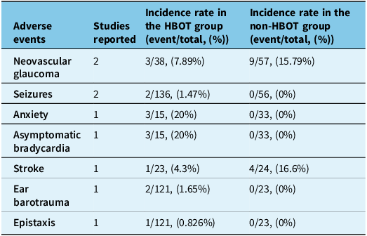
We summarize the AEs in Table 3, which outlines the number of studies reporting each AE, the incident rate of events and the totals in the HBOT group compared to the non-HBOT group.
Summary of adverse events (AEs) in the hyperbaric oxygen therapy (HBOT) group and non-HBOT group

Table 3 Long description
A table comparing adverse events in HBOT and non-HBOT groups. The table has 7 rows and 4 columns. Column headers are Adverse events, Studies reported, Incidence rate in the HBOT group (event/total, (%)), and Incidence rate in the non-HBOT group (event/total, (%)). Row 1: Neovascular glaucoma, 2, 3/38 (7.89 percent), 9/57 (15.79 percent). Row 2: Seizures, 2, 2/136 (1.47 percent), 0/56 (0 percent). Row 3: Anxiety, 1, 3/15 (20 percent), 0/33 (0 percent). Row 4: Asymptomatic bradycardia, 1, 3/15 (20 percent), 0/33 (0 percent). Row 5: Stroke, 1, 1/23 (4.3 percent), 4/24 (16.6 percent). Row 6: Ear barotrauma, 1, 2/121 (1.65 percent), 0/23 (0 percent). Row 7: Epistaxis, 1, 1/121 (0.826 percent), 0/23 (0 percent).
-
Neovascular glaucoma: Higher incidence in the non-HBOT group (15.79%) compared to the HBOT group (7.89%).
-
Seizures: Occurred only in the HBOT group (1.47%), with no events in the non-HBOT group.
-
Anxiety and asymptomatic bradycardia: Both were reported in 20% of HBOT group patients but were absent in the non-HBOT group.
-
Stroke: Higher incidence in the non-HBOT group (16.6%) compared to the HBOT group (4.3%).
-
Ear barotrauma and epistaxis: Reported only in the HBOT group (1.65% and 0.826%, respectively), with no cases reported in the non-HBOT group. No further data were available in the included studies regarding the potential interventions that are typically pursued in case of ear barotrauma.
Discussion
This systematic review and meta-analysis aim to provide important insights into the efficacy and safety of HBOT for RAO. Our analysis included nine studies with a total of 499 patients (286 HBOT, 213 non-HBOT). The results show that HBOT might be associated with better BCVA in patients with RAO when heterogeneity is accounted for, although no significant differences were found in uncorrected VA between HBOT and non-HBOT. Serious AEs reported with HBOT were rare and included seizures, ear barotrauma and epistaxis. Interestingly, HBOT was notably associated with lower rates of neovascular glaucoma and stroke. Our results highlight the need for high-quality comparative prospective studies with standardized outcome measures to better guide RAO clinical management.
This meta-analysis provides nuanced insights into the potential role of HBOT in managing RAO in the acute setting. While our analysis showed a significant improvement in BCVA after removing one heterogeneous study, the overall evidence remains controversial. The heterogeneity observed in our analysis underscores the complexity of RAO management. Factors such as time from symptom onset, patient age and underlying comorbidities likely influence treatment outcomes. The varied baseline VA measurements across studies further complicate direct comparisons.
HBOT functions through a unique mechanism of action, primarily by increasing tissue oxygenation, which might ultimately mitigate ischemic damage to the retinal tissue.Reference Hadanny, Maliar and Fishlev17 It increases tissue oxygenation by delivering 100% oxygen under elevated atmospheric pressure, substantially raising the partial pressure of oxygen in the blood. In RAO, perfusion to the inner retinal layers is compromised due to obstruction of the central or branch retinal artery. However, the outer retina remains perfused through the choroidal circulation, which is supplied by the posterior ciliary arteries. HBOT facilitates enhanced diffusion of oxygen from the well-perfused outer retina toward the ischemic inner retina. This compensatory oxygen delivery may help sustain retinal metabolism, delay irreversible cellular damage and preserve visual function while awaiting spontaneous or therapeutic reperfusion.Reference Beiran, Goldenberg, Adir, Tamir, Shupak and Miller18–Reference Celebi20 Ultimately, by saturating tissues with high-concentration oxygen, HBOT aims to reduce hypoxia, support cellular metabolism and potentially limit the irreversible damage associated with RAO.Reference Celebi20 Therefore, HBOT has a distinctive mechanism of action from thrombolytic interventions considered for RAO patients, including systemic intravenous thrombolysis (IVT)Reference Huang, Wang and Zhang21 and intra-arterial fibrinolysis.Reference Page, Khattar and White22 One of the main limitations of HBOT in the treatment of RAO is the limited availability of hyperbaric chambers in the hyperacute setting. HBOT requires specialized equipment and trained personnel and is often located in tertiary centers or facilities with limited hours of operation, making timely access challenging for many patients. This logistical barrier limits the widespread implementation of HBOT as a first-line treatment despite its physiologic rationale and potential benefit when administered early.Reference Butler and Moon23
It is crucial to understand the current evidence in the literature regarding the available acute treatment modalities for RAO other than HBOT. Systemic IVT, primarily using tissue plasminogen activator, presents another potential treatment strategy for RAO.Reference Mac Grory, Schrag and Biousse24 Several studies have shown that while systemic IVT might theoretically restore retinal blood flow, the permanent visual damage often occurs within the first few hours of occlusion, rendering late interventions largely ineffective.Reference Kozner, Eichenmann, Ceska Burdova, Pavlikova, Hlozanek and Dotrelova25,Reference Schultheiss, Hartig and Spitzer26 However, its application remains highly debated due to significant limitations.Reference Stretz, Paddock and Burton27 The narrow therapeutic window – typically less than 4.5 hours from symptom onset – severely restricts its clinical utility. Moreover, the risk of systemic complications, particularly hemorrhagic transformation, makes clinicians hesitant to routinely employ this approach.Reference Huang, Wang and Zhang21
On the other hand, the EAGLE trial provided valuable insights into the use of local intra-arterial thrombolysis (IAT) for central RAO (CRAO). This approach directly targets the occlusive thrombus through localized intervention, offering a mechanistic approach to addressing the underlying pathophysiology of CRAO. Although the trial was terminated early after the first interim analysis due to similar efficacy between IAT and conservative management and a higher rate of AEs in the intervention group, it underscored critical safety considerations and the challenges of translating promising interventional approaches into clinical benefit. Rather than representing a therapeutic advancement per se, the EAGLE trial highlights the necessity of well-designed, adequately powered trials to evaluate interventions for CRAO.Reference Schumacher, Schmidt and Jurklies28
To summarize, while our primary analysis focused on the efficacy and safety of HBOT, it is essential to contextualize its potential role among other acute interventions for RAO. IVT, typically administered within 4.5 hours of symptom onset, has demonstrated modest visual recovery rates ranging from 20% to 35% in observational studies but carries a risk of systemic bleeding and is often underutilized due to delayed presentation.Reference Mac Grory, Lavin, Kirshner and Schrag29,Reference Schrag, Youn, Schindler, Kirshner and Greer30 IAT, explored in the EAGLE trial, showed no superiority to conservative therapy and was associated with higher complication rates, leading to early termination of the study.Reference Schumacher, Schmidt and Jurklies28 In contrast, HBOT may offer visual improvement in 30%–45% of treated patients when initiated within 12–24 hours. In one of the largest retrospective studies, minor and reversible AEs occurred in approximately 5.5% of patients.Reference Hadanny, Maliar and Fishlev17 However, access to HBOT is often restricted by the limited availability of hyperbaric chambers, which constrains timely intervention.Reference Butler and Moon23 The current guidelines from the American Heart Association suggest that while IVT may be considered in select CRAO patients, evidence remains limited, and HBOT is not yet formally endorsed as standard of care.Reference Mac Grory, Schrag and Biousse24 Therefore, the optimal management strategy continues to evolve, and head-to-head comparative trials are needed to guide treatment selection.
Strengths and limitations
To our knowledge, our meta-analysis is the largest pooled analysis on the efficacy and safety of HBOT for RAO patients, including 9 studies with a total of 499 patients. However, limitations in the current body of research on the efficacy and safety of HBOT for RAO are notable. The small number of studies, relatively limited patient populations, and methodological variations suggest the need for more robust, standardized research. Prospective, randomized controlled trials with larger sample sizes and consistent outcome measures are essential to definitively establish the role of HBOT in RAO management. Regarding the reporting of the outcomes, VA data extracted from the included studies were not explicitly stated as best corrected in any of the studies. Therefore, we interpreted these as uncorrected VA unless otherwise specified, which is a limitation inherent to the retrospective nature of these studies. Furthermore, due to the lack of availability of granular data in the included studies, we were not able to conduct a subgroup analysis according to the severity of RAO (branch of retinal artery occlusion [BRAO] vs. CRAO), taking into consideration that a few included studies did not include BRAO patients,Reference Lee, Choi, Jeon, Chang, Wang and Hong2,Reference Kim, Nam and Park10,Reference Lifson, Salloum, Kurochkin, Bivona, Yin and Alpert13–Reference Rozenberg, Hadad and Peled16,Reference Yucel, Bodur and Yucel31 while others included both CRAO and BRAO patients.Reference Aisenbrey, Krott, Heller, Krauss, Rossler and Heimann9,Reference Kalaw, Chartrand and Wedekind11 Also, for the same reason, no subgroup analysis was conducted according to the time interval between the onset of symptoms and the HBOT initiation. Lastly, while HBOT is typically considered for non-arteritic RAO, only a few of the included studies specifically stated this in their inclusion criteria.Reference Lee, Choi, Jeon, Chang, Wang and Hong2,Reference Menzel-Severing, Siekmann, Weinberger, Roessler, Walter and Mazinani14,Reference Rozenberg, Hadad and Peled16,Reference Yucel, Bodur and Yucel31
Conclusion
Clinical decision-making regarding the use of HBOT for RAO remains challenging. While our analysis might suggest potential benefits of HBOT, it should not be considered a standalone solution. Clinicians must continue to evaluate individual patient factors, consider multiple treatment modalities and recognize the critical importance of rapid intervention.
Supplementary material
The supplementary material for this article can be found at https://doi.org/10.1017/cjn.2025.10364.
Acknowledgments
None.
Author contributions
-
ME: contributed to the conception and design of the study, acquisition, analysis and interpretation of data, drafting, revising and final approval of the article.
-
HS: contributed to the acquisition and extraction of data, conducting statistical analysis and drafting the article.
-
AA: contributed to the extraction of data and final approval of the article.
-
AB: contributed to the extraction of data and final approval of the article.
-
HSG: contributed to the extraction of data and final approval of the article.
-
MHG: contributed to the extraction of data and final approval of the article.
-
AS: contributed to the extraction of data and final approval of the article.
-
MIK: contributed to the extraction of data and final approval of the article.
-
MA: contributed to the extraction of data and final approval of the article.
-
ZN: contributed to the extraction of data and final approval of the article.
-
CM: contributed to the extraction of data and final approval of the article.
-
DAS: contributed to the extraction of data and final approval of the article.
-
KAH: contributed to the extraction of data and final approval of the article.
-
DE: contributed to the extraction of data and final approval of the article.
-
GK: contributed to the extraction of data and final approval of the article.
-
CDG: contributed to the extraction of data and final approval of the article.
-
FA: contributed to the conception and design of the study, drafting, revising and final approval of the article.
Funding statement
The authors received no funding for any part of this work.
Competing interests
The authors report no conflicts of interest.