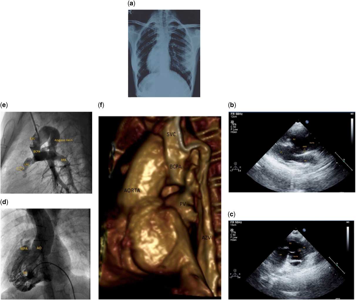
A 17-year-old female presented with progressive effort intolerance and cyanosis, with oxygen saturation of 65%. She had been diagnosed in infancy as having situs inversus totalis with double-outlet right ventricle, large subaortic ventricular septal defect, and severe pulmonic stenosis. A bidirectional cavopulmonary anastomosis was performed at the age of 9 months with subsequent improvement of cyanosis, with oxygen saturation of 88%, and effort tolerance. Echocardiography (Fig 1b and c) confirmed the intracardiac anatomy and demonstrated patency of bidirectional cavopulmonary anastomosis. Cardiac catheterisation (Fig 1d) (Supplementary video 1) demonstrated the classical features of double-outlet right ventricle and left pulmonary artery stenosis. Shunt angiogram was remarkable for left pulmonary artery stenosis and a humongously dilated azygous vein draining the cavopulmonary junction into the inferior caval vein (Fig 1e) (Supplementary video 2). Cardiac CT with three-dimensional reconstruction confirmed the diagnosis (Fig 1f) (Supplementary video 3).
( a ) Chest X-ray posterior-anterior PA view demonstrating situs inversus totalis. ( b ) Two-dimensional (2D) echocardiogram (PLAX view) showing subaortic ventricular septal defect with an overriding dilated aorta with aorto-mitral discontinuity (AMD). ( c ) 2D echocardiogram (modified high parasternal short-axis view) demonstrating two great vessels originating from the right ventricle (RV) with subaortic conus (marked with asterisks). ( d ) Right ventricular angiogram (anterior-posterior view) showing the origin of both great vessels from the RV. ( e ) Shunt angiogram demonstrating patent bidirectional cavopulmonary anastomosis with left pulmonary artery stenosis (marked with arrow) and humongously dilated azygous vein draining into the inferior caval vein. ( f ) Cardiac CT with 3D reconstruction showing dilated azygous vein draining the cavopulmonary junction into the inferior caval vein. SVC=superior vena cava; BCPA=bidirectional cavopulmonary anastomosis; Ao=aota; MPA=main pulmonary artery; LPA=left pulmonary artery; RPA=right pulmonary artery; PV=pulmonary veins; AZV=azygous vein; RA=right atrium; RV=right ventricle; AMD=aorto-mitral discontinuity.

The development of systemic venous anomalies can be found frequently after bidirectional cavopulmonary anastomosis. The incidence ranges from 17Reference Gross, Jonas, Castaneda, Hanley, Mayer and Bridges 1 to 31%Reference Magee, McCrindle, Mawson, Benson, Williams and Freedom.2 A late manifestation as in our patient, however, is extremely rare. Common causes for the development of such collaterals are reduction in caval or pulmonary artery cross-sectional area, presence of pulmonary artery hypoplasia, distortion or obstruction of the anastomosis. The opening of previously existing channels is the presumed aetiology for the development of such collaterals. Collateralisation takes two forms – either systemic venous-to-systemic venous connections or systemic venous-to-pulmonary venous or left atrial connections. Increased cyanosis hence could be a result from either a reduced effective pulmonary blood flow or an increased admixture of pulmonary venous return. Large collaterals as in our case result in clinically unacceptable desaturation and will require either transcatheter-based intervention with coils or surgical ligation.
Acknowledgements
None.
Financial Support
This research received no specific grant from any funding agency, commercial, or not-for-profit sectors.
Conflicts of Interest
None.
Supplementary Material
To view supplementary material for this article, please visit http://dx.doi.org/10.1017/S1047951115002607