INTRODUCTION
This investigation is part of a wider programme examining the role of altered drug metabolism in the development of resistance of Fasciola hepatica to triclabendazole (TCBZ). Biochemical studies have demonstrated that the flavin monooxygenase (FMO) and cytochrome P450 (CYP 450) enzyme pathways are involved in the metabolism of TCBZ by the fluke and are upregulated in TCBZ-resistant flukes (Mottier et al. Reference Mottier, Virkel, Solana, Alvarez, Salles and Lanusse2004; Robinson et al. Reference Robinson, Lawson, Trudgett, Hoey and Fairweather2004; Alvarez et al. Reference Alvarez, Solana, Mottier, Virkel, Fairweather and Lanusse2005). Complementary scanning electron microscope (SEM) studies have shown that co-incubation of TCBZ or its sulphoxide metabolite with metabolic inhibitors, such as methimazole (MTZ), piperonyl butoxide (PB) and ketoconazole (KTZ), leads to greater surface disruption of TCBZ-resistant than TCBZ-susceptible flukes (Devine et al. Reference Devine, Brennan, Lanusse, Alvarez, Trudgett, Hoey and Fairweather2009, Reference Devine, Brennan, Lanusse, Alvarez, Trudgett, Hoey and Fairweather2010a, Reference Devine, Brennan, Lanusse, Alvarez, Trudgett, Hoey and Fairweatherc). This article is a follow-on from the previous SEM paper with PB (Devine et al. Reference Devine, Brennan, Lanusse, Alvarez, Trudgett, Hoey and Fairweather2010a). It examines internal changes to the tegument of the fluke, as visualized by transmission electron microscopy (TEM). While surface changes represent the sum of a number of drug effects, TEM will be more informative with regard to the mechanisms contributing to them and thus will extend understanding of drug action. PB is an established CYP 450 inhibitor and it has been shown to inhibit the metabolism of TCBZ by F. hepatica (Alvarez et al. Reference Alvarez, Solana, Mottier, Virkel, Fairweather and Lanusse2005).
MATERIALS AND METHODS
The protocol for this investigation is the same as that used for the SEM study involving PB (Devine et al. Reference Devine, Brennan, Lanusse, Alvarez, Trudgett, Hoey and Fairweather2010a); the reader is referred to that publication for full details. Briefly, adult flukes of the Cullompton TCBZ-susceptible and the Oberon TCBZ-resistant fluke isolates were pre-incubated in PB at a concentration of 1×10⁻⁴ m for 2 h at 37°C, before transfer to fresh NCTC 135 culture medium for 22 h at 37°C. The fresh medium contained either PB; PB+NADPH (1 nm); PB+NADPH+TCBZ (15 μg/ml); or PB+NADPH+TCBZ.SO (15 μg/ml). A stock solution of PB was initially prepared at a concentration of 1×10⁻¹ m in methanol. Controls at 0 h and 24 h were also prepared. After incubation, the flukes were fixed and processed for transmission electron microscopy (TEM), as described by Devine et al. (Reference Devine, Brennan, Lanusse, Alvarez, Trudgett, Hoey and Fairweather2010b). A minimum of 4 flukes were prepared for each treatment.
RESULTS
After incubation in all drug and inhibitor combinations for 24 h in vitro, the flukes were alive prior to fixation. Sections for TEM were taken from the midbody region of the flukes as this area was shown to be highly affected by drug and inhibitor action in a previous SEM study (Devine et al. Reference Devine, Brennan, Lanusse, Alvarez, Trudgett, Hoey and Fairweather2010a).
Controls
The tegumental ultrastructure of the control specimens was normal. For images of normal morphology, the reader is referred to the papers by Halferty et al. (Reference Halferty, Brennan, Trudgett, Hoey and Fairweather2009, Fig. 4A–C) and Fairweather et al. (Reference Fairweather, Threadgold, Hanna and Dalton1999, Figs 3.3 and 3.4).
Oberon and Cullompton isolates treated with PB and PB+NADPH
The tegumental syncytium retained a relatively normal morphology (Fig. 1A). Below the apical plasma membrane, T1 and T2 secretory bodies were present and existed in normal numbers (Fig. 1B). There was some swelling of the mucopolysaccharide masses surrounding the basal infolds in the basal region of the syncytium (Fig. 1C). The muscle blocks below the basal lamina remained unchanged (Fig. 1C). Within the tegumental cells, there were numerous T1 secretory bodies (Fig. 1D). The cells also contained numerous active and well-developed Golgi complexes and the nuclei and mitochondria retained a normal morphology (Fig. 1D and 1E).
Transmission electron micrographs (TEMs) of the tegumental syncytium, underlying musculature and tegumental cells of adult Fasciola hepatica (Cullompton and Oberon isolates) treated in vitro with PB and PB+NADPH for 24 h. (A) TEM showing the full depth of the tegumental syncytium, from the apical plasma membrane (APM) to the basal lamina (BL). The muscle blocks (Mu) beneath the basal lamina, like the syncytium, retain a normal morphology. m, mitochondrion. (B) A high-power micrograph of the apical region of the tegumental syncytium. Numerous T1 (T1) and T2 (T2) secretory bodies are present below the apical plasma membrane (APM). m, mitochondrion. (C) Basal region of the tegumental syncytium, showing swelling of the mucopolysaccharide masses (arrow). T1 (T1) and T2 (T2) secretory bodies are present in the syncytium. Below the basal lamina (BL), the muscle blocks (Mu) retain a normal morphology. BI, basal infolds; m, mitochondrion. (D) TEM of a T1-type of tegumental cell. Well-developed Golgi complexes (GC) and numerous T1 secretory bodies (T1) are present within the cell. The mitochondria (m) retain a relatively normal morphology. N, nucleus. (E) A high-power image of a well-developed Golgi complex (GC). T1 secretory bodies (T1) are present within the cell and the mitochondria (m) retain a relatively normal morphology.

Cullompton isolate treated with TCBZ and TCBZ.SO
Descriptions of ultrastructural changes brought about by treatment with the two drugs have been published elsewhere and will not be repeated here. The changes observed in the present study matched these descriptions. Therefore, the reader is referred to the paper by Halferty et al. (Reference Halferty, Brennan, Trudgett, Hoey and Fairweather2009) (Fig. 4D–F for TCBZ and Fig. 5A–C for TCBZ.SO).
Cullompton isolate treated with PB+NADPH+TCBZ
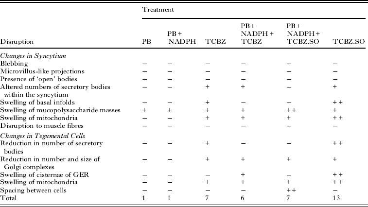
Within the tegumental syncytium, mild swelling of the mucopolysaccharide masses surrounding the basal infolds was observed (Fig. 2A). In the apical region, T1 and T2 secretory bodies were present in normal numbers. The mitochondria present throughout the syncytium were swollen and rounded, with distinct cristae (Fig. 2B and C). There was some swelling of the mucopolysaccharide masses in the basal region, although the basal infolds remained tightly closed. Numerous T1 secretory bodies were present at the base of the syncytium (Fig. 2C). Within Type-1 tegumental cells, T1 secretory bodies were abundant and Golgi complexes were present (Fig. 2D and E). The mitochondria and cisternae of the granular endoplasmic reticulum (GER) were swollen (Fig. 2D). Within the Type-2 tegumental cells, the nucleus and mitochondria appeared relatively normal, and numerous T2 secretory bodies were present (Fig. 2F).
Transmission electron micrographs (TEMs) of the tegumental syncytium, underlying musculature and tegumental cells of adult Fasciola hepatica (Cullompton isolate) following 24 h in vitro treatment with PB+NADPH+TCBZ. (A) TEM showing the full depth of the tegumental syncytium, from the apical plasma membrane (APM) to the basal lamina (BL). Some swelling of the mucopolysaccaride masses (arrow) can be seen in the basal region. m, mitochondrion. (B) A high-power micrograph of the apical region of the syncytium. Numerous T1 (T1) and T2 (T2) secretory bodies are present below the apical plasma membrane (APM). The mitochondria (m) are swollen with distinct cristae (arrow). (C) TEM showing the basal region of the syncytium. The mucopolysaccharide masses (arrow) are slightly swollen just above the basal lamina (BL). The mitochondria (m) are swollen and rounded in appearance. Numerous T1 (T1) secretory bodies are present. BI, basal infolds. (D) TEM showing a T1-type of tegumental cell. There are many T1 secretory bodies (T1) in the cell; Golgi complexes (GC) are also present. Some mitochondria (m) are swollen in appearance, as are the cisternae of the granular endoplasmic reticulum (arrow). (E) A high-power micrograph of a Golgi complex (GC), which retains a normal morphology. N, nucleus. (F) A high-power TEM of a T2-type of tegumental cell. T2 (T2) secretory bodies are present within the cell. The mitochondria (m) present within the cell retain a relatively normal morphology. N, nucleus.

Cullompton isolate treated with PB+NADPH+TCBZ.SO
The tegumental syncytium appeared relatively normal, with only very minor swelling of the mucopolysaccharide masses (Fig. 3A). In the apical region of the tegument, T1 and T2 secretory bodies were present in normal numbers (Fig. 3B). Swelling of the mucopolysaccharide masses was most evident in the basal region of the syncytium, although the membranes of the basal infolds were tightly apposed. T1 secretions were abundant throughout the basal region and the mitochondria present throughout the syncytium were swollen and rounded in appearance (Fig. 3C). There was evidence of spacing between tegumental cells (Fig. 3D). The tegumental cells contained numerous T1 secretory bodies and some Golgi complexes were present, but appeared reduced in size. The nuclei appeared normal but many mitochondria were swollen and rounded in appearance (Fig. 3D).
Transmission electron micrographs (TEMs) of the tegumental syncytium, underlying musculature and tegumental cells of adult Fasciola hepatica (Cullompton isolate) following in vitro treatment for 24 h with PB+NADPH+TCBZ.SO. (A) TEM showing the full depth of the tegumental syncytium, from the apical plasma membrane (APM) to the basal lamina (BL). The mitochondria (m) in the syncytium are rounded in appearance. Slight swelling of the mucopolysaccharide masses (arrow) can be seen above the basal lamina, although the basal infolds (BI) remain tightly closed. (B) A high-power micrograph of the apical region of the tegumental syncytium. Numerous T1 (T1) and T2 (T2) secretory bodies are present below the apical plasma membrane (APM). (C) TEM showing the basal region of the syncytium. Above the basal lamina (BL), the mucopolysaccharide masses (arrow) are swollen, but the basal infolds (BI) remain closed. The mitochondria (m) appear slightly swollen. T1, T1 secretory bodies. (D) TEM showing T1-type tegumental cells. There is spacing between the cells (arrows). T1 secretory bodies (T1) are abundant in number within the cells, but Golgi complexes (GC) appear reduced in size. Some mitochondria (m) have become swollen in appearance. The nuclei (N) of the cells retain a normal morphology.

Oberon isolate treated with TCBZ
Descriptions of ultrastructural changes brought about by drug treatment have been published elsewhere and will not be repeated here. The changes observed in the present study matched these descriptions. Therefore, the reader is referred to the paper by Devine et al. (Reference Devine, Brennan, Lanusse, Alvarez, Trudgett, Hoey and Fairweather2010b) (Figs 12–16).
Oberon isolate treated with TCBZ.SO
The changes to the tegumental syncytium following incubation in TCBZ.SO have been described by McKinstry (Reference McKinstry2008; Chapter 7, Figs 15–20) and will not be repeated in detail here. A brief summary has been given by Devine et al. (Reference Devine, Brennan, Lanusse, Alvarez, Trudgett, Hoey and Fairweather2010b).
Oberon isolate treated with PB+NADPH+TCBZ
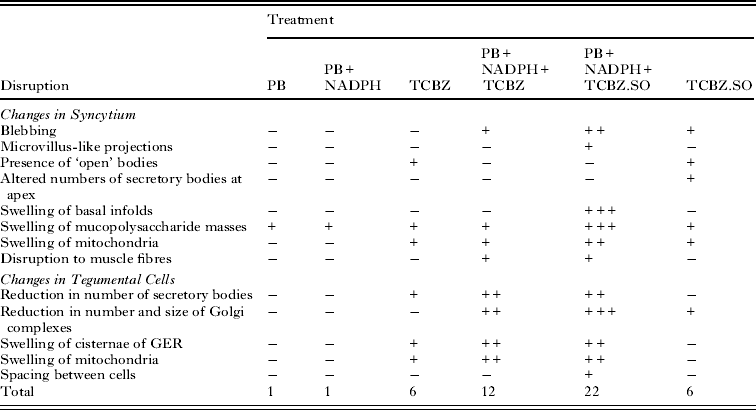
Minimal changes were observed within the tegumental syncytium. Swelling of the mucopolysaccharide masses was limited, although the mitochondria present within the syncytium appeared swollen (Fig. 4A). Small blebs were present along the apical plasma membrane (Fig. 4B). T1 and T2 secretory bodies were present but were abnormal, in that they appeared swollen and electron-lucent (Fig. 4B). The basal infolds above the basal lamina were swollen at their bases (Fig. 4C). Below the tegumental syncytium, the fibres of the muscle blocks appeared more loosely-packed than normal (Fig. 4D). Within the T1-type tegumental cells, T1 secretory bodies were present within the cells, although their numbers were severely reduced and Golgi complexes, if present, were reduced in size (Fig. 4E). The mitochondria and the cisternae of the GER were swollen, but the nuclei retained a normal appearance (Fig. 4E). T2 secretory bodies were present within Type-2 tegumental cells, although they were few in number (Fig. 4F). The mitochondria were swollen, with distinct cristae, and the cisternae of the GER were also swollen (Fig. 4F).
Transmission electron micrographs (TEMs) of the tegumental syncytium, underlying musculature and tegumental cells of adult Fasciola hepatica (Oberon) isolate treated in vitro for 24 h with PB+NADPH+TCBZ. (A) TEM of the tegumental syncytium, showing limited swelling of the mucopolysaccharide masses (arrows) at the distal ends of the basal infolds (BI). The mitochondria (m) appear slightly swollen. APM, apical plasma membrane. (B) High-power micrograph of the apical region of the tegument showing blebs (B) on the apical plasma membrane (APM). Numerous T1 (T1) and T2 (T2) secretory bodies are present within this region, but appear swollen and electron-lucent. (C) TEM showing the basal region of the tegumental syncytium. Above the basal lamina (BL), the amino mucopolysaccharide masses (arrow) are swollen. The mitochondria (m) are also swollen. BI, basal infolds. (D) TEM showing the basal region of the tegumental syncytium and the underlying muscle blocks (Mu). The muscle fibres in the muscle blocks (Mu) are not as tightly-packed as normal. BL, basal lamina. (E) TEM showing a T1-type of tegumental cell. The Golgi complexes (GC) present in the cell appear abnormal in shape and reduced in size. T1 (T1) secretory bodies are reduced in number within the cell. The cisternae of granular endoplasmic reticulum (arrow) are severely swollen and the mitochondria (m) are swollen and rounded in appearance. The nucleus (N) retains a normal morphology. Inset shows a Golgi complex (GC) which is reduced in size. (F) A high-power TEM of a T2-type of tegumental cell. A small number of T2 (T2) secretory bodies are present within the cell. The mitochondria (m) are rounded with swollen cristae (arrow). N, nucleus.

Oberon isolate treated with PB+NADPH+TCBZ.SO
The major feature of the tegumental syncytium was the severe swelling of the basal infolds, accompanied by swelling of the mucopolysaccharide masses surrounding them (Fig. 5A). Along the apex of the syncytium, blebbing of the tegumental surface was observed (Fig. 5B). At higher magnifications, microvillus-like projections were seen along the apical plasma membrane (Fig. 5C). T1 and T2 secretory bodies were present within the syncytium, but appeared swollen and electron-lucent (Fig. 5C). The mitochondria present within the syncytium were swollen and assumed a rounded appearance, rather than the typical cylindrical shape (Fig. 5D). The sub-tegumental muscle blocks contained fewer fibres than normal and the fibres were less tightly packed than normal (Fig. 5D). Spaces were observed between the tegumental cells. T1 and T2 secretory bodies were present in their respective tegumental cells, although the number of secretory bodies was fewer than normal in both cell types (Fig. 5E and F). Within the Type-1 tegumental cells, the T1 secretory bodies appeared swollen and were severely reduced in number (Fig. 5E). Golgi complexes were not observed in the cells. The mitochondria were swollen, with distinct cristae, but the nuclei retained a normal appearance (Fig. 5E). In the Type-2 tegumental cells, the perinuclear space between the inner and outer nuclear membranes was enlarged (Fig. 5F). Within both cell types, the cisternae of the GER were swollen (Fig. 5E and F).
Transmission electron micrographs (TEMs) of the tegumental syncytium, underlying musculature and tegumental cells of adult Fasciola hepatica (Oberon) isolate treated in vitro for 24 h with PB+NADPH+TCBZ.SO. (A) TEM showing the full depth of the tegumental syncytium from the apical plasma membrane (APM) to the basal lamina (BL). The basal infolds (BI) are severely swollen and swelling of the mucopolysaccharide masses (arrow) at the proximal end can also be seen. Mitochondria (m) are present throughout the syncytium, but appear slightly swollen. (B) A high-power micrograph of the apex of the tegumental syncytium. Blebbing (B) of the apical plasma membrane (APM) is evident and the mucopolysaccharide masses are slightly swollen (arrow) towards the distal end of the basal infolds (BI). (C) High-power micrograph of the apical region of the tegument showing microvillus-like projections (MV) and blebbing (B) along the apical plasma membrane. T1 (T1) and T2 (T2) secretory bodies are present within this region and appear swollen and electron-lucent. (D) TEM showing the basal region of the tegumental syncytium. Above the basal lamina (BL), the basal infolds (BI) are severely swollen. The mitochondria (m) are also swollen. The fibres within the subtegumental muscle blocks (Mu) are fewer and appear more loosely packed than normal. (E) TEM showing a T1-type of tegumental cell. A small number of swollen T1 (T1) secretory bodies can be seen within the cell. The mitochondria (m) are rounded with swollen cristae (arrow) and the cisternae of granular endoplasmic reticulum (double arrow) are severely swollen. The nucleus (N) appears normal. (F) A high-power TEM of a T2-type of tegumental cell. A small number of T2 (T2) secretory bodies are present within the cell. The perinuclear space around the nucleus is swollen (arrow).

The main changes brought about by drug action and the relative severity of the changes are summarized in Tables 1 and 2.
Oberon isolate of Fasciola hepatica. Summary of changes to the tegumental system following different drug treatments

−, No noticeable disruption; +, general/mild disruption; ++, severe disruption; +++, very severe disruption. PB, piperonyl butoxide; NADPH, nicotinamide adenine dinucleotide phosphate; TCBZ, triclabendazole; TCBZ.SO, triclabendazole sulphoxide; GER, granular endoplasmic reticulum.
Cullompton isolate of Fasciola hepatica. Summary of changes to the tegumental system following different drug treatments

−, No noticeable disruption; +, general/mild disruption; ++, severe disruption; +++, very severe disruption. PB, piperonyl butoxide; NADPH, nicotinamide adenine dinucleotide phosphate; TCBZ, triclabendazole; TCBZ.SO, triclabendazole sulphoxide; GER, granular endoplasmic reticulum.
DISCUSSION
The results of this study have shown that co-administration of PB, a CYP 450 inhibitor, with either TCBZ or TCBZ.SO can potentiate drug action against the TCBZ-resistant Oberon isolate of Fasciola hepatica. While treatment with TCBZ or TCBZ.SO alone resulted in more severe disruption to the TCBZ-susceptible Cullompton isolate, co-treatment with PB did not enhance the action of either drug against this isolate.
Incubation in PB alone or PB+NADPH caused limited disruption to the ultrastructure of both fluke isolates. Very minor swelling of the mucopolysaccharide masses associated with the basal infolds, was observed in a few specimens but this was a limited phenomenon. The syncytium of the Oberon isolate treated with TCBZ alone retained a relatively normal morphology. Only limited swelling of the mucopolysaccharide masses and mitochondria within the syncytium was observed. ‘Open’ bodies were present at the apex of the syncytium. They occur in a stress situation, such as that induced by drug action, and are caused by the accelerated release of secretory bodies from the tegumental surface (Rogan and Threadgold, Reference Rogan and Threadgold1984). Within the tegumental cells, numerous secretory bodies and active Golgi complexes were observed, indicating that the production of the secretory bodies had not been disrupted. When TCBZ was co-incubated with PB, blebbing was observed along the apical plasma membrane of the Oberon isolate. Blebbing is a stress response to drug treatment: it is a survival mechanism for shedding surface membrane damaged by drug action. It has been observed in a number of studies using fasciolicides (Stitt and Fairweather, Reference Stitt and Fairweather1993; Buchanan et al. Reference Buchanan, Fairweather, Brennan, Trudgett and Hoey2003; Mc Kinstry et al. Reference McKinstry, Fairweather, Brennan and Forbes2003; Meaney et al. Reference Meaney, Fairweather, Brennan, McDowell and Forbes2003, Reference Meaney, Fairweather, Brennan and Forbes2004, Reference Meaney, Allister, McKinstry, McLaughlin, Brennan, Forbes and Fairweather2006; Walker et al. Reference Walker, McKinstry, Boray, Brennan, Trudgett, Hoey, Fletcher and Fairweather2004). T1 and T2 secretory bodies were present in the apical region of the syncytium in relatively normal numbers, but appeared abnormal in that they were swollen and electron-lucent. The fibres within the muscle blocks appeared more loosely packed than those seen in control specimens. Production of secretory bodies in the tegumental cells appeared to be affected, as indicated by the reduced size and number of Golgi complexes and the reduced number and abnormality of the secretory bodies. In addition, swelling of the mitochondria and the cisternae of GER was greater following co-incubation of TCBZ with PB.
Treatment of the Oberon isolate with TCBZ.SO led to a similar level of disruption as that induced by TCBZ. However, co-incubation with PB led to a dramatic increase in the degree of disruption observed. The major altered feature of the tegumental syncytium was the severe swelling of the basal infolds and their associated mucopolysaccharide masses, which is indicative of disruption of the osmoregulatory function of the tegument (Fairweather et al. Reference Fairweather, Threadgold, Hanna and Dalton1999). This swelling would account for the general swelling of the tegument observed in the previous SEM study (Devine et al. Reference Devine, Brennan, Lanusse, Alvarez, Trudgett, Hoey and Fairweather2010a). If the basal infolds continued to swell, this would lead to the separation of the syncytium from the underlying basal lamina and the eventual sloughing of the tegument. This progression of changes has been seen in previous studies involving TCBZ.SO (Stitt and Fairweather, Reference Stitt and Fairweather1994; Halferty et al. Reference Halferty, Brennan, Trudgett, Hoey and Fairweather2009) and other fasciolicides, including clorsulon, closantel, diamphenethide, nitroxynil and compound alpha (Fairweather et al. Reference Fairweather, Anderson and Threadgold1986; Skuce et al. Reference Skuce, Anderson and Fairweather1987; Skuce and Fairweather, Reference Skuce and Fairweather1990; Meaney et al. Reference Meaney, Fairweather, Brennan and Forbes2004, Reference Meaney, Allister, McKinstry, McLauglin, Brennan, Forbes and Fairweather2007; McConville et al. Reference McConville, Brennan, Flanagan, Edgar, McCoy, Castillo, Hernández-Campos and Fairweather2008, Reference McConville, Brennan, Flanagan, Edgar, Castillo, Hernández-Campos and Fairweather2009; McKinstry et al. Reference McKinstry, Brennan, Halferty, Forbes and Fairweather2007). The blebbing and microvillus-like projections present along the apical plasma membrane are indicative of the accelerated release of damaged surface membrane. The secretory bodies are abnormal and are few in number in the cells; this can be attributed to the lack of Golgi complexes. The GER and mitochondria were disrupted, too. So, the production of secretory bodies is severely affected and this will have a knock-on effect on the ability of the fluke to maintain the integrity of the surface membrane.
With the Cullompton isolate, incubation in TCBZ alone resulted in limited swelling of the basal infolds, mucopolysaccharide masses and mitochondria in the syncytium (Halferty et al. Reference Halferty, Brennan, Trudgett, Hoey and Fairweather2009). The decline in the number of secretory bodies in the syncytium could be attributed to the disruption of the Golgi complexes and the reduced numbers of secretory bodies within the tegumental cells (Halferty et al. Reference Halferty, Brennan, Trudgett, Hoey and Fairweather2009). Co-incubation of PB with TCBZ led to no potentiation of drug action in the Cullompton isolate. While there was greater swelling of the GER with the drug combination, there was no swelling of the basal infolds or reduction in number of secretory bodies in the tegumental cells, as seen following treatment with TCBZ.
Following treatment in TCBZ.SO alone, there was greater disruption to the Cullompton isolate than was seen with TCBZ treatment alone. That is, there was greater swelling of the basal infolds, mitochondria and GER, along with fewer secretory bodies present in the tegumental cells (Halferty et al. Reference Halferty, Brennan, Trudgett, Hoey and Fairweather2009). When TCBZ.SO was combined with PB, there was little potentiation of drug action: in fact, there was less disruption than with TCBZ.SO alone.
The internal changes in the tegument observed in this study are compatible with the surface changes described in a previous investigation using the same drug and inhibitor incubations (Devine et al. Reference Devine, Brennan, Lanusse, Alvarez, Trudgett, Hoey and Fairweather2010a). That is, with the Oberon isolate following incubation in TCBZ.SO and PB, surface swelling and furrowing were observed, while severe swelling of the basal infolds and mucopolysaccharide masses was seen internally. This can be linked to the disruption of the osmoregulatory system in the syncytium. Energy-dependent ion pumps are located along the apical and basal plasma membranes and their disruption would lead to the influx of water into the fluke and cause the basal infolds to swell (Threadgold and Brennan, Reference Threadgold and Brennan1978; Skuce et al. Reference Skuce, Anderson and Fairweather1987). It would also account for the spacing, or flooding, between cells seen in the subtegumental region. The presence of blebs and microvillus-like projections indicates that the fluke is releasing surface membrane damaged by drug action, in order to facilitate its replacement and repair by fresh membrane, but will be unable to continue to do so. As indicated above, this is probably due to the sharp decrease in the number of secretory bodies in the tegumental system generally and the absence of Golgi complexes in the tegumental cells. The inability to synthesize, produce and transport secretory bodies would have a major impact on the maintenance of the apical plasma membrane and lead to the blebbing seen in this study and in the previous SEM study using PB (Devine et al. Reference Devine, Brennan, Lanusse, Alvarez, Trudgett, Hoey and Fairweather2010a).
The combined SEM and TEM results show that the inhibition of drug metabolism by the CYP 450 inhibitor PB has a greater effect in the TCBZ-resistant than TCBZ–susceptible isolates. It is known that the TCBZ-resistant Sligo isolate has a greater ability to metabolize TCBZ to TCBZ.SO than the TCBZ–susceptible Cullompton isolate (Alvarez et al. Reference Alvarez, Solana, Mottier, Virkel, Fairweather and Lanusse2005) and TCBZ.SO to TCBZ.SO2 (Robinson et al. Reference Robinson, Lawson, Trudgett, Hoey and Fairweather2004). This could possibly be due to the over-expression of the CYP 450 enzyme pathway within the TCBZ-resistant isolates, making them particularly susceptible to enzyme inhibition. It seems as though a similar mechanism is operating in the Oberon flukes. The changes following combination treatment with PB and the FMO inhibitor, MTZ are similar: namely, the severe swelling of the basal infolds and mucopolysaccharide masses and the swelling of the mitochondria and cisternae of GER (Devine et al. Reference Devine, Brennan, Lanusse, Alvarez, Trudgett, Hoey and Fairweather2010b; present study). However, there are a number of changes that are more specific to PB, namely, the absence of Golgi complexes, the abnormal secretory bodies and the spacing seen between the tegumental cells. Ketoconazole (KTZ), another CYP 450 inhibitor, is more disruptive than PB, in that it caused the loss of the apical plasma membrane, which would greatly exacerbate drug action. The combined data from the studies published to date indicate that enhanced metabolism is involved in resistance to TCBZ, because inhibition of the FMO and CYP 450 pathways in the fluke seriously affects the ability of TCBZ-resistant flukes to deal with TCBZ and TCBZ.SO and maintain their resistant status (Alvarez et al. Reference Alvarez, Solana, Mottier, Virkel, Fairweather and Lanusse2005; Devine et al. Reference Devine, Brennan, Lanusse, Alvarez, Trudgett, Hoey and Fairweather2009, Reference Devine, Brennan, Lanusse, Alvarez, Trudgett, Hoey and Fairweather2010a, Reference Devine, Brennan, Lanusse, Alvarez, Trudgett, Hoey and Fairweatherb, Reference Devine, Brennan, Lanusse, Alvarez, Trudgett, Hoey and Fairweatherc).
Recently, the co-administration of benzimidazole anthelmintics with metabolic inhibitors has been investigated as a possible strategy to treat drug-resistant parasites. The idea behind this is to extend the active lifespan of the drug by altering its pharmacokinetic profile and bioavailability, with the aim of increasing its efficacy. Cytochrome P450 enzymes are known to be involved in the metabolism of benzimidazole drugs in mammals (Gottschall et al. Reference Gottschall, Theodorides and Wang1990; Velík et al. Reference Velík, Baliharová, Fink-Gremmels, Bull, Lamka and Skálová2004). Co-administration of PB with fenbendazole has been shown to increase the pharmacokinetic profile of the drug and enhance its nematodicidal activity in sheep (Benchaoui and McKellar, Reference Benchaoui and McKellar1996) and horses (Sanchez-Bruni et al. Reference Sanchez-Bruni, Fusé, Moreno, Saumell, Álvarez, Fiel, McKellar and Lanusse2005). Studies with another benzimidazole compound, oxfendazole have also shown that significantly higher plasma concentrations are reached in the presence of PB (McKellar et al. Reference McKellar, Gokbulut, Muzandu and Benchaoui2002; Sanchez et al. Reference Sanchez, Small, Jones and McKellar2002). More recently, the pharmacokinetics of TCBZ in sheep has been shown to be enhanced by co-administration with PB and KTZ, but not MTZ (Virkel et al. Reference Virkel, Lifschitz, Sallovitz, Ballent, Scarcella and Lanusse2009). Comparing the impact of the inhibitors on the pharmacokinetics of TCBZ in the host and their action in the fluke, from the evidence obtained to date, the CYP 450 pathway may be a more promising target for drug manipulation than the FMO pathway.
In conclusion, this study has shown that it is possible to alter the susceptibility of a TCBZ-resistant isolate of F. hepatica and change it to a more TCBZ-susceptible state by co-treatment with the CYP 450 inhibitor, PB. In doing so, it has reinforced the view that enhanced metabolism is involved in the resistance mechanism. With TCBZ-resistant populations emerging and no new drugs currently in development for F. hepatica, co-treatment with metabolic inhibitors may offer one practical solution to modify drug metabolism both within the host and the parasite and maintain the efficacy of the drug. The proof-of-concept data obtained to date with 3 metabolic inhibitors, MTZ, PB and KTZ, suggest that this approach has some potential and needs to be explored further in vivo. PB itself may not be used, although it is used as a synergist for the delivery of insecticides in veterinary and human medicine. However, inhibitors with similar action could be adapted or developed for use, in suitable drug+inhibitor combinations.
ACKNOWLEDGEMENTS
This investigation was supported by a DARDNI Post-graduate Studentship to Catherine Devine. It was also partly supported by a grant from the European Union (DELIVER grant, no. FOOD-CT-200X-023025) and by a BBSRC/Defra grant (C00082X/1).