Introduction
Toxocara cati, Toxocara canis and Toxascaris leonina are common ascaridoid nematodes of cats and dogs; adult worms live within the lumen of the small intestine. Toxocariasis is a zoonotic parasitic disease caused by the migration of second-stage larvae of T. cati and T. canis (Despommier, Reference Despommier2003). Toxascaris leonina may also be considered as a causative agent of toxocariasis (Cho et al., Reference Cho, Lee, Lee, Kang, Ock, Hong, Lee and Yu2009). Humans become infected through the ingestion of viable, embryonated eggs from the environment. After hatching, emerging larvae are unable to complete their life cycle and develop into mature adult worms in the human host; they may, however, migrate in the body for several months or years. The clinical symptoms of toxocariasis depend on the location of the larvae, and there are several forms of toxocariasis, namely, visceral larva migrans, ocular larva migrans, covert toxocariasis and neurotoxocariasis (Rubinsky-Elefant et al., Reference Rubinsky-Elefant, Hirata, Yamamoto and Ferreira2010).
Genetic diversity plays an important role in the survival and adaptability of a parasite when its environment changes. Accurate analysis of this variation is applicable for studies on pathogenesis, epidemiology, population biology, taxonomy and evolutionary biology of parasites. Different methods, such as polymerase chain reaction (PCR)-based mutation scanning and sequencing of partial or complete genes, have been used for genetic analysis. The existence of genetic variation and phylogenetic relationships based on the ribosomal and mitochondrial gene sequences among parasite populations have been reported in different studies (Betson et al., Reference Betson, Nejsum, Llewellyn-Hughes, Griffin, Atuhaire, Arinaitwe, Adriko, Ruggiana, Turyakira, Kabatereine and Stothard2012; Wang et al., Reference Wang, Li, Jiang, Liu and Cui2012). There are also some studies on genetic characterization of T. cati, T. canis and T. leonina (Jacobs et al., Reference Jacobs, Zhu, Gasser and Chilton1997; Zhu et al., Reference Zhu, Gasser, Chilton and Jacobs2001; Fogt, Reference Fogt2006; Li et al., Reference Li, Zhu, Gasser, Lin, Sani, Lun and Jacobs2006, Reference Li, Lin, Chen, Sani, Song and Zhu2007, Reference Li, Lin, Song, Wu and Zhu2008a; Chen et al., Reference Chen, Zhou, Nisbet, Xu, Huang, Li, Wang and Zhu2012). The ribosomal (Zhu & Gasser, Reference Zhu and Gasser1998; Zhu et al., Reference Zhu, Jacobs, Chilton, Sani, Cheng and Gasser1998; Fogt-Wyrwas et al., Reference Fogt-Wyrwas, Mizgajska-Wiktor, Pacon and Jarosz2013) and mitochondrial (Li et al., Reference Li, Lin, Song, Sani, Wu and Zhu2008b) gene sequences have been used to analyse genetic variations of ascaridoid nematodes in different parts of the world. However, while there are some reports on seroprevalence of toxocariasis and prevalence of ascaridoid nematodes in Iran, no data are available from genetic analysis of these nematodes.
Epidemiological studies have indicated that the prevalence of T. cati in cats ranges from 8 to 52.8%, and the prevalence of T. canis and T. leonina in dogs from different parts of the country ranges from 4.3 to 37% and 1.4 to 32.53%, respectively (Borji et al., Reference Borji, Razmi, Karami, Yaghfoori, Ahmadi and Abedi2011; Mirzaei & Fooladi, Reference Mirzaei and Fooladi2012). In respect of the high prevalence of T. cati, T. canis and T. leonina in cats and dogs in Iran, and their medical importance, the present study was designed for characterization and analysis of genetic variation within and among Iranian isolates of T. cati, T. canis and T. leonina by sequencing partial mitochondrial cytochrome c oxidase subunit 1 (pcox1) and NADH dehydrogenase subunit 1 (pnad1) genes. Using these sequences, phylogenetic relationships of Iranian isolates compared with isolates from different areas of the world were studied.
Materials and methods
Collection of nematodes
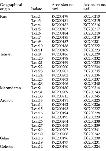
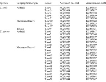
Sixty adult nematodes, including 32 isolates of T. cati from 32 cats, 9 isolates of T. canis from 9 dogs and 19 isolates of T. leonina from 19 dogs were collected from different geographical areas in Iran (tables 1 and 2). Individual adult worms removed from infected cats and dogs were washed extensively in physiological saline. After identification to species level according to their morphological features, using existing keys and descriptions (Yamaguti, Reference Yamaguti1961), they were preserved in 70% (v/v) ethanol until extraction of genomic DNA.
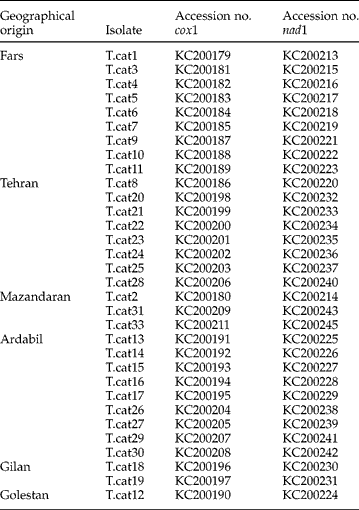
Geographical origin and accession numbers of pcox1 and pnad1 sequences of Toxocara cati isolates from cats in Iran.
Geographical origin and accession numbers of pcox1 and pnad1 sequences of Toxocara canis and Toxascaris leonina isolates from dogs in Iran.

Molecular and phylogenetic analyses
Samples were thoroughly washed in distilled water to remove ethanol. Total genomic DNA was extracted using QIAamp DNA Mini Kit (Qiagen, Hilden, Germany) according to the manufacturer's instructions and the DNAs were stored at − 20°C until PCR amplification. Two mitochondrial genes, partial cox1 (pcox1) and partial nad1 (pnad1), were subjected to PCR amplification. The forward JB3 (5′-TTTTTTGGGCATCCTGAGGTTTAT-3′) and reverse JB4.5 (5′-TAAAGAAAGAACATAATGAAAATG-3′) primers were used to amplify a portion of the cox1 gene; and the forward ND1F (5′-TTCTTATGAGATTGCTTTT-3′) and reverse ND1R (5′-TATCATAACGAAAACGAGG-3′) primers were used for amplification of the partial nad1 gene (Li et al., Reference Li, Lin, Song, Sani, Wu and Zhu2008b). All PCR reactions were carried out in a 25 μl reaction mix, containing 12.5 μl of PCR premix (2 × Master Mix RED Ampliqon, Denmark), which included 1.25 U Taq DNA polymerase, 200 μm dNTPs and 1.5 mm MgCl2; 25 pmol of each primer and 5 μl of template DNA. The temperature profile was one cycle of 94°C for 5 min (primary denaturation), followed by 35 cycles of 94°C for 30 s (denaturation), 50°C for 30 s (annealing), and 72°C for 30 s (extension), and a final extension of 72°C for 5 min. A sample containing water instead of template DNA was included in each run as a negative control. PCR products were separated by electrophoresis on a 1.5% agarose gel in TBE (Tris 0.09 m, borate 0.09 m, EDTA 0.02 m) at 80 V for 1 h. Gels were stained with 0.5 μg/ml ethidium bromide (Roche, Germany) and the bands were visualized using a UV transilluminator and digitally photographed. PCR products were purified using the AccuPrep® PCR purification Kit (Bioneer, Korea), according to the manufacturer's instructions, and were sequenced in both directions, using the same primers as used in the PCR. Sequence results were edited and analysed by the Geneious software (www.geneious.com) and the consensus sequences were compared with each other and GenBank reference sequences using the BioEdit software (http://www.mbio.ncsu.edu/bioedit/bioedit.html) and BLAST program (http://www.ncbi.nlm.nih.gov/). Three separate phylogenetic analyses of sequence data were conducted: (1) using pcox1 sequences determined in the present study only; (2) using pnad1 sequences determined in the present study only; and (3) using concatenated pcox1+ pnad1 sequences obtained in this study along with relevant sequences deposited in GenBank. Phylogenetic analyses were carried out employing Bayesian inference (BI) and maximum likelihood (ML) methods. BI was conducted using the program MrBayes v.3.1.2 (http://mrbayes.csit.fsu.edu/index.php). Posterior probabilities (pp) were set for 2,000,000 generations (ngen = 2,000,000). The program TreeviewX v.0.5.0 (Page, Reference Page1996) was used for trees. A maximum likelihood tree was constructed and pairwise comparisons were made of the level of sequence differences within and among species using the MEGA 5.0 software (Tamura et al., Reference Tamura, Peterson, Peterson, Stecher, Nei and Kumar2011). Bootstrap analyses (using 1000 replicates) were carried out to determine the robustness of the finding.
Results and discussion
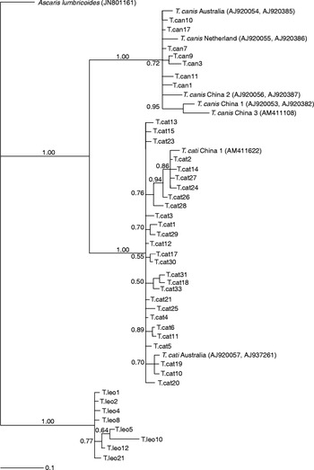
For all isolates, amplicons of about 450 and 370 base pairs (bp) were successfully produced by PCR for pcox1 and pnad1, respectively. Sequences of the Iranian isolates obtained in this study were deposited in GenBank and the accession numbers of these sequences are given in tables 1 and 2. Phylogenetic studies based on the analysis of DNA sequence differences represent a useful tool to gain information on an organism's evolutionary relationships. Previous studies have reported the existence of genetic variation among nematode populations. However, few studies have been done on the molecular characterization of Toxocara and Toxascaris species. For the first time, Jacobs et al. (Reference Jacobs, Zhu, Gasser and Chilton1997) reported the differentiation of nematodes of T. canis, T. cati and T. leonina based on internal transcribed spacer (ITS)-2 sequences. In addition to ribosomal gene sequences, sequences of the mitochondrial genome have been used for identification, systematic and phylogenetic relationship analyses of ascaridoid nematodes (Li et al., Reference Li, Lin, Song, Sani, Wu and Zhu2008b). The present study was designed for analysis of genetic variation within and among Iranian isolates of T. cati, T. canis and T. leonina by sequencing partial mitochondrial genes. Intra-species variation within Iranian isolates of T. cati, T. canis and T. leonina amounted to 0–2.3%, 0–1.3% and 0–1.0% for pcox1 and 0–2.0%, 0–1.7% and 0–2.6% for pnad1, respectively; meanwhile, inter-species sequence differences among the three ascaridoid nematodes were significantly higher, being 9.5–16.6% for pcox1 and 11.9–26.7% for pnad1. Concatenated pcox1+ pnad1 sequences revealed 27, 7 and 8 different haplotypes among Iranian isolates of T. cati, T. canis and T. leonina, respectively (fig. 1). The sequences of some samples collected from different provinces in Iran were identical; for example, pnad1 sequences of seven isolates (T.cat4, T.cat6, T.cat7, T.cat8, T.cat9, T.cat12 and T.cat21) collected from three provinces in Iran were identical. Based on these results, there is no correlation between homogeny of these isolates and their geographical origin.
Phylogenetic relationship of concatenated pcox1+ pnad1 sequences of isolates of Toxocara cati, Toxocara canis and Toxascaris leonina from Iran, using the Bayesian inference method with Ascaris lumbricoides (AN: JN801161) as the outgroup; nodal support is given as a pp value.

For the first time, Zhu et al. (Reference Zhu, Jacobs, Chilton, Sani, Cheng and Gasser1998) used ITS-1 and ITS-2 sequences for characterization of an ascaridoid nematode of cats from Kuala Lumpur, Malaysia. This nematode was previously identified morphologically as T. canis (Lee et al., Reference Lee, Cheng and Bohari1993), but a molecular approach using the ribosomal DNA (rDNA) sequences characterized it as Toxocara sp. cf. canis (Zhu et al., Reference Zhu, Jacobs, Chilton, Sani, Cheng and Gasser1998). A detailed morphological study of this nematode was carried out by Gibbons et al. (Reference Gibbons, Jacobs and Sani2001) and the parasite was described and named Toxocara malaysiensis. The first report of T. malaysiensis in cats outside of Malaysia was from China; the nematodes collected from cats in China were morphologically and genetically consistent with T. malaysiensis (Li et al., Reference Li, Zhu, Gasser, Lin, Sani, Lun and Jacobs2006). These studies showed that this species has a broader geographical distribution. In the present study, using the BLAST system, all 32 isolates from cats were identified as T. cati.
The pcox1 and pnad1 sequence analysis of T. cati, T. canis and T. leonina has not been done before in Iran, and the present study was designed for characterization and analysis of genetic variation among and within Iranian isolates and comparison with data available from other countries. Based on pairwise comparisons, there was sequence variation within and among the three ascaridoid species. Li et al. (Reference Li, Lin, Song, Sani, Wu and Zhu2008b) described genetic variation among and within T. canis, T. cati, T. malaysiensis, Toxocara vitulorum and T. leonina from different geographical origins using an single-strand conformation polymorphism (SSCP)-sequencing approach. In the present study, BLAST analysis of pcox1 sequences indicated that eight isolates of T. cati (T.cat4, T.cat7, T.cat8, T.cat9, T.cat15, T.cat16, T.cat19 and T.cat23) from four provinces in Iran were identical and exhibiting 100% homology with an isolate from Australia (accession no. AJ920057). The pcox1 sequence of two isolates of T. cati (T.cat2 and T.cat27) showed 100% homology with two reference sequences of T. cati from China (accession nos. AM411622, NC_010773). Seven isolates of T. leonina collected from dogs in Iran (T.leo5, T.leo7, T.leo11, T.leo12, T.leo13, T.leo16 and T.leo17) did not differ from an isolate of T. leonina collected from a grey wolf (Canis lupus) in China (accession no. JF780946), across the pcox1 gene. None of the pnad1 sequences of T. cati, T. canis and T. leonina from Iran had 100% homology with reference sequences in GenBank, except for an isolate of T. canis (T.can9) which showed 100% homology to an isolate from Australia (accession no. AJ920383). Phylogenetic analysis not only confirmed the differences within Iranian isolates of T. cati, but showed a large difference between isolates of T. cati from Iran and three isolates of T. cati from China (accession nos. JF780941, JF780942, JF780945 for pcox1 and JF833957–JF833959 for pnad1); the three isolates of T. cati from China were collected from different hosts (jungle cat, leopard cat and Asian golden cat).
In conclusion, this study demonstrated the existence of genetic diversity in mitochondrial genes within and among isolates of T. cati, T. canis and T. leonina in Iran. The results can be used for studies of genetic variability, specific identification of ascaridoid nematodes and diagnosis of toxocariasis, and the results clearly highlight the applicability of mitochondrial gene sequences as genetic markers. For a deeper understanding of genetic diversity among populations of T. cati, T. canis and T. leonina, analysis of more isolates from various geographical areas, other felid hosts and variable genetic markers are recommended.
Acknowledgements
The authors would like to thank Mrs N. Jalalizand from the molecular biology lab in Isfahan Station of National Institute of Health Research for kind assistance. Also, thanks are due to Mr M. Sharifdini and Mrs S. Charehdar for assisting with sample collection.
Financial support
This research was supported by the Tehran University of Medical Sciences; Project No. 90-02-27-14163.
Conflict of interest
None.
Ethical standards
This research project was reviewed and approved by the Ethics Committee of Tehran University of Medical Sciences, Iran.