Diastolic dysfunction of the left ventricle is one of the major determinants in the prognosis and management of cardiomyopathies in the paediatric age group.Reference Friedberg, Roche, Mohammed, Balasingam, Atenafu and Kantor 1 , Reference Dragulescu, Mertens and Friedberg 2 Currently, the gold standard for assessment of the left ventricular diastolic function is measurement of the left ventricular end-diastolic pressure and/or pulmonary capillary wedge pressure via cardiac catheterisation. Pulmonary capillary wedge pressure is used as a surrogate for left atrial pressure. This measurement obtained via cardiac catheterisation indirectly estimates the left ventricular compliance.
Left atrial enlargement has been generally used as a non-invasive marker of the severity and chronicity of the left ventricular diastolic dysfunction.Reference Lang, Bierig and Devereux 3 – Reference Pritchett, Mahoney, Jacobsen, Rodeheffer, Karon and Redfield 7 The left atrium not only acts as a conduit for passage of blood from the pulmonary vein to the left ventricle, but also as a reservoir to collect pulmonary venous return during ventricular systole and as a contractile pump that delivers 15–30% of the left ventricular filling. The left atrial volume measured by two-dimensional echocardiography is considered as one of the most accurate measures of the left atrial size and therefore is frequently used as a non-invasive measure of left atrial size.Reference Lang, Bierig and Devereux 3 , Reference Taggart, Cetta, O’Leary, Seward and Eidem 8 , Reference Khankirawatana, Khankirawatana and Porter 9 The left atrial volume indexed to the body surface area has been shown to reflect chronically elevated left ventricular filling pressure in children.Reference Dragulescu, Mertens and Friedberg 2 , Reference Nagueh, Appleton and Gillebert 10 Normative data for paediatric left atrial volumes have been well established over the recent years.Reference Taggart, Cetta, O’Leary, Seward and Eidem 8 , Reference Bhatla, Nielsen, Ko, Doucette, Lytrivi and Srivastava 11 There are a few studies in the adult literature reporting the relationship between left atrial volume and left atrial pressure.Reference Sauter, Dodge, Johnston and Graham 12 , Reference Geske, Sorajja, Nishimura and Ommen 13 However, to the authors’ knowledge, there are no paediatric studies evaluating the relationship between the two-dimensional left atrial volume calculated by echocardiogram and left atrial pressure obtained at the same time by cardiac catheterisation in children with cardiomyopathy.
B-type natriuretic peptides are hormones released in response to increased cardiac volume and resultant chamber distension.Reference Nishikimi, Minamino and Nakao 14 N-terminal prohormone brain natriuretic peptide is known to be a valuable prognostic marker for management of patients with heart failure. In children with chronic left ventricular systolic dysfunction, studies have shown that N-terminal prohormone brain natriuretic peptide can be a useful predictor of adverse cardiovascular outcomes.Reference Price, Thomas and Grenier 15 , Reference Mangat, Carter, Riley, Foo and Burch 16 The purpose of this study was to assess the correlation between the indexed left atrial volume and left atrial pressure and to evaluate the correlation between left atrial pressure and N-terminal prohormone brain natriuretic peptide levels in children with cardiomyopathy and acute myocarditis.
Materials and methods
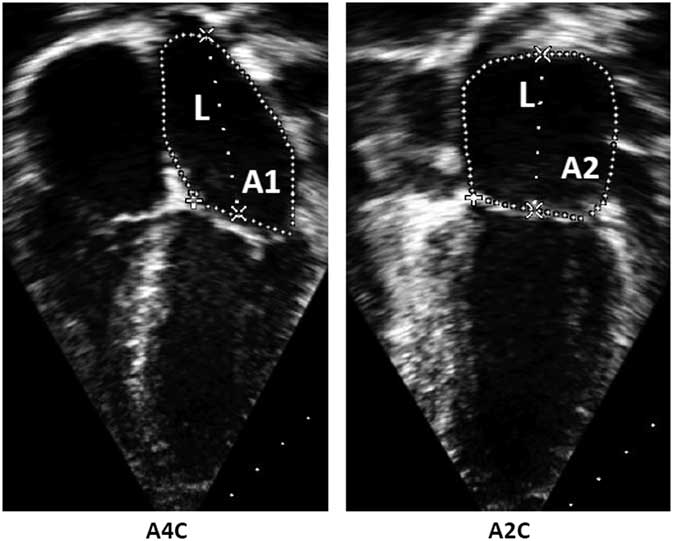
We retrospectively reviewed the data from the medical records for symptomatic patients, with the majority belonging to NYHA Classes II and III, <18 years of age, with a diagnosis of cardiomyopathy and acute myocarditis, who underwent cardiac catheterisation, and had a transthoracic echocardiogram performed around the time of the cardiac catheterisation, over a 11-year period between October, 2003 and December, 2014. The Institutional Review Board of the University of Miami, Miller School of Medicine and Holtz Children’s Hospital, approved the study. The echocardiographic left atrial measurements were performed by a single observer – a board certified-paediatric cardiologist – from the digitally archived studies. The left atrial volume was calculated using a standardised biplane area–length method recommended by the American Society of Echocardiography: left atrial volume=8/3π [(A1) (A2)/(L)]. The left atrial area was measured at the end-ventricular systole when the left atrial chamber is at its largest dimension in apical four-chamber view (A1) and apical two-chamber view (A2) (Fig 1). The confluences of the pulmonary veins and left atrial appendage were excluded. The length (L) was measured from the back wall of the left atrium to a line across hinge points of the mitral valve. The shorter of the lengths from either the apical four-chamber or two-chamber view was used in the equation. When the apical two-chamber view was not available or the image quality was not adequate for measurement of the left atrium in that view, we used the following formula to calculate left atrial volume: 8 (A12/3π (L)), assuming A1=A2.Reference Lang, Bierig and Devereux 3 , Reference Lang, Badano and Mor-Avi 17 , Reference Lopez, Colan and Frommelt 18 The left atrial volume obtained by the echocardiogram was indexed to the body surface area (BSA). Another set of data was obtained in the same cohort using the method suggested by Bhatla et alReference Bhatla, Nielsen, Ko, Doucette, Lytrivi and Srivastava 11 , in which left atrial volume is indexed to BSA1.48 for patients with a body surface area <1 m2 and to BSA1.08 for patients with a BSA≥1 m2. z-Scores of the indexed left atrial volume were obtained using the graphs provided in that study. Cardiac catheterisation-derived pulmonary capillary wedge pressure data were obtained to estimate the left atrial pressure. N-terminal prohormone brain natriuretic peptide values obtained around the time of cardiac catheterisation were analysed. Demographic data such as age, gender, and diagnosis were reviewed.
Measurement of the left atrial volume by standardised biplane area–length method. A1=left atrial area measured in apical 4-chamber view (A4C); A2=left atrial area measured in apical 2-chamber view (A2C).

Descriptive data analyses were used and categorical variables were explored and reported using frequencies and proportions, whereas continuous variables were explored and reported using means and standard deviations or median with range. Correlation analysis was used for the measurement of the association between variables – left atrial pressure, left atrial volume z-score, and N-terminal pro-brain natriuretic peptide. Statistical analyses were performed using SAS software version 9.2 (SAS Institute Inc., Cary, North Carolina, United States of America). All statistical significance in the current study was based on an α level of 0.05.
Results
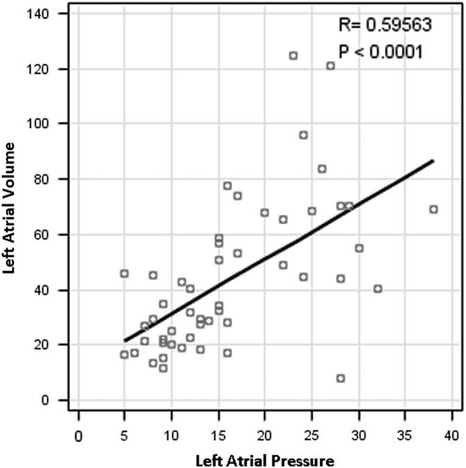
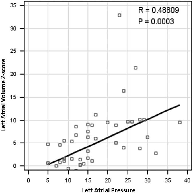
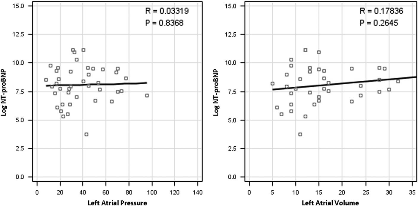
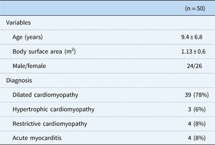
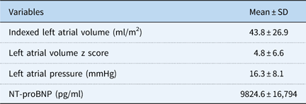
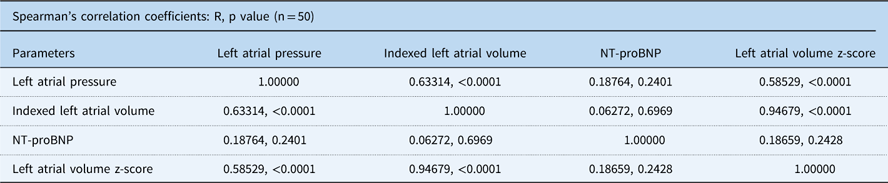
A total of 50 patients met the inclusion criteria. Of these, 26 (52%) were girls; the study age range was 0.2–17 years (mean±SD, 9.4±6.8). Table 1 shows the demographic data for the study cohort. Among the 50 patients, 39 had dilated cardiomyopathy (78%), three had hypertrophic cardiomyopathy (6%), four had restrictive cardiomyopathy (8%), and four had acute myocarditis (8%), who eventually progressed to dilated cardiomyopathy. The mean body surface area ±SD was 1.13±0.6 m2. The indexed left atrial volume in the entire group using the biplane area–length method ranged from 8.1 to 124.4 ml/m2 (mean±SD, 43.8±26.9 ml/m2). Table 2 shows the data regarding estimated left atrial pressure, left atrial volume, and N-terminal prohormone brain natriuretic peptide in the patients. The mean z-score in the study patients for left atrial volume was 4.8±6.6. The mean±SD of the pulmonary capillary wedge pressure was 16.3±8.1 mmHg, ranging from 5 to 38 mmHg. The mean N-terminal prohormone brain natriuretic peptide was 9824.6 pg/ml, ranging from 41.7 to 70,000. The median time between the date of cardiac catheterisation and transthoracic echocardiogram was 3 days. In our patient population, the estimated left atrial pressure and indexed left atrial volume were significantly correlated (r=0.63; p<0.001) (Fig 2). There was a linear correlation between estimated left atrial pressure and indexed left atrial volume z-score for the entire cohort (r=0.59; p<0.001) (Fig 3). Both the left atrial pressure and volume were not correlated with the log N-terminal prohormone brain natriuretic peptide levels (r=0.19; p=0.24) (Fig 4). Table 3 demonstrates the correlation analysis of the variables in the study.
Correlation between left atrial volume and left atrial pressure (n=50).

Correlation between left atrial volume z-score and left atrial pressure (n=50).

Correlation between log-pro brain natriuretic peptide (BNP) level and left atrial pressure; log-proBNP and left atrial volume. NT=N-terminal.

Demographic data.

Echocardiographic and haemodynamic data (n=50).

NT-proBNP, N-terminal prohormone brain natriuretic peptide
Correlation analysis of the variables.

NT-proBNP, N-terminal prohormone brain natriuretic peptide
Discussion
To our knowledge, this is the first study that explores the correlation of left atrial pressure estimated by measuring pulmonary capillary wedge pressure in the cardiac catheterisation laboratory, which is considered the gold standard, and left atrial volume obtained via transthoracic echocardiography in children with cardiomyopathy and myocarditis. We found that there was a strong correlation between the estimated left atrial pressure – obtained by cardiac catheterisation – and the indexed left atrial volume. In addition, we found a correlation between estimated left atrial pressure and the left atrial volume z-scores obtained using the proposed allometric model. We hypothesise that the less muscular and more compliant nature of the left atrium is probably responsible for significant dilatation of the atrium with minimal increases in the left ventricular end-diastolic pressure, which is a surrogate of left atrial pressure. In a large paediatric study by Bhatla et al,Reference Bhatla, Nielsen, Ko, Doucette, Lytrivi and Srivastava 11 the authors noted that the conventional technique of indexing left atrial volume using “per m2 BSA” model fails to eliminate the residual relationship of indexed left atrial volume to BSA. Therefore, the use of a validated allometric model was recommended to allow for an accurate diagnosis of left atrial enlargement that will be independent of patient’s body size, age, and gender.Reference Appleton, Galloway, Gonzalez, Gaballa and Basnight 6 In patients with cardiomyopathy, who will need repetitive evaluation of haemodynamics to evaluate progression and prognosis of their disease; echocardiography remains the simplest, least invasive, and the most cost-effective method for cardiac evaluation. Serial measurements of left atrial volumes in these patients might be reflective of worsening diastolic left ventricular function. Several studies have evaluated cardiac CT and cardiac MRI for the evaluation of left atrial volume.Reference Lang, Badano and Mor-Avi 17 – Reference Pritchett, Jacobsen, Mahoney, Rodeheffer, Bailey and Redfield 19 Although cardiac MRI provides accurate measurements of left atrial volume with acceptable temporal resolution, it is limited by increased cost, an inability to measure phasic volumes with gated three-dimensional sequences, problems related to gadolinium contrast, need for sedation in children, and a relative inability to scan patients with intracardiac devices.
Studies with serial N-terminal prohormone brain natriuretic peptide measurement have shown that elevated N-terminal prohormone brain natriuretic peptide level is strongly associated with worse outcomes in chronic paediatric heart failure and dilated cardiomyopathy, but its levels should be cautiously interpreted in the setting of acute paediatric heart failure.Reference Price, Thomas and Grenier 15 , Reference Mangat, Carter, Riley, Foo and Burch 16 , Reference Lopez, Colan and Frommelt 18 In this study, we did not find any significant correlation between estimated left atrial pressure and log N-terminal prohormone brain natriuretic peptide levels (r=0.19; p=0.24). This can be explained by the fact that we had a mixed patient population including both with acute myocarditis and chronic cardiomyopathy. In addition, in our study, we took single-point measurement of N-terminal prohormone brain natriuretic peptide for correlation with left atrial pressure rather than serial measurement. The value of N-terminal prohormone brain natriuretic peptide in assessment of diastolic dysfunction is still debatable. Price et alReference Price, Thomas and Grenier 15 found no significant correlation between left ventricular end-diastolic dimension and the log N-terminal prohormone brain natriuretic peptide levels, whereas Mangat et al.Reference Mangat, Carter, Riley, Foo and Burch 16 reported significant correlation between log N-terminal prohormone brain natriuretic peptide and left ventricular end-diastolic dimension z score (p<0.001) in a similar patient population.
N-terminal prohormone brain natriuretic peptide levels has been described to have a strong correlation with tissue Doppler imaging in the adult population and can reliably provide estimation of elevated left ventricular filling pressures in patients with documented diastolic dysfunction.Reference Lopez, Colan and Frommelt 18 Few studies have attempted to correlate brain natriuretic peptide with left atrial volume in children with volume overload. Sakata et alReference Sakata, Hayabuchi, Inoue, Onishi and Kagami 20 reported a significant correlation between left atrial volumetric parameters at each phase of the cardiac cycle and N-terminal prohormone brain natriuretic peptide. Tschope et alReference Tschope, Kasner, Westermann, Gaub, Poller and Schultheiss 21 reported that when the left ventricular ejection fraction is preserved but diastolic function is impaired, the correlation between N-terminal prohormone brain natriuretic peptide levels and invasively measured filling pressure is weaker (r=0.45), but the negative predictive values (~90%) for elevated left ventricular end-diastolic pressure remain high. The variable relationship between N-terminal prohormone brain natriuretic peptide and left ventricular filling pressures is consistent with the complexity of factors influencing secretion and clearance. With such conflicting data, it is difficult to come to a conclusion with confidence about the utility of N-terminal prohormone brain natriuretic peptide in children with heart failure and preserved systolic function. Future randomised studies are needed to validate the use of N-terminal prohormone brain natriuretic peptide in children with diastolic heart failure.
Limitations
The main limitations of this study are its retrospective design, potential variability in the error of measurements due to limitations in acoustic window, and the small study cohort.
Conclusions
Our study demonstrates that the echocardiogram-derived left atrial volume significantly correlated with estimated left atrial pressure in the cardiac catheterisation laboratory. Left atrial volume can therefore be used as a non-invasive surrogate to estimate left atrial pressure and may decrease the need for, and the frequency of, more invasive studies such as cardiac catheterisations in patients with cardiomyopathy. Log N-terminal prohormone brain natriuretic peptide did not correlate with left atrial pressure in this study. The utility of N-terminal prohormone brain natriuretic peptide as a biomarker to assess severity of diastolic dysfunction needs further evaluation. Prospective long-term studies with larger patient sample are needed to confirm the results from this study.
Acknowledgements
Authors’ contribution: S.L. and A.A. performed data collection and drafted the initial manuscript and approved the final manuscript as submitted. S.R.G., P.G.R., and H.L. critically reviewed and approved the final manuscript as submitted. N.S. and S.S. conceptualised and designed the study, drafted the protocol, supervised data collection and statistical analysis, and edited, approved, and revised the final manuscript as submitted. All authors approved the final manuscript as submitted and agree to be accountable for all aspects of the work.
Financial Support
This research received no specific grant from any funding agency or from commercial or not-for-profit sectors.
Conflicts of Interest
None.