Introduction
Peltigera is a widely distributed genus of primarily muscicolous and terricolous macrolichens with more than 90 species recognized worldwide (Goward et al. Reference Goward, Goffinet and Vitikainen1995; Martinez et al. Reference Martinez, Burgaz, Vitikainen and Escudero2003; Vitikainen Reference Vitikainen2006; Kirk et al. Reference Kirk, Cannon, Minter and Stalpers2008; Han et al. Reference Han, Zhang and Guo2013, Reference Han, Zheng and Guo2015). Most of them are bipartite symbioses, with one mycobiont and a Nostoc cyanobacterial photobiont. These species have thalli that range in colour from dark green to bluish grey and brown when moist and are grouped into five sections: Horizontales, Hydrothyriae, Peltigera, Polydactylon, and Retifoveatae (Miadlikowska & Lutzoni Reference Miadlikowska and Lutzoni2000). Thalli of different colours within a species (colourmorphs) are notable in some bipartite taxa, and in one case this has been correlated with different strains of the Nostoc photobiont (Miao et al. Reference Miao, Rabenau and Lee1997). The remaining members of the genus are classified in sections Chloropeltigera, Peltidea and Phlebia. With two exceptions (P. malacea and P. frippii), the members of those three sections are tripartite symbioses, with one mycobiont and two photobionts, and are ‘emerald’ or ‘grass’ green. These consort with a green alga, Coccomyxa, as the main photobiont in the thallus while a secondary Nostoc photobiont is compartmentalized in external cephalodia in the form of bluish or brownish ‘warts’.
At the genus level, Peltigera are among the most recognizable of terrestrial macrolichens and easily distinguished from others owing to their broad lobes and veined, ecorticate lower surfaces. However, identification of some species may be difficult due in part to intrinsic variation and high phenotypic plasticity (Vitikainen Reference Vitikainen1994; Goward et al. Reference Goward, Goffinet and Vitikainen1995; Goffinet & Bayer Reference Goffinet and Bayer1997; Miadlikowska & Lutzoni Reference Miadlikowska and Lutzoni2000, Reference Miadlikowska and Lutzoni2004; Miadlikowska et al. Reference Miadlikowska, Lutzoni, Goward, Zoller and Posada2003; O’Brien et al. Reference O’Brien, Miadlikowska and Lutzoni2009; Sérusiaux et al. Reference Sérusiaux, Goffinet, Miadlikowska and Vitikainen2009). Cryptic species and species complexes present special challenges, and previously unrecognized taxa may be discovered only after molecular data have been examined. Examples include undescribed lichens from western North America reported by Miadlikowska et al. (Reference Miadlikowska, Lutzoni, Goward, Zoller and Posada2003) in section Peltigera, viz. P. “neorufescens”, P. “fuscopraetextata”, and P. “neocanina”. More recently, O’Brien et al. (Reference O’Brien, Miadlikowska and Lutzoni2009), working with material from British Columbia, Canada, suggested yet another potential undescribed species in the same section, referred to simply as P. sp. A.
In 2010, the first author collected an emerald green but non-cephalodiate Peltigera specimen at Öskjuhlíd, a hill in Reykjavík in south-western Iceland. The specimen did not correspond to any of the 22 Peltigera species reported from Iceland (Kristinsson & Heiðmarsson Reference Kristinsson and Heiðmarsson2009). Analysis of the nuclear ribosomal internal transcribed spacer region (ITS) revealed a match to P. sp. A sensu O’Brien et al. (Reference O’Brien, Miadlikowska and Lutzoni2009). In light of this finding, Öskjuhlíd was revisited, mainly in 2013, to obtain additional specimens and to more closely examine the community of lichens in this habitat.
The main purposes of this contribution are to formally describe Peltigera islandica sp. nov., based on collections from Iceland, with an updated phylogeny of the mycobiont, and to characterize its photobiont, particularly in relation to the Peltigera community at Öskjuhlíd. Additionally, P. “neorufescens”, a second undescribed taxon collected during fieldwork in 2013, is reported as new to Iceland.
Materials and Methods
Sample collection and identification
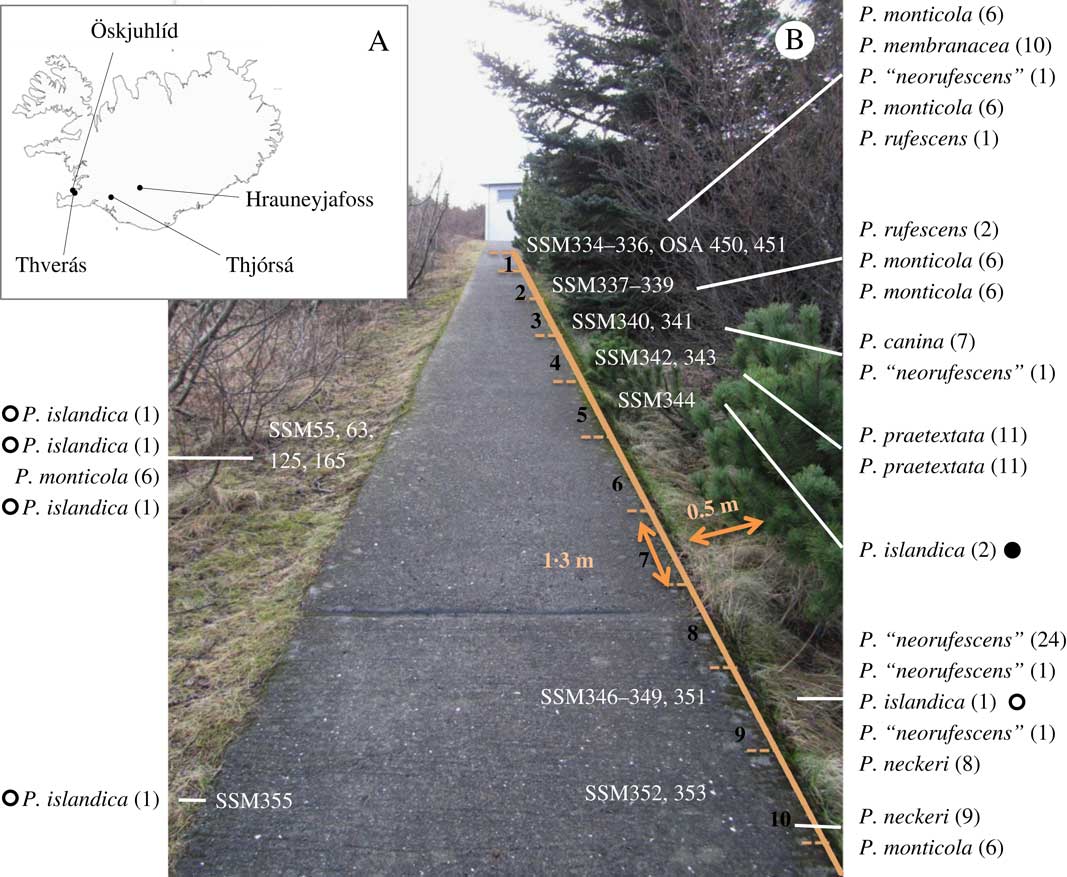
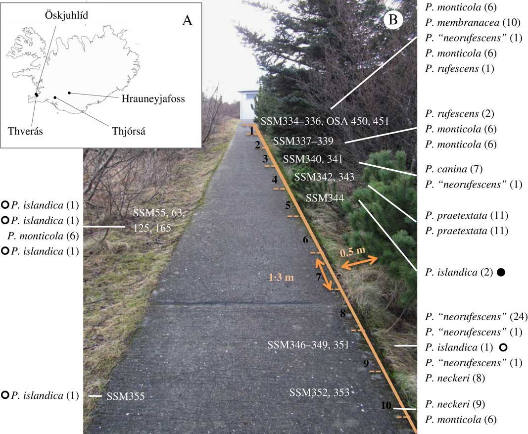
Peltigera islandica was collected from four localities in south-west Iceland (Fig. 1A, Table 1): Öskjuhlíd (elevation 40 m a.s.l. 64·131683°N, 21·920017°W), Thverás (64·104183°N, 21·780667°W), Thjórsá (64·03603°N, 20·3152°W), and near Hrauneyjafoss (2 sites: 64·16659°N, 19·1536°W and 64·17036°N, 19·1296°W). At Öskjuhlíd, the area near where the first specimen, accession SSM55, was found in 2010 was marked off in 1·3 m×0·5 m sectors (Fig. 1B) to support more systematic collecting, as well as to facilitate description and recording of the spatial relationship among samples. In addition to P. islandica, representatives of every apparent morphotype in each sector were also collected.
Collection sites for Peltigera islandica. A, location of collection sites in Iceland; B, collection site at Öskjuhlíd. Cover vegetation (April 2013) and sectors along the path where samples were collected are shown. The order of voucher numbers corresponds to the order of the species listed. Appended to each name is the corresponding rbcLX allele (Table 1). ○=emerald green; ●=dark green colourmorphs of P. islandica. In colour online.
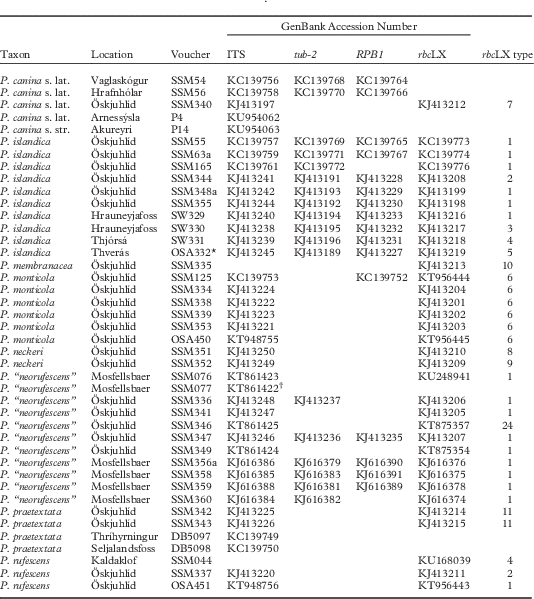
Specimen information and GenBank accession numbers for the species of Peltigera from Iceland used in this study
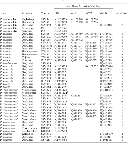
* P. islandica OSA392, collected in the summer of 2015, is not listed separately above because it was part of the same thallus sampled and accessioned as OSA332 in 2013. The ITS sequence of OSA392 was confirmed as identical to that of OSA332.
† While other P. “neorufescens” are identical to each other and similar to AY257910, SSM077 is similar to AY257916.
Samples were sorted, cleaned of field debris and soil, and examined under a hand lens or a dissecting microscope for identification. Fresh thalli of P. islandica were hand-sectioned using a sterile razor blade and examined through a compound microscope using autofluorescence (Leica model DM2500) and phase contrast (Nikon model Labophot Trinocular Microscope) microscopy, and standard C, K, KC, and PD spot tests were performed on the cortex and medulla (Orange et al. Reference Orange, James and White2001). After identification, a piece of each specimen was used for DNA extraction (to corroborate identifications, see below) and the rest was saved for herbarium vouchers. Except for P4 and P14 (Miadlikowska & Lutzoni s. n.) deposited at Duke University herbarium (DUKE), vouchers were saved in the Andrésson laboratory at the University of Iceland, with a portion of some accessions (P. islandica SSM55, SSM63, SSM165, SSM344, SSM348, SSM355, SW329, SW330, SW331, OSA332, and “P. neorufescens” SSM347, SSM359) also deposited at the Icelandic Institute of Natural History (AMNH).
Isolation and culture of cyanobacterial photobionts
Fresh, washed thallus fragments of accession SSM165 were macerated and spread under sterile conditions on BG110 plates and incubated under low light intensities at 25 °C. After three weeks, the cyanobacteria that grew away from the mycobiont were transferred to fresh plates using a sterile needle under a dissecting microscope, and purified cultures were maintained on BG110 (Hainfried & Schenk Reference Hainfried and Schenk2013).
Isolation of DNA, PCR amplification and sequencing
Total genomic DNA was isolated from thalli as described previously (Sinnemann et al. Reference Sinnemann, Andresson, Brown and Miao2000) for use as PCR templates for mycobiont and photobiont markers. DNA was prepared from replicate portions (‘a’ and ‘b’) of accessions SSM63, SSM348 and SSM356 to assess potential genetic heterogeneity of the thallus. For PCR from cultured photobionts, a lysate was first made (a few cells transferred from agar plates into 5% Chelex (Sigma), heated to 100°C for 2 min, and clarified by centrifugation), and an aliquot of the supernatant was used as the template for PCR.
The mycobiont ITS region was amplified with primers ITS1 and ITS4 (White et al. Reference White, Bruns, Lee and Taylor1990), while part of the RNA polymerase II largest subunit (RPB1) and β−tubulin (tub-2) loci were amplified using newly designed and/or modified primers: forward R1 (5'-AGAGTGTCCCGGGCATTTTGGTCA-3') and reverse R2 (5'-TGGCCGGCAATCTCATTATCCATGTA-3') based on Stiller & Hall (Reference Stiller and Hall1997) and Matheny et al. (2002), and forward T1 (5'-ACCTTCGTGCGGATACACACT-3') and reverse T2 (5'-ACCCTCAGTGTARTGACCCTTGGC-3') based on Glass & Donaldson (Reference Glass and Donaldson1995). The photobiont rbcLX locus was amplified using primers CW and CX (Rudi et al. Reference Rudi, Skulberg and Jakobsen1998), while the 16S rDNA gene was amplified from SSM55 using primer 8F (Turner et al. Reference Turner, Pryer, Miao and Palmer1999) and a new 16S rDNA reverse primer, PiR (5'-AAAGGAGGTGATCCAGCCAC-3'). Reactions were performed in 25 μl containing 1×PCR buffer (Fermentas), 2·5 mM MgSO4, 0·2 mM each dNTP, 0·24 μM primers and 0·2 U Taq DNA polymerase, except for tub-2 for which 1×Eppendorf Master Mix was used. Amplifications were performed with an initial heating step of 94°C for 2 min (5 min when using lysates) and 33 cycles of 94°C for 20 s, 55°C for 20 s and 72°C for 50 s (90 s when amplifying tub-2). PCR products were treated with exonuclease I and then alkaline phosphatase (ExoSAP, New England BioLabs) to degrade single stranded DNA and inactivate remaining nucleotides, and subjected to cycle sequencing using ABI Big Dye Terminator v3.1. PCR amplification primers were used for sequencing, except for the 16S amplicon for which internal primers from the literature (e.g. 515F, 519R; Turner et al. Reference Turner, Pryer, Miao and Palmer1999) were used to complete primer walking. Reaction products were run on an ABI3500XL genetic analyzer, and automated base calls were manually confirmed after inspection of chromatograms. Sequences, including 41 ITS, 19 RPB1, 15 tub-2 and 35 rbcLX, were compared by BLAST searches in public databases to corroborate species identification, then annotated and deposited in GenBank (Table 1) as part of this study ahead of manuscript completion. Comparison to public databases was last conducted on 24 April 2016.
Sequence alignment and phylogenetic analyses
The ITS dataset used for phylogenetic analysis of the mycobiont included 19 newly generated sequences (Table 1) and 22 sequences from GenBank. The latter represented P. sp. A and seven species closely related to P. sp. A (all members of section Peltigera (‘P. canina group’) including three undescribed taxa, P. “boreorufescens”, P. “fuscopraetextata” and P. “pallidorufescens”), selected based on published phylogenies (Miadlikowska et al. Reference Miadlikowska, Lutzoni, Goward, Zoller and Posada2003; O’Brien et al. Reference O’Brien, Miadlikowska and Lutzoni2009; Sérusiaux et al. Reference Sérusiaux, Goffinet, Miadlikowska and Vitikainen2009) and GenBank BLAST results (highest similarity to P. islandica), and two outgroup species (P. tereziana and P. degenii). Identical haplotypes were collapsed and a total of 22 ITS sequences were aligned manually with MacClade 4.07 (Maddison & Maddison Reference Maddison and Maddison2003). Ambiguously aligned regions, sensu Lutzoni et al. (Reference Lutzoni, Wagner, Reeb and Zoller2000), were delimited manually (105 of a total of 619 characters) and excluded. Phylogenetic analyses of the remaining 514 characters were performed using the maximum likelihood (ML) optimization criterion as implemented in RAxML (Stamatakis Reference Stamatakis2006; Stamatakis et al. Reference Stamatakis, Hoover and Rougemont2008) and Bayesian analysis with BAli-Phy (Suchard & Redelings Reference Suchard and Redelings2006). A maximum likelihood search for the most likely tree (1000 replicates) and bootstrap analysis (1000 bootstrap pseudoreplicates) were performed using RAxMLHPC-MPI-SSE3 as implemented in the Mobyle SNAP Workbench version 1.0.5, a portal for evolutionary and population genetics analyses (North Carolina State University online facilities, Monacell & Carbone (Reference Monacell and Carbone2014)) with the six parameter substitution model and gamma distribution approximated with four categories (GTRGAMMA; Rodriguez et al. Reference Rodriguez, Oliver, Marin and Medina1990) applied independently to the ITS1 and ITS2 regions (one partition) and 5.8S region (second partition). The nexus file for the ML analysis was deposited in TreeBASE (http://purl.org/phylo/treebase/phylows/study/19185). Bayesian analysis was performed on a non-censored alignment sampled from the joint posterior distribution of phylogeny, alignment, and model parameter; for detailed information see Redelings & Suchard (Reference Redelings and Suchard2005), Gaya et al. (Reference Gaya, Redelings, Navarro-Rosines, Llimona, De Cáceres and Lutzoni2011) and Sliwa et al. (Reference Sliwa, Miadlikowska, Redelings, Molnar and Lutzoni2012). The alignment for 5.8S sequences was held constant, while the alignment for ITS1 and ITS2 was allowed to vary through the Markov chain Monte Carlo (MCMC) process. The substitution model parameters were estimated for the ITS1 and ITS2 regions using the Akaike Information Criterion (AIC) as implemented in MrModeltest 2.3 (Nylander Reference Nylander2004). A six-rate parameter model for nucleotide substitutions (GTR+INV) with a proportion of invariable sites was used for ITS1, and the HKY model with a gamma distribution (Hasegawa et al. Reference Hasegawa, Kishino and Yano1985) was implemented for 5.8S and ITS2. The relative substitution rate for each partition was allowed to vary independently. The alignment algorithm for the ITS1 and ITS2 partitions followed the RS07 indel model (Redelings & Suchard Reference Redelings and Suchard2007) with the same parameters; 100000 iterations were sampled from each of eight MCMC chains, and the first 45000 iterations were discarded as burn-in (assessed using the web tool Are We There Yet (AWTY; Wilgenbusch et al. Reference Wilgenbusch, Warren and Swofford2004) as implemented on the website http://king2.scs.fsu.edu/CEBProjects/awty/awty_start.php).
The Nostoc locus used for determining photobiont haplotypes designated in Table 1 corresponded to rbcLX nucleotide positions 13–794 of GenBank Z94891 (Rudi et al. Reference Rudi, Skulberg and Jakobsen1998) and was processed using alignment tools and ClustalW as implemented in MacVector 12.0.02. This locus includes sequences encoding the last 82 amino acids of the RUBISCO large subunit (rbcL), a putative chaperone gene (rbcX) and two intergenic spacers (Li & Tabita Reference Li and Tabita1997). For phylogenetic analysis, the dataset of 295 OTUs comprised published rbcLX sequences of free-living and symbiotic strains from Nostoc clades I and II (Otalora et al. Reference Otalora, Martinez, O’Brien, Molina, Aragon and Lutzoni2010; O’Brien et al. Reference O’Brien, Miadlikowska and Lutzoni2013; Miadlikowska et al. Reference Miadlikowska, Richardson, Magain, Ball, Anderson, Cameron, Lendemer, Truong and Lutzoni2014a), symbionts from Gunnera magellanica, as well as published (Manoharan et al. Reference Manoharan, Miao and Andrésson2012) or new (this study) sequences from P. islandica and other Peltigera species from Iceland. In this more diverse dataset, both spacers were aligned poorly (positional homology could not be determined unambiguously) and therefore were removed, leaving 636 coding characters. RAxML-HPC2 v. 7.2.8, as implemented on the CIPRES portal (Miller et al. Reference Miller, Pfeiffer and Schwartz2010) was used for analysis. Optimal tree and bootstrap searches were conducted with the rapid hill-climbing algorithm for 1000 replicates with the GTRGAMMA substitution model estimated independently for three partitions corresponding to the first, second, and third codon positions of the rbcLX.
Results
Collection of P. islandica sp. nov. and Öskjuhlíd Peltigera community
Öskjuhlíd, the hill where most of the material in this study was collected, was cleared in the 1940s for installation of above-ground municipal hot water tanks. After these were demolished in the 1960s, recreational trails were laid on top of the buried pipes, and the adjacent area has grown up to various herbs and shrubs, as well as to downy birch (Betula pubescens), Sitka spruce (Picea sitchensis), and mountain pine (Pinus mugo), introduced through afforestation (Sigurðsson Reference Sigurðsson1993). Terricolous lichens (Cladonia spp., Peltigera membranacea and crustose species) were sparse in the open, grassy area NE of the study site, and somewhat more abundant in the wooded area towards the SW. Except for infrequent, scattered occurrences of Cladonia species, lichens were generally absent next to paths, but the first specimen, SSM55, was found in 2010 in a small area with an unusually large number of Peltigera thalli. Most of this Peltigera community was distributed on the SW side of the path, with some sectors supporting one to several thalli or patches and other sectors having none (e.g. sector 1 vs 7, Fig. 1B). Thus, while the focus of the 2013 fieldwork was to obtain more specimens such as SSM55, samples of the other Peltigera in the vicinity were also collected in order to complement the morphological description of the new lichen with information on its ecology.
In total, 25 samples (including SSM55) representing eight morphospecies were collected (Table 1). Six were previously reported from Iceland: P. canina, P. membranacea, P. monticola, P. praetextata and P. rufescens in section Peltigera, and P. neckeri in section Horizontales (Kristinsson & Heiðmarsson Reference Kristinsson and Heiðmarsson2009). The seventh species is currently informally designated P. “neorufescens”, and represents an undescribed member of the P. rufescens complex first recognized by Miadlikowska et al. (Reference Miadlikowska, Lutzoni, Goward, Zoller and Posada2003) through ITS and ribosomal gene sequence data. This species was found at one other location in Iceland (Table 1), and will be formally described elsewhere, once its morphological circumscription and geographical distribution are better understood (T. Goward & J. Miadlikowska, unpublished data).
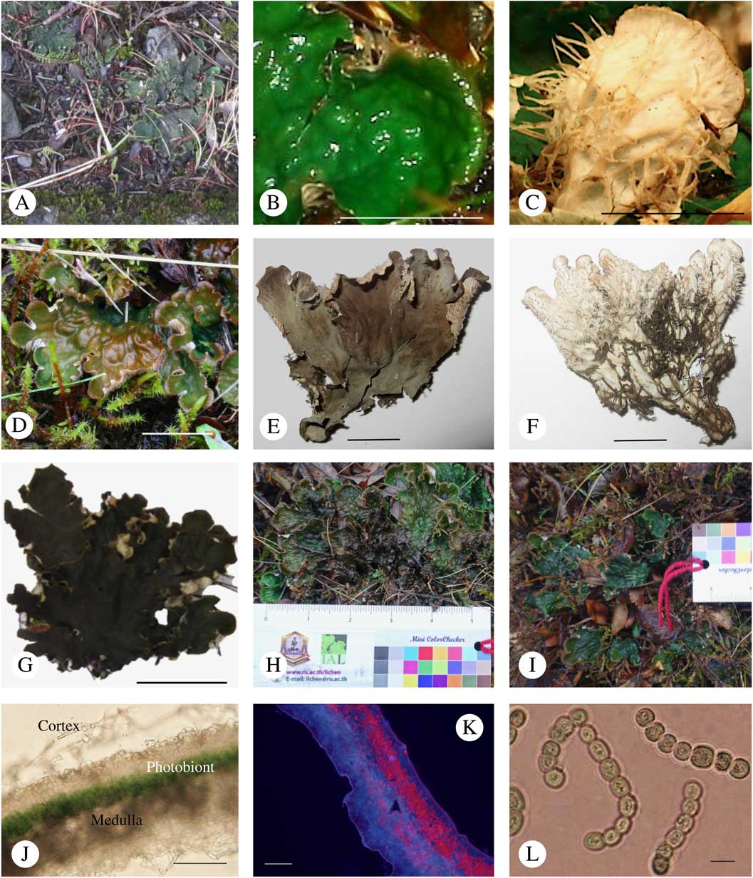
The final taxon collected at Öskjuhlíd comprised six representatives of an undescribed species, formally described here as Peltigera islandica sp. nov. (see below). Five specimens (SSM55, SSM63, SSM165, SSM348, SSM355) were distinctly emerald green when wet (Fig. 2A–C), but the sixth (SSM344), which shared an identical ITS sequence, was dark green (Fig. 2G) and is interpreted as a variant (colourmorph) of the species. These lichens occurred as small patches of a few lobes directly on soil and were found on both sides of the path, alone or with other species in the same or adjacent sectors (Fig. 1B).
Peltigera islandica. A, voucher SSM348; B & C, voucher SSM63, upper and lower surface, respectively; D–F, voucher OSA392, in situ and upper and lower surface of a portion of the dried holotype; G, voucher SSM344; H & I, thallus from which voucher OSA392 was collected (April 2016), and a new patch nearby; J & K, SSM63 thallus cross-sections viewed under white and UV light, respectively; L, Nostoc SSM165-N2a. Scales: B–F=1 cm; G=2cm; J & K=50 µm; L=10 µm. A ruler with colour card (7th International Association of Lichenology Symposium, Bangkok, 2012) was used as a standard to cross reference between images H and I.

Additional bright green-coloured examples of the new species were collected from three other locations in south-western Iceland (Fig. 1A, Table 1). In each instance, the collecting locality had been subject to recent disturbance: on a residential property in a neighbourhood (Thverás) in Reykjavík (OSA332, OSA392; Fig. 2D–F), beside a major road near Hrauneyjafoss (SW329, SW330) in the south highlands, and on the bank of the River Thjórsá (SW331). The ITS sequences from all these specimens were identical to that of SSM55.
Phylogenetic delimitation of P. islandica mycobiont
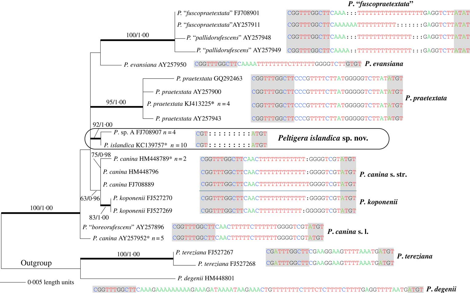
The ITS has proved to be a very informative molecular marker for species delimitation in the genus Peltigera (Goffinet et al. Reference Goffinet, Miadlikowska and Goward2003; Miadlikowska et al. Reference Miadlikowska, Kauff, Hognabba, Oliver, Molnar, Fraker, Gaya, Hafellner, Hofstetter and Gueidan2014b; Han et al. Reference Han, Zheng and Guo2015), and the 8–126 bp hypervariable region in the ITS1 (ITS1–HR) has been especially useful for species distinction in the taxonomically very challenging section Peltigera (Miadlikowska et al. Reference Miadlikowska, Lutzoni, Goward, Zoller and Posada2003). As the ITS sequence of accession SSM55 was 99% identical (546/547 nucleotides) to that deposited for P. sp. A (GenBank FJ708907, undescribed Canadian material represented by HOB020708–62–1–1; O’Brien et al. Reference O’Brien, Miadlikowska and Lutzoni2009), a context for the phylogenetic validation of P. islandica could be established based on the strong bootstrap support that P. sp. A had received for placement in the section Peltigera, Group C (‘P. canina group’; O’Brien et al. Reference O’Brien, Miadlikowska and Lutzoni2009) (Fig. 3).
Phylogenetic relationships among selected members (7 taxa, 18 OTUs) of the P. canina complex (section Peltigera) based on ITS sequences. Names in quotation marks indicate putative undescribed species. Sequences of the hypervariable ITS1 region (absent from P. islandica) are aligned within each putative species; conserved flanking motifs are shaded in grey. Values associated with internodes represent bootstrap support (ML-BP; before the slash) and posterior probability support (PP; after the slash). Only values >50% for ML-BP and 0·95 for PP are shown. Thick branches indicate relationships with strong bootstrap (ML-BP ≥70%) and posterior probability (PP ≥0·95%) support. Peltigera degenii and P. tereziana were used to root the tree. *=includes newly generated sequences from specimens collected in Iceland. In colour online.
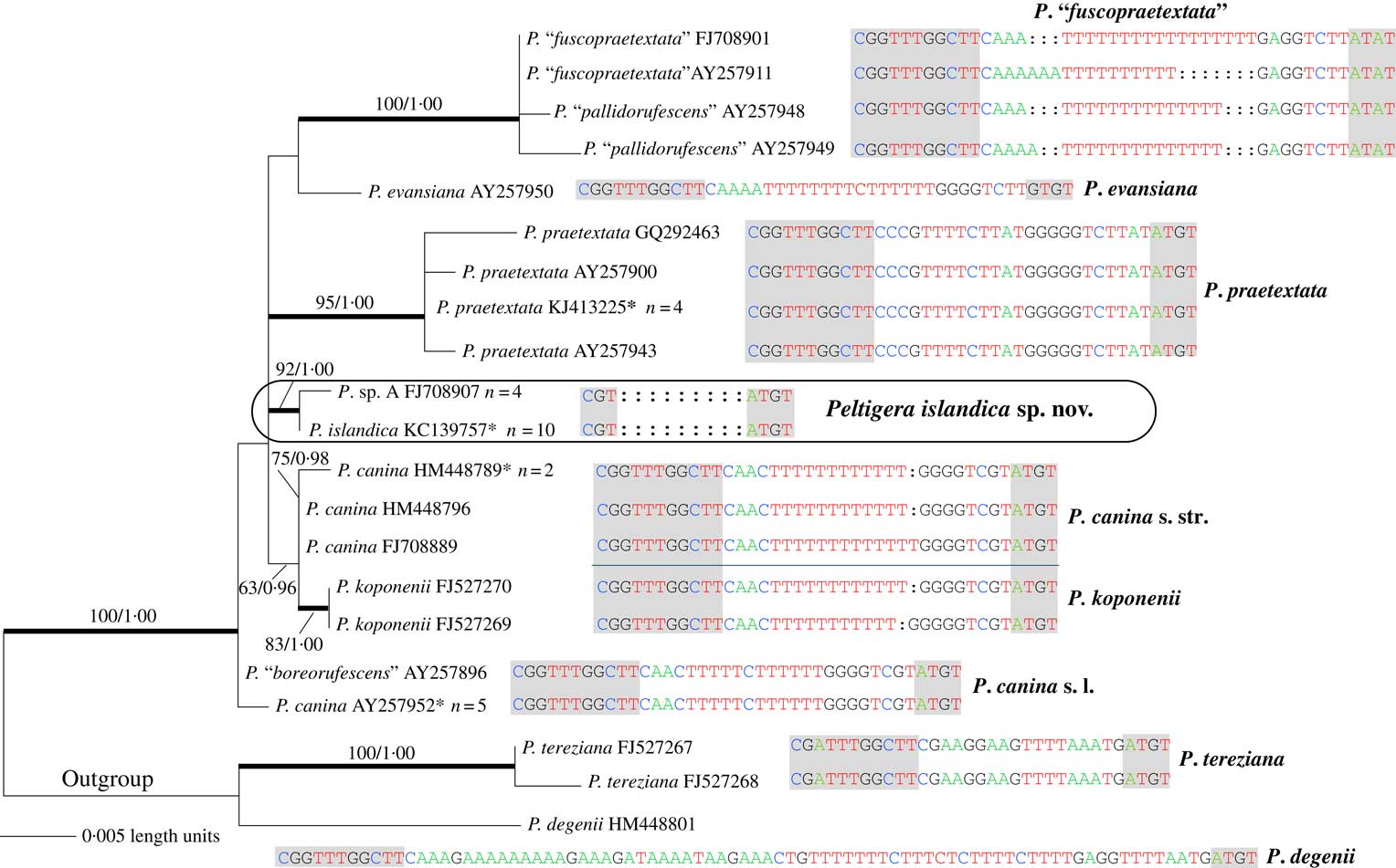
Although not all relationships among species in the present phylogeny of Group C are settled, most of the species represented by multiple individuals were significantly supported as monophyletic (Fig. 3): P. praetextata, P. koponenii, and the putative P. “fuscopraetextata” (including P. “pallidorufescens”) (Miadlikowska et al. Reference Miadlikowska, Lutzoni, Goward, Zoller and Posada2003; O’Brien et al. Reference O’Brien, Miadlikowska and Lutzoni2009). Length variation of homopolymeric tracts in the ITS1-HR region within the P. “fuscopraetaxtata”/P. “pallidorufescens” clade may indicate the occurrence of multiple species or a single broadly defined taxon (see O’Brien et al. Reference O’Brien, Miadlikowska and Lutzoni2009). Similarly, shared C/T substitutions in ITS1-HR between some individuals of P. canina (e.g. AY257952) and P. “boreorufescens” (e.g. AY257896), and their position outside the P. canina s. str./P. koponenii clade, may indicate a new species (P. “boreorufescens”) or intraspecific variation within a broadly defined P. canina, if its monophyly is confirmed (see Miadlikowska et al. Reference Miadlikowska, Lutzoni, Goward, Zoller and Posada2003). The validity of P. koponenii (known only from Papua New Guinea; Sérusiaux et al. Reference Sérusiaux, Goffinet, Miadlikowska and Vitikainen2009) as an independent species versus a morphotype within P. canina s. str., as suggested by its identical ITS1-HR with members of P. canina s. str., should also be further evaluated.
Against this background, all individuals of P. islandica and P. sp. A examined stand together for their lack of the ITS1-HR, which is present in all other members of the P. canina complex. As expected, P. islandica and P. sp. A form a monophyletic, highly supported separate lineage, although here a sister relationship with P. praetextata (see O’Brien et al. Reference O’Brien, Miadlikowska and Lutzoni2009) was not recovered. Overall bootstrap support was in agreement with posterior probability support (both or neither significant) for all internodes except for the one in which P. koponenii was sister to P. canina s. str., with the exclusion of P. canina s. lat. (significant support by posterior probability only; Fig. 3). Contrary to other studies (Gaya et al. Reference Gaya, Redelings, Navarro-Rosines, Llimona, De Cáceres and Lutzoni2011; Sliwa et al. Reference Sliwa, Miadlikowska, Redelings, Molnar and Lutzoni2012), incorporating signal from ambiguously aligned regions in the ITS and using an alignment-free method (BAli-Phy) did not increase the resolution and robustness of the ITS phylogeny, perhaps due to an insufficient number of variable characters provided by the ITS locus, even if ambiguously aligned regions (delimited manually and excluded from the RAxML analyses) were incorporated in the Bayesian analysis.
Two other mycobiont loci were also examined. RPB1 was the same among all accessions of P. islandica (Table 1) and P. sp. A (represented by GenBank FJ709247 from HOB020708–62–1–1). All P. islandica samples shared the same tub-2 allele, but this differed by two or three silent mutations from the alleles reported for P. sp. A (represented by GenBank FJ709454 and FJ709453 from HOB020708–62–1-4 and HOB020708–62–1–1, respectively). Nonetheless, all three were distinct from alleles of other members of the ‘P. canina’ group, including those newly obtained for this study.
The sister relationship of P. islandica and P. sp. A revealed by the ITS phylogeny (Fig. 3) and identity or close similarity of other genes indicate that the emerald green specimens, as well as the dark green forms most likely including P. sp. A, represent a single new species. This new member of the P. canina species complex (section Peltigera) is described here as P. islandica.
Characterization of photobiont
Hand-sectioned water mounts of P. islandica SSM55 and SSM63 (Fig. 2J & K) examined by light microscopy revealed the stratified interior expected in bipartite Peltigera species. The photobiont layer (150–175µm) appeared green under white light, but autofluoresced red under UV illumination, consistent with the presence of chlorophylls and/or phycobilins. Nostoc-like cells, 6–10 µm in diameter, were present in short chains of several cells. The 16S rRNA gene sequences amplified from whole lichen DNA, extracted from accessions SSM55 (KC139748) and SSM63, were identical and shared 99% identity with sequences from symbiotic Nostoc sp. from plants (e.g. 1271/1274 nucleotide identity to Nostoc from Gunnera tinctoria, GenBank AM711527; Papaefthimiou et al. Reference Papaefthimiou, Hrouzek, Mugnai, Lukesova, Turicchia, Rasmussen and Ventura2008) and lichens (e.g. 1270/1274 identity to Nostoc sp. from P. extenuata, GenBank KF359710.1).
Results from the examination of whole lichens were supported by characterization of cyanobacteria cultivated from P. islandica SSM165. The isolates, SSM165-N2a and SSM165-N2b, were consistent with Nostoc sp. in morphology (Fig. 2L), and their rbcLX sequences (KC139777, KC139778), identical to that of the source lichen SSM165 (haplotype rbcLX-1, Table 1), shared high similarity to reported Nostoc photobionts (below).
The rbcLX locus was useful not only for supporting identification of the photobiont to Nostoc sp., but also for distinguishing strains and placing the photobiont of P. islandica in a larger biological and phylogenetic context. Of the nine rbcLX haplotypes associated with the Peltigera community at Öskjuhlíd (Table 1, Fig. 1), all five emerald green forms of P. islandica had rbcLX-1, while the dark green form, SSM344, had rbcLX-2, a quite different allele with a 24-nucleotide insertion and >10 substitutions compared to rbcLX-1. These haplotypes were not exclusive to P. islandica: rbcLX-1, the most common allele at Öskjuhlíd, was present in P. rufescens OSA451 as well as four of five P. “neorufescens”, while rbcLX-2 was also found with P. rufescens SSM337 nearby. At Hrauneyjafoss, one P. islandica (SW329) shared rbcLX-1 with the green morphotypes at Öskjuhlíd, but the second (SW330), and those from the final two localities were different, with changes at 2, 4 and 9 positions in rbcLX, respectively (Table 1).
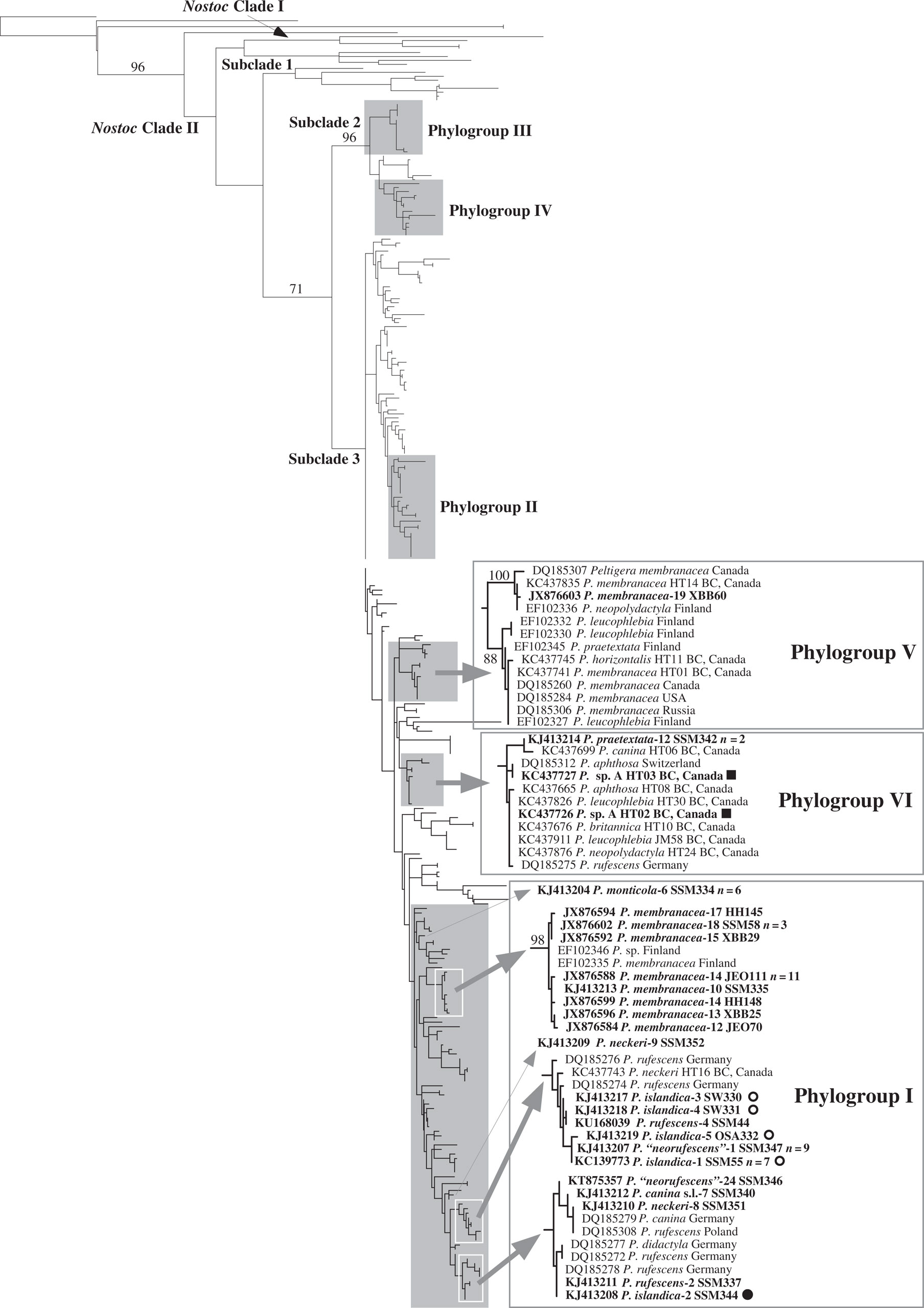
Despite the diversity of rbcLX types associated with colourmorphs and locations, when compared to a large worldwide dataset that included a significant proportion of Peltigera symbionts (O’Brien et al. Reference O’Brien, Miadlikowska and Lutzoni2013), all photobionts of P. islandica grouped together in a large clade (phylogroup I; Fig. 4) that also contained the majority of Peltigera rbcLX types known from Iceland, including many from P. membranacea (Manoharan et al. Reference Manoharan, Miao and Andrésson2012). Within this clade, cyanobionts from the emerald green morphs of P. islandica were clustered, regardless of geographical origin, whereas Nostoc from the dark green morph of P. islandica and P. rufescens SSM337, in a different subclade, was more closely related to strains from geographically distant (e.g. from Germany and Poland) species of section Peltigera. In contrast, cyanobionts of P. membranacea (with one exception) seem to be associated with a unique (highly supported), potentially widely distributed (across Iceland and beyond, e.g. Finland) clade of Nostoc. Notably, the photobionts reported with the Canadian specimens of P. sp. A were placed in a different cluster (phylogroup VI; Fig. 4), despite availability, albeit in low abundance (2%), of phylogroup I photobionts at their collection sites in Canada (O’Brien et al. Reference O’Brien, Miadlikowska and Lutzoni2013).
Phylogenetic placement of cyanobionts associated with P. islandica (including P. sp. A) and other Peltigera species from Iceland revealed by maximum likelihood analysis of 295 individuals based on the rbcLX locus. Names of all Icelandic specimens and P. sp. A are in bold. Boxes identify phylogroups (I, VI, and V) that include the Nostoc symbionts from Iceland (a complete phylogeny is available upon request). Names of Icelandic specimens are appended with a hyphen and corresponding rbcLX allele designation. n=1 unless otherwise stated. ●=cyanobionts from dark green thalli of P. islandica from Iceland; ■=cyanobionts from dark green thalli of P. islandica (including P. sp. A) from Canada; ○=P. islandica cyanobionts from emerald green thalli collected in Iceland. Bootstrap support values ≥70% are provided above internodes. Delimitation of clades, subclades, and phylogroups (clusters) follows Otálora et al. (2010) and O’Brien et al. (Reference O’Brien, Miadlikowska and Lutzoni2013). Fischerella muscicola (Stigonematales) was used to root the tree.

Taxonomic Section
Peltigera islandica Goward & Manoharan-Basil sp. nov.
MycoBank No.: MB 817007
Thallus foliose, some forms similar to P. membranacea, others to P. rufescens, but lobes narrower than the first and lobe tips predominately distinctly downturned, not exclusively upturned as in the second. The hypervariable region in the internal transcribed spacer 1 (ITS1-HR; Miadlikowska et al. Reference Miadlikowska, Lutzoni, Goward, Zoller and Posada2003) is absent, contrary to the remaining species currently recognized in the section Peltigera.
Type: Iceland, Thverás, terricolous, 64·104183°N, 21·780667°W, with grass and moss, under Salix arctica and Calluna vulgaris by a driveway in a residential neighbourhood, 5 August 2015, Ó. S. Andrésson OSA392 (AMNH—holotype; UBC, H—isotypes).
Thallus foliose, loosely appressed, usually small, 1·5–3·0 cm across (see below); lobes thin, brittle, 5–10(–15) mm wide, loosely overlapping, short to somewhat elongate, irregularly branching; lobe tips rounded, predominantly downturned; lobe margins mostly even, not crisped; upper surface bright emerald green to apparently rarely khaki green (moist) or pale bluish grey to purplish brown (dry), dull, often distinctly billowed, tomentose near lobe tips, the tomentum becoming sparse or absent toward thallus centre, appressed to occasionally in part erect; soredia, isidia and marginal lobules lacking; lower surface distinctly veined; veins whitish except grading inward to medium brown, not appearing to overlap, narrow to somewhat broad, low to more often raised, usually lacking tomentum, but sometimes distinctly erect-tomentose; interstices white, lenticular to occasionally in part polygonal, moderately deeply set; rhizines concolourous with veins, variable, ranging from discrete to flocculent or hedgerow-forming towards lobe tips, inwards becoming mostly threadlike and unbranched, 2–3(–5) mm long, lacking tomentum or sometimes distinctly erect-tomentose; cortex c. 250–300 µm thick; photobiont layer 150–175 µm thick, containing Nostoc; medulla white, c. 450–550 µm thick.
Apothecia not seen.
Chemistry. All spot tests negative.
Distribution. Peltigera islandica is known from Iceland and from southern intermontane British Columbia, Canada.
Discussion. Peltigera islandica is characterized by the presence of a cyanobacterial photobiont (Nostoc) in combination with a usually bright emerald green upper surface when wet, narrow lobes 5–10 mm wide, laminal tomentum, and predominantly downturned lobe tips. Peltigera membranacea, P. praetextata and to some extent P. rufescens, are similar but the first species differs in having broader lobes (10–20(–30) mm wide) and the latter two species differ in having upturned lobe tips.
This is perhaps one of the most variable members of section Peltigera (the ‘P. canina group’), especially with respect to the morphology of the lower surface. In some thalli, for example, the veins and rhizines are densely covered in erect tomentum, whereas tomentum is lacking altogether in other thalli, with every gradation between. The outermost rhizines too can be hedgerow-forming (as in P. rufescens), to threadlike/non-tomentose (as in P. praetextata), flocculent (as in P. canina), or threadlike/erect-tomentose (as in P. membranacea), even in different portions of a single thallus. Even so, these species are unlikely to be confused with P. islandica owing to the presence of upturned lobe tips in P. rufescens and P. praetextata, and broader lobes in P. canina and P. membranacea. In contrast to P. islandica, all of these currently recognized species in section Peltigera contain ITS1-HR, as defined in Miadlikowska et al. (Reference Miadlikowska, Lutzoni, Goward, Zoller and Posada2003).
The material of P. islandica from which lobes were sampled for accessions OSA332 and OSA392 (the holotype) was found on previously bare soil beside a residential driveway built in 1989. Those vouchers are from a larger colony than those at Öskjuhlíd, but apparently still represent one thallus that was first noted in 2013 for its emerald green colour. The same thallus was re-photographed in situ most recently in April 2016 (Fig. 2H), and estimated to be c. 12 cm across. The oldest (central) part has deteriorated, with lobes that are broken and brown; growth at the periphery, however, is emerald green. No apothecia were noted, but another smaller, emerald green patch was found nearby (Fig. 2I). We made the decision not to collect all of the original patch for use as holotype material in order to maintain the type strain of the mycobiont (and possibly the photobiont) in nature, leaving open the possibility of obtaining additional clonally-related material for archival as well as for future studies.
Selected specimens examined. Iceland: Öskjuhlíd: 64·131683°N, 21·920017°W, terricolous with Hylocomium splendens and other mosses, beside a paved path in an afforested area with Betula pubescens, Picea sitchensis and Pinus mugo, 2013, S. S. Manoharan-Basil SSM344, SSM348 and SSM355 (IMNH). Thverás: 64·104183°N, 21·780667°W, terricolous with grass and moss, under Salix arctica and Calluna vulgaris, by a driveway in a residential neighbourhood, 2013, Ó. S. Andrésson OSA332 (IMNH).—Canada: British Columbia: Spahats Falls, 2400 m, 51·738889°N–120·016111°W, on soil above trail along W-facing slope, N of waterfall in Abies-Betula forest, with Thuja and Rosa understorey, 2002, P. sp. A H. O’Brien HOB020708-66-1-4 (DUKE).
Discussion
The lichen flora of Iceland has been surveyed since 1870 and was noted to comprise over 755 species by 2009 (Kristinsson & Heiðmarsson Reference Kristinsson and Heiðmarsson2009). While it is considered fairly well studied by lichenologists, new species continue to be added. Hansen (Reference Hansen2009) conducted floristic studies of nine localities in 2007 and 2008 and documented 12 species new to Iceland. Kristinsson et al. (Reference Kristinsson, Heiðmarsson and Hansen2014) reviewed the herbarium of Svanhildur Svane and added two more, on top of the 15 from this collection already provisionally incorporated (Kristinsson & Heiðmarsson Reference Kristinsson and Heiðmarsson2009). The present study adds a further two new entries to the list of Icelandic lichens, and brings the number of Peltigera species recorded there to 24.
Peltigera species worldwide are often considered colonizers of areas with man-made or natural disturbance (Topham Reference Topham1977; Louwhoff Reference Louwhoff2009), and this seems generally consistent with where the new taxa in this study were found. The locations in south-west Iceland are geographically separated so dispersal has occurred, although the mechanism(s) are presently unknown. Clonal reproduction (vertical transmission of the photobiont) as a means to account for the presence of rbcLX-1 throughout the population of P. islandica with emerald green thalli at Öskjuhlíd is an attractive hypothesis for its simplicity, although formally, high photobiont selectivity for the species cannot be ruled out (Otalora et al. Reference Otalora, Martinez, O’Brien, Molina, Aragon and Lutzoni2010; O’Brien et al. Reference O’Brien, Miadlikowska and Lutzoni2013). The latter may be unlikely, however, as the mycobiont is also associated with Nostoc rbcLX-2 at Öskjuhlíd, as well as other rbcLX types at each of the other locations (whether by de novo synthesis or “photobiont sharing” (Rikkinen et al. Reference Rikkinen, Oksanen and Lohtander2002; Rikkinen Reference Rikkinen2003) is unclear). Low selectivity may characterize this species more generally, as the specimens originally designated P. sp. A from Canada carry Nostoc strains from a different phylogroup to those in Iceland (O’Brien et al. Reference O’Brien, Miadlikowska and Lutzoni2013).
It may be more relevant to note that in each case the association appeared to be with a dominant local photobiont: at Öskjuhlíd rbcLX-1 was common, while in Canada the haplotype that included the P. sp. A photobionts comprised about two-thirds of all Nostoc determined from the sites (O’Brien et al. Reference O’Brien, Miadlikowska and Lutzoni2013). The capacity to adapt to locally abundant and presumably successful photobionts could confer an advantage for exploiting a variety of environments, perhaps facilitating expansion of the geographical range of the mycobionts. In connection with this, a 99% match to an ITS sequence (GenBank accession no. KC965216) from a metagenomic study suggests that P. islandica may also exist in the Canadian Arctic (Timling et al. Reference Timling, Walker, Nusbaum, Lennon and Taylor2014). Overall, occurrence in localities separated by >100 km and association with different local cyanobionts suggests that P. islandica is well established in Iceland; however, its status in climatically similar regions of western North America needs further work, and it should also be looked for in Europe.
Characterization of a new species from the onset with consideration of its community as a broader framework has a number of benefits, because a more comprehensive dataset might lead to additional insights into the biology and ecology of the lichen (Seaward Reference Seaward1977; Nash Reference Nash2008). With adequate sampling, co-occurrence of closely-related species could offer opportunities to explore species boundaries by searching for evidence of gene flow, as was done for mixed communities in British Columbia, wherein P. sp. A and P. canina were first suggested to be distinct (O’Brien et al. Reference O’Brien, Miadlikowska and Lutzoni2009). Given that P. canina s. lat. from Vaglaskógur and Hrafnhólar characterized in this study have the same RBP1 and tub-2 alleles as P. canina in British Columbia, both of which are different from those of P. islandica, further examination of P. canina at Öskjuhlíd would allow comparable analyses to be made not only within site in Iceland, but also across continents.
From an ecological perspective, the presence of multiple Peltigera species in the study area at Öskjuhlíd might represent an initial stage of colonization by ruderals (Topham Reference Topham1977; Scheidegger Reference Scheidegger1995; Clayden Reference Clayden2010), in which P. islandica would seem at least as adept as the others. Alternatively, it may be interpreted as indicating that P. islandica can be an efficient competitor in later stages of ecological succession, insofar as one study has shown that mature forests hold a higher diversity of Peltigera species than young forests (Ramirez-Fernandez et al. Reference Ramirez-Fernandez, Zuniga, Mendez, Caru and Orlando2013). In Iceland, the original forests were felled centuries ago and nearly all large tracts with trees today result from afforestation since the mid-1900s (Eysteinsson Reference Eysteinsson2013). Relative to stand age, the concrete path at Öskjuhlíd is therefore not a recent disturbance, but as much a fixture as the trees, and the ‘snapshot’ of the consortia of Peltigera beside it might represent a seral stage of transition. Further characterization and monitoring of this and other terrestrial lichen communities in modern Icelandic forests might offer an additional window into this emerging landscape.
A second important realization is that P. islandica is probably more common in Iceland than implied by its recent recognition as a new species. Although colour is not typically a reliable character for bipartite Peltigera species, the distinctive emerald green hue, coupled with the absence of cephalodia, was nonetheless part of the original search criteria, and not surprisingly only green morphs were recovered from the other locations. The dark green morph would have been overlooked, except in studies where the goal was to examine the community. While this form is a minority at Öskjuhlíd, a more thorough survey is needed to accurately determine the abundance and distribution of P. islandica in all its forms in Iceland. The prevalence of the green form might be related to a humid microhabitat and/or association with particular cyanobionts (e.g. Nostoc rbcLX-1, rbcLX-6), as there were a number of other somewhat greenish bipartite Peltigera species at Öskjuhlíd. Among them, the finding of P. “neorufescens”, another taxon new to Iceland, was interesting, given that some thalli were located within a metre of P. islandica. The detection of two ITS haplotypes of P. “neorufescens” in Iceland suggests that it also might be relatively common.
The number and the circumscriptions of Peltigera species worldwide are likely to change as a result of recent lichen inventories (e.g. China and South America) and ongoing revisionary studies with extended sampling and new Peltigera-specific markers. With the use of a more integrated approach to complement traditional morphology-based investigations of new species, exploration of even small spatial scale Peltigera communities in interesting habitats might also offer opportunities to discover and study yet more additions to this genus.
We thank S. Werth for the gift of samples SW329–331, B. Redeling for help in performing BAli-Phy analysis, and N. Magain and C. Truong for providing a broad phylogenetic context (unpublished data) for the validation of P. islandica. We are also very grateful to our reviewers from the journal for their useful suggestions. JM was supported by the National Science Foundation through a REVSYS award on the genus Peltigera (DEB-1025930); SSM and ÓSA were supported by the Icelandic Research Fund.
The authors dedicate this paper to Dr Irwin M. Brodo, in recognition of his many important contributions to lichen taxonomy and biology over the past half century.