Clostridium difficile infection (CDI) molecular epidemiology, as well as the clinical implications of particular C. difficile strains, has been well described in adult patients. The dramatic increase in the incidence and severity of CDI observed in adults over the past decade is largely attributed to the emergence of a highly fluoroquinolone-resistant epidemic strain identified as group BI by restriction endonuclease analysis (REA), NAP1 by pulsed-field gel electrophoresis (PFGE), and ribotype 027 by polymerase chain reaction (PCR) ribotyping.Reference Kelly and Lamont 1 , Reference McDonald, Killgore and Thompson 2 The incidence of pediatric CDI is also increasing, particularly among those thought to be at low risk for CDI, such as previously healthy children without recent hospitalization or antibiotic exposure.Reference Benson, Song, Campos and Singh 3 – Reference Tamma and Sandora 8 Although an increase in CDI severity has not yet been reported in children, recurrence complicates up to 20%–25% of pediatric CDIs.Reference Kim, Shaklee and Smathers 9 – Reference Tschudin-Sutter, Tamma, Milstone and Perl 11 The C. difficile strain types causing CDI in children, including BI/NAP1/027, have been previously described,Reference Toltzis, Kim, Dul, Zoltanski, Smathers and Zaoutis 12 , Reference Wendt, Cohen and Mu 13 but risk factors and outcomes related to infection with particular C. difficile strains among children are not well understood. Our objectives were to define the C. difficile REA groups causing CDI in a pediatric cohort and to determine risk factors and outcomes associated with CDI caused by epidemic C. difficile strains in children.
Methods
Study Design and Setting
We performed a molecular epidemiologic survey and retrospective observational cohort study of all patients with CDI at the Ann & Robert H. Lurie Children’s Hospital of Chicago, a 288-bed, university-affiliated hospital that provides tertiary care to children in the greater Chicago area. The clinical microbiology laboratory restricts testing for toxigenic C. difficile by the tcdB (toxin B gene) PCR Xpert C. difficile assay (Cepheid, Sunnyvale, CA)Reference Shin, Kim and Kim 14 to unformed stools obtained from children ≥12 months old. The Institutional Review Board at Lurie Children’s waived informed consent.
Patients and Data Collection
The study cohort included all patients ≥12 months old who had an unformed stool collected between December 9, 2012, and December 8, 2013, who tested positive by tcdB PCR. Cases were identified through Medmined (CareFusion, San Diego, CA), an electronic infection surveillance tool, and confirmed by manual review of all clinical and demographic data extracted from the electronic medical record. Patients were included if they had documented diarrhea or ileus, had a diagnosis of CDI documented by the primary healthcare provider or hospital infection control personnel, and/or had received treatment for CDI. CDI cases representing a second positive tcdB PCR test within 2 weeks of the original infection were also excluded unless the patient experienced resolution and then recurrence of CDI symptoms. Patient follow-up was ascertained by manual review of subsequent inpatient, outpatient, and telephone encounter notes documented in the medical record. The records of all patients were reviewed for at least 8 weeks from their most recent CDI.
Microbiology
After completion of the tcdB PCR assay, stools were stored at −70°C and batch processed for isolation of C. difficile, as previously described,Reference Wilson, Kennedy and Fekety 15 at the Microbiology Research Laboratory at Hines VA Hospital in Hines, Illinois. Stool was cultured onto prereduced taurocholate-cycloserine-cefoxitin-fructose agar (TCCFA) in an anaerobic chamber and incubated at 36°C for 48 hours. C. difficile was identified based on typical morphology. When C. difficile was detected on TCCFA, a single colony was subcultured on blood agar. Negative cultures on TCCFA at 48 hours were recultured after an alcohol shock procedure with 95% ethanol as previously described.Reference Clabots, Gerding, Olson, Peterson and Gerding 16 Negative cultures at 48 hours after alcohol shock were not evaluated further. All C. difficile isolates underwent REA. As previously described,Reference Clabots, Johnson and Bettin 17 bacterial DNA was isolated from a single C. difficile colony sampled from blood agar. DNA underwent digestion using HindIII restriction endonuclease, and DNA bands were visualized by agarose gel electrophoresis. REA group was assigned to each isolate based on comparison of the DNA band pattern to a library of reference REA groups available through the Microbiology Research Laboratory.
Risk Factor and Outcome Analyses
After the REA group causing each CDI was identified, risk factors and outcomes associated with select epidemic strains of C. difficile were assessed. For patients with multiple CDIs caused by the same strain, only the first infection with that particular strain was included. The primary outcome measure was first CDI recurrence within 8 weeks of the previous episode. Potential CDI risk factors assessed included age, gender, patient comorbidities, antibiotic receipt in the previous 30 days, and proton pump inhibitor use at the time of CDI diagnosis. CDI cases were classified as hospital-onset (HO) healthcare facility-associated (HCFA), community-onset (CO) HCFA, indeterminate, community-associated (CA), and recurrent using standard definitions.Reference McDonald, Coignard and Dubberke 18 Comorbidities included inflammatory bowel disease, malignancy, solid organ transplant, and gastrostomy tube dependence.
Relapse versus Reinfection in Children with Multiple CDIs
In children with multiple CDIs, REA band patterns of consecutive C. difficile isolates from those patients were compared to classify the subsequent CDI as a relapse or reinfection irrespective of the duration of time between CDIs. A relapse was defined as a subsequent CDI caused by a C. difficile strain with the identical REA DNA band pattern as the previous CDI. A reinfection was defined as a subsequent CDI caused by a different C. difficile strain as the previous CDI. Of note, the definitions for relapse and reinfection in the present study are independent of time from previous infection as previously described.Reference Kamboj, Khosa, Kaltsas, Babady, Son and Sepkowitz 19 Therefore, these definitions deviate from the standard 8-week recurrence interval. For this analysis, in addition to including cases from the pediatric cohort described above, cases were also selected from a convenience sample of C. difficile isolates from saved tcdB PCR-positive stools collected between March 2011 and October 2012. This permitted analysis of a larger sample of patients with multiple CDIs. Patients with >2 CDIs had multiple pairs of isolates compared. For example, in a patient with 3 CDIs, the first and second CDIs were compared to identify the second CDI as a relapse or reinfection; the second and third CDIs were compared to identify the third CDI as a relapse or reinfection. To assess the relationship between strain type and risk of relapse, only the first CDI per patient was included in this analysis.
Statistical Analysis
Medians of continuous variables were compared using the Wilcoxon rank-sum test. Proportions were compared using the χ2 test (or Fisher’s exact test when appropriate). Risk ratios (RRs) and 95% confidence intervals (CIs) were calculated using log-binomial regression analysis, and variables with statistical significance on bivariate analysis were included in multivariate regression analysis. Two-sided P values <.05 were considered statistically significant. Analyses were performed using Stata/IC statistical software, version 12.1 (StataCorp LP, College Station, TX).
Results
C. difficile REA Groups causing CDI in Children
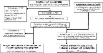
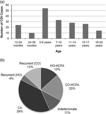
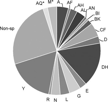
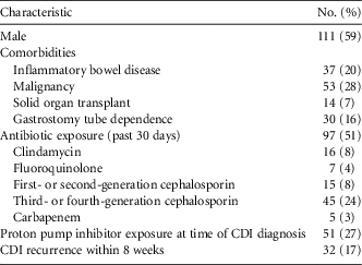
Between December 9, 2012, and December 8, 2013, 203 tcdB PCR-positive stool specimens were collected from 151 patients. Of these 203 eligible CDI cases, a review of the medical records confirmed 189 CDIs among 145 patients (Figure 1). Table 1 and Figure 2 show the demographics/clinical characteristics and the age distribution/CDI classification, respectively, of these CDI cases. Of the 189 included CDI cases, 156 stool specimens (83%) were available for culture. Among those, C. difficile was isolated from 132 stool specimens (85%). Of these isolates, 15 were from patients from whom the same strain was identified from a subsequent CDI during the cohort period. REA of the remaining 117 isolates revealed a highly diverse distribution of REA groups (Figure 3). REA group Y, a common and highly diverse endemic strain, was the most common REA group identified. REA group DH was the predominant epidemic strain isolated, occurring in 19 of 117 CDIs (16%). REA group BI caused CDI in only 1 patient. Nonspecific REA groups (ie, DNA gel patterns that are not consistent with a recognized REA group in the reference library) were identified in 29 CDIs. DNA gel patterns of these 29 isolates demonstrated 28 unique nonspecific REA groups. In total, 46 unique REA groups (ie, 18 assigned and 28 unique nonspecific REA groups) were identified.
Flow chart of patients included in study analyses.

(A) Age distribution and (B) classification of 189 cases of pediatric C. difficile infection over a 1-year period. HO: hospital-onset; HCFA: healthcare facility-associated; CO: community-onset; CA: community-associated.

Restriction endonuclease analysis of 117 C. difficile isolates from pediatric patients over a 1-year period. *Non-toxigenic REA group. Non-sp, 28 unique non-specific REA groups.

Demographic and Clinical Characteristics of 189 Cases of Pediatric C. difficile Infection (CDI) over a 1-Year Period

Risk Factors Associated with REA Group DH
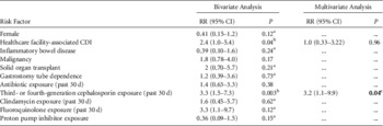
Risk factors and frequency of recurrence within 8 weeks were compared between cases of CDI caused by REA group DH and those caused by non-DH strains. Of the 132 CDI cases with isolates available for REA, 15 were omitted because they represented a subsequent case of CDI caused by the same strain as the previous case during the cohort period. The final cohort analysis compared 19 cases of DH CDI to 98 cases of non-DH CDI (Figure 1). Median ages of patients with DH and non-DH CDI were similar (8.4 vs 9.8, P=.43). Median tcdB PCR cycle threshold (Ct) was significantly lower with DH CDI (23.2 vs 25.9, P=.041). Compared with non-DH CDI, risk of recurrence within 8 weeks following DH CDI was similar (RR, 1.7; 95% CI, 0.71–4.2, P=.31). DH CDI was more likely to occur in patients exposed to third- or fourth-generation cephalosporins in the previous 30 days (RR, 3.2; 95% CI, 1.1–9.9, P=.04; Table 2).
Risk Factors Associated with Pediatric C. difficile Infection (CDI) Caused by Restriction Endonuclease Analysis (REA) Group DH: Comparison of 19 Cases of DH Strain Type CDI to 98 Cases of Non-DH CDI

a Fisher’s exact test.
b Risk factors with P<0.05 included in multivariate analysis.
c Bold P value indicates statistical significance.
Relapse versus Reinfection in Children with Multiple CDIs
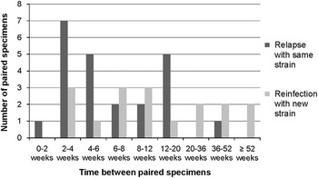
In addition to the 132 available C. difficile isolates from the cohort, REA was also performed on 21 C. difficile isolates that were available from the convenience sample (Figure 1). In total, 2 or more C. difficile isolates from children with multiple CDIs were available in 30 patients (23 patients with 2 isolates; 4 patients with 3 isolates; and 3 patients with 4 isolates). This permitted comparison of 40 pairs of consecutive isolates. REA identified 23 relapses (ie, CDI with same strain) and 17 reinfections (ie, CDI with a new strain). Duration of time between all CDIs classified as either relapse or reinfection is illustrated in Figure 4. The proportion of CDI relapses occurring within 8 weeks versus after 8 weeks of the previous infection was not significantly different (68% of repeat CDIs were relapses within 8 weeks vs 44% of repeat CDIs after 8 weeks, P=.13). Among patients with CDI relapse, 20 of 23 (87%) were discharged from the hospital and spent time in the community between relapsed CDIs.
Duration of time between collection of paired stool specimens among individual pediatric patients with Clostridium difficile infection complicated by subsequent relapse versus reinfection.

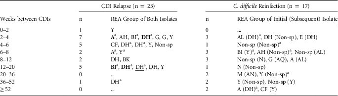
The particular REA groups causing relapses and reinfections are listed in Table 3. To assess the relationship between strain type and risk of relapse, only the first CDI per patient was included in this analysis. Compared with those whose initial infection was caused by a non-DH strain, those whose initial infection was caused by REA group DH had a similar rate of CDI relapse (80% vs 52%; P=.25) but were significantly more likely to experience multiple CDI relapses (40% vs 8%; P=.05).
Restriction Endonuclease Analysis of Isolates from Cases of Pediatric C. difficile Infection Complicated by Relapse vs. Reinfection

NOTE. Non-sp, non-specific REA group.
a CDIs in patients with multiple relapses or reinfections. Bolded and underlined REA groups indicate the second and third relapses, respectively, with the same strain as a previous relapse in an individual patient.
Discussion
In the present study, a wide variety of C. difficile strains representing 46 different REA groups was identified. REA group DH (also known as NAP11 by PFGE and ribotype 106 by PCR) was the predominant epidemic strain identified. Compared with CDI caused by non-DH/NAP11/106 strains, CDI caused by DH/NAP11/106 was associated with third- or fourth-generation cephalosporin exposure in the previous 30 days. Among children with multiple CDIs, relapses commonly occurred up to 20 weeks after the initial CDI, suggesting that the current definition for recurrent CDI may lead to misclassification of some recurrent CDIs as new CDIs in children. CDI caused by DH/NAP11/106 was associated with an increased risk of experiencing multiple subsequent CDI relapses.
In our cohort, BI/NAP1/027 was isolated from only 1 patient. This was surprising because in recent studies, BI/NAP1/027 prevalence was 61% in Chicago area primarily adult acute care hospitals in 2009;Reference Black, Weaver and Jones 20 19% among children in Philadelphia and Cleveland in 2006–2007;Reference Toltzis, Kim, Dul, Zoltanski, Smathers and Zaoutis 12 and 23% in a pediatric cohort collectively representing 10 diverse US geographic locations in 2010–2011.Reference Wendt, Cohen and Mu 13 The molecular epidemiology of CDI is a dynamic process,Reference Belmares, Johnson and Parada 21 and it is unknown whether our more recent data presented here represent a regional/national trend in declining prevalence of BI/NAP1/027 or if our cohort is anomalous. Declining prevalence of BI/NAP1/027 has been reported recently in Europe but not yet in the United States.Reference Johnson 22 Less frequent fluoroquinolone use in pediatrics alone does not appear to explain this finding because of the notable proportion of BI/NAP1/027 in other pediatric populations in recent years.Reference Toltzis, Kim, Dul, Zoltanski, Smathers and Zaoutis 12 , Reference Wendt, Cohen and Mu 13
The DH/NAP11/106 strain is common in the United Kingdom, causing the second most CDI cases after the BI/NAP1/027 strain, but it has been relatively uncommonly reported in the United States.Reference Freeman, Bauer and Baines 7 However, in a pediatric cohort collectively representing 10 diverse US geographic locations, DH/NAP11/106 accounted for 13% of 132 CDI cases, which suggests that DH/NAP11/106 infection may be increasing in the United States.Reference Wendt, Cohen and Mu 13 Compared to BI/NAP1/027, studies of outcomes related to DH/NAP11/106 CDI are limited. Mortality related to CDI caused by DH/NAP11/106 was reported to be significantly lower than mortality related to CDI caused by BI/NAP1/027 in adults.Reference Sundram, Guyot, Carboo, Green, Lilaonitkul and Scourfield 23
Because DH/NAP11/106 CDI was associated with a lower tcdB PCR Ct value, DH/NAP11/106 CDI was likely associated with a higher fecal bacterial burden.Reference Dionne, Raymond, Corbeil, Longtin, Gervais and Longtin 24 However, it is unclear whether the potentially higher bacterial burden contributed to more prolonged DH/NAP11/106 carriage and increased risk of multiple relapses. In the present study, DH/NAP11/106 acquisition was associated with third- or fourth-generation cephalosporin exposure in the previous 30 days. Because third-generation cephalosporins confer a higher CDI risk for 3 months after exposure compared with other classes of antibiotics,Reference Hensgens, Goorhuis, Dekkers and Kuijper 25 the CDI risk with potentially prolonged DH/NAP11/106 carriage may be exacerbated following broad-spectrum cephalosporin exposure. The clinical significance of tcdB PCR Ct requires further investigation.
The proportion of relapsed CDI in the present pediatric study was lower than that reported in adults (relapse in 88% and 65% of repeat infections within and after 8 weeks, respectively).Reference Kamboj, Khosa, Kaltsas, Babady, Son and Sepkowitz 19 The differences may be explained by our use of REA, which has significantly greater discriminatory power than PCR ribotyping (index of discrimination 0.93 vs 0.70).Reference Killgore, Thompson and Johnson 26 Similar to previous studies in adults,Reference Kamboj, Khosa, Kaltsas, Babady, Son and Sepkowitz 19 , Reference Marsh, Arora, Schlackman, Shutt, Curry and Harrison 27 our data also suggest that classifying a subsequent CDI as a new, unrelated infection when it occurs between 8 and 20 weeks could lead to overestimation of the burden of new CDI determined by healthcare-associated infection surveillance. Therefore, our data provide additional evidence for extending the definition of recurrent CDI to as long as 20 weeks after the initial CDI. Notably, we cannot definitively rule out misclassification of a reinfection with the same strain as relapse. However, in the majority (87%) of our cases, the patients were out of the hospital between CDI episodes. Therefore, reinfection with the same strain related to contamination of the patient’s healthcare environment was unlikely.
Strengths of this study include the use of REA, a highly discriminatory C. difficile typing method, and its focus on CDI in the pediatric population, which is understudied compared to adult CDI. Because this study was conducted at a single center, the generalizability of the molecular epidemiology to children in other geographic areas may be limited. The retrospective design may have limited the quality of data collected. Exposure and follow-up data were collected from the electronic medical record, and healthcare and medication exposures outside of the institution may not have been entirely captured. This also may have precluded capture of information about each patient’s subsequent CDI outcome. However, our hospital provides primary to tertiary care to a large patient population, and many affiliated community pediatricians utilize our integrated electronic medical record system. In addition, an outpatient electronic medication prescribing system is utilized. Therefore, the impact of missing data is likely minimal. The present study did not evaluate CDI severity as an outcome related to strain type because of recent data suggesting poor predictive value of current definitions of CDI severity in children.Reference Tschudin-Sutter, Tamma, Milstone and Perl 28 , Reference Kociolek, Patel, Shulman and Gerding 29 Definitions for pediatric CDI severity must be validated before accurate conclusions can be drawn regarding the association of C. difficile strain type and CDI severity in children.
In summary, DH/NAP11/106, a common epidemic strain in the UK that is less often reported in the United States, was the predominant epidemic strain causing CDI in our pediatric cohort. DH/NAP11/106 CDI was associated with recent exposure to third- or fourth-generation cephalosporins. CDI relapses were common and often occurred >8 weeks from the previous CDI. DH/NAP11/106 was associated with multiple relapses among patients with multiple CDIs. Additional studies are also needed to clarify the pathophysiology of DH/NAP1/106 and the trends in the molecular epidemiology of pediatric CDI over time and in other geographical regions.
Acknowledgments
Assistance with REA performance and interpretation was provided by Adam Cheknis, Laurica Petrella, Eric Purdue, and Susan Sambol at the Microbiology Research Laboratory at Hines VA Hospital under the direction of Dale Gerding, MD and Stuart Johnson, MD. The project was supported by the National Center for Research Resources (grant no. 5UL1RR025741), and is now at the National Center for Advancing Translational Sciences (grant no. 8UL1TR000150). The content is solely the responsibility of the authors and does not necessarily represent the official views of the NIH.
Financial support: Thrasher Research Fund (Early Career Award #11854) and Merck Sharp & Dohme Corp. (Merck Investigators Studies Program #50366) to LKK.
Potential conflicts of interest: LKK reports receiving laboratory assays from Alere. DNG reports serving on the boards of Sanofi Pasteur, Actelion, Merck, and Rebiotix, and he has consulted for Summit and Viropharma. DNG holds patents licensed to Viropharma/Shire.