The bidirectional cavopulmonary shunt is performed as a staged approach to create the Fontan circulation in patients with a functionally single ventricle. This shunt reduces the volume load on the systemic ventricle, and allows a smoother postoperative course after conversion to the Fontan circulation. In patients with bidirectional cavopulmonary shunting, the superior caval venous flow provides the major supply of blood to the lungs. Aortopulmonary collateral vessels, however, frequently develop before and after bidirectional cavopulmonary shunting, then providing an additional source of flow of blood to the lungs.Reference Spicer, Uzark, Moore, Mainwaring and Lamberti1
There are obvious potential disadvantages to the presence of aortopulmonary collateral vessels in patients undergoing conversion to the Fontan circulation. These collateral vessels should increase the volume load in the functionally single ventricle, with concomitant increase in flow of blood to the lungs, thereby elevating pulmonary arterial pressure. Increased ventricular volume and elevated pulmonary arterial pressures have been identified as risk factors for conversion to the Fontan circulation.Reference Kirklin, Blackstone, Kirklin, Pacifico and Bargeron2–Reference McElhinney, Reddy, Tworetzky, Petrossian, Hanley and Moore8 Previous reports have described an increased incidence of pleural effusions, and even increased mortality, after conversion in children with significant aortopulmonary collaterals.Reference Spicer, Uzark, Moore, Mainwaring and Lamberti1, Reference Ichikawa, Yagihara and Kishimoto9–Reference Hsu, Wang and Lin11 Despite these disadvantages, to the best of our knowledge, as yet there is no method established for accurate preoperative measurement of aortopulmonary collateral flow. The presence of aortopulmonary collaterals could also affect the calculation of haemodynamic parameters by the Fick method at cardiac catheterization in patients after bidirectional cavopulmonary shunting, since the saturation of blood in the pulmonary artery will vary with the site of sampling. Conventionally, even when collaterals are present, the Fick method is used to calculate the amount of pulmonary flow, the assumption being made that collateral flow is negligible.Reference Salim, Case, Sade, Watson, Alpert and DiSessa12 This conventional method may be inaccurate when there is substantial collateral flow.
By combining lung perfusion scintigraphy and cardiac catheterization, we have now devised a new method to assess aortopulmonary collateral flow quantitatively in patients after bidirectional cavopulmonary shunting. The aims of this study were threefold:
• to introduce the basic principles of the new method
• to demonstrate the consistency between quantified collateral flow and angiographic collateral scale
• and to examine the impact of collateral flow on the calculation of pulmonary vascular resistance.
Materials and methods
Basic principles
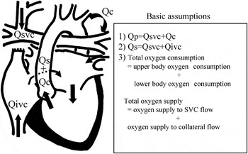
A standard model of the circulation in patients after construction of a bidirectional cavopulmonary shunt with aortopulmonary collateral vessels is shown in Figure 1. There are three basic assumptions in this study:
Model of the circulation in patients after bidirectional cavopulmonary shunting in the presence of aortopulmonary collateral vessels. Qc = the amount of collateral blood flow; Qivc = the amount of inferior vena caval blood flow; Qp = the amount of pulmonary blood flow; Qs = the amount of systemic blood flow; Qsvc = the amount of superior vena caval blood flow; SVC = the superior caval vein.

• the amount of pulmonary flow, in litres per minute, equals the amount of superior caval venous flow (Qsvc) together with the amount of collateral flow (Qc)
• the amount of systemic flow, in litres per minute, equals the amount of superior caval venous flow (Qsvc) plus the amount of inferior caval venous flow (Qivc)
• total consumption of oxygen equals consumption of oxygen by the upper body together with consumption of oxygen by the lower body, and total supply of oxygen equals supply of oxygen to the superior caval venous flow together with supply to the aortopulmonary collateral flow.
First, to quantify the collateral flow after cavopulmonary shunting, we performed lung perfusion scintigraphy with 99m-Tc-labeled macroaggregated albumin and determined the ratio of collateral to systemic flow (M) by whole-body imaging of the scintigraphy. When macroaggregated particles are administered transvenously from the leg in patients after bidirectional cavopulmonary shunting, they directly enter the systemic circulation via the inferior caval vein. Macroaggregated particles spread in accordance with the distribution of blood. The lung count on the scintigram reflects the amount of collateral flow, while the total count minus the lung count reflects the amount of systemic flow. Then, the ratio of collateral to systemic flow (Qc/Qs) was obtained by the following equation:

Second, we performed cardiac catheterization and calculated the ratio of pulmonary to systemic flow (Qp/Qs = K) using a formula derived from the Fick principle and the three basic assumptions mentioned above (see Appendix for details):

where M is the ratio of collateral to systemic flow, and Sao, Sivc, and Spv are the saturations of oxygen saturation in the aorta, the inferior caval vein, and pulmonary veins, respectively.
Finally, we calculated the absolute amount of each flow using the ratio of pulmonary to systemic flow (K) (see Appendix for details):

where O2cap is oxygen-carrying capacity and Ssvc is saturation of oxygen in the superior caval vein. In this way, we obtained the amount of collateral flow by integrating lung perfusion scintigraphy and cardiac catheterization data.
Patients
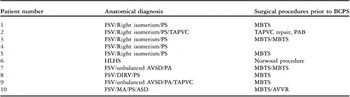
We enrolled 10 consecutive patients after construction of a bidirectional cavopulmonary shunt who required cardiac catheterization in Chiba Children’s Hospital from May, 2006, to February, 2007. Indications for the examination were evaluation prior to completion of the Fontan circulation, or interventional catheter procedures. The anatomical diagnoses, and surgical procedures prior to bidirectional cavopulmonary shunting, are listed in Table 1. In 5 patients, there were bilateral superior caval veins, so bilateral cavopulmonary anastomoses were performed. In 2 patients, bidirectional cavopulmonary shunt was associated with additional flow from the systemic ventricle subsequent to banding of the pulmonary trunk or placement of a conduit from the right ventricle to the pulmonary arteries. In these cases, the additional flow could be calculated as a part of aortopulmonary collateral flow, because both flows were considered to have the same levels of saturation of oxygen. The median age at construction of the bidirectional cavopulmonary shunt was 1.41 years, with a range from 0.75 to 15.3 years, and the median age at catheterization was 2.33 years, with a range from 1.32 to 15.8 years. Informed consent was obtained in accordance with the rules of the Institutional Ethics Committee.
Summary of anatomical diagnoses and surgical procedures prior to creation of a bidirectional cavopulmonary shunt.

ASD: atrial septal defect; AVSD: atrioventricular septal defect; AVVR: atrioventricular valvar replacement; BCPS: bidirectional cavopulmonary shunt; DIRV: double inlet right ventricle; HLHS: hypoplastic left heart syndrome; MA: mitral atresia; MBTS: Modified Blalock-Taussig shunt; PA: pulmonary atresia; PAB: pulmonary artery banding; PS: pulmonary stenosis; FSV: functionally single ventricle; TAPVC: totally anomalous pulmonary venous connection.
Lung perfusion scintigraphy
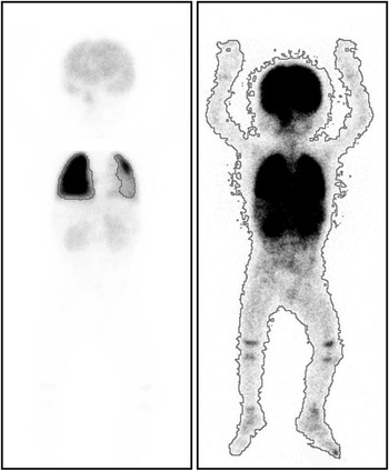
Lung perfusion scintigraphy was performed within two days before cardiac catheterization under sedation with midazolam infusion. Scanning was performed soon after intravenous injection of 99m-Tc-labeled macroaggregated albumin from the leg. Whole-body imaging required approximately 5 to 10 minutes. After isolating the lungs as areas of interest on the computer, total body and lung counts were determined separately (Fig. 2).
Whole-body images of macroaggregated albumin scintigraphy. Lung count (left) and total body count (right) were traced separately.

Cardiac catheterization
Cardiac catheterization was performed via femoral and jugular approaches under general anaesthesia. Saturations of oxygen were obtained from the aorta, the superior and inferior caval veins, and the pulmonary veins. In patients with bilateral superior caval veins, data was averaged for both veins. Aortography was performed to visualize aortopulmonary collateral vessels. If visualized, collaterals were graded by two observers according to the 4-point scale reported by Spicer et al.Reference Spicer, Uzark, Moore, Mainwaring and Lamberti1 Ventricular volume was measured with the area-length method without correction for trabeculations or papillary muscles.Reference Sano, Ogawa and Taniguchi13 Cardiac index by angiography was defined as follows:

In contrast, cardiac index by our combined scintigraphy-catheterization method was defined as:

Pulmonary vascular resistance by our method (PVR) was determined as follows:

In addition, we calculated the amount of pulmonary flow and pulmonary vascular resistance using conventional methods.Reference Salim, Case, Sade, Watson, Alpert and DiSessa12 The amount of pulmonary blood flow obtained by the conventional method was denoted as Qp′, in distinction to Qp by our method:

Then, pulmonary vascular resistance by the conventional method (PVR′) was defined:

In this study, we assumed the consumption of oxygen to calculate each flow.Reference LaFarge and Miettinen14
Statistics
Data analyses were performed on JMP Version 6.0. Regression analysis was used to evaluate the relationship between independent variables. Spearman’s Rank Correlation Coefficient (rs) was applied to test the correlation of ordinal data. A p-value < 0.05 was considered significant, and data are presented as mean ± standard deviation.
Results
Lung perfusion scintigraphy
The mean collateral to systemic flow ratio obtained by lung perfusion scintigraphy was 0.53 ± 0.17.
Cardiac catheterization
No patient had significant pulmonary arteriovenous fistulas. Neither aortic nor atrioventricular valvar regurgitation greater than mild was shown. In all 10 patients, aortopulmonary collateral vessels were visualized, being graded as 2 in 1 patient, 3 in 5 patients, and 4 in 4 patients. Coil embolization of the collateral vessels was needed in 3 patients.
Calculations
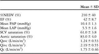
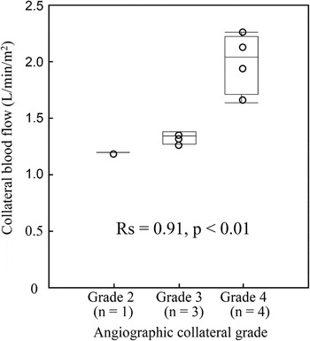
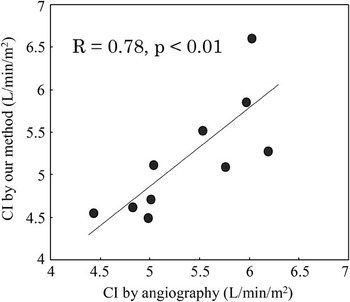
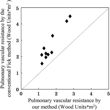
By our new method, the amount of the pulmonary, systemic and collateral flows were 3.00 ± 0.49, 3.43 ± 0.52 liters/min/m2, and 1.75 ± 0.46 liters/min/m2, respectively. The haemodynamic data are summarized in Table 2. The amount of collateral flow in patients in each angiographic scale is shown in Figure 3, excluding 2 patients with additional sources of pulmonary flow. The aortographic collateral scale was significantly correlated with our new method (rs = 0.91, p < 0.01). Cardiac index obtained by our combined scintigraphy-catheterization method was 5.18 ± 0.67 litres/min/m2, and was significantly correlated with the cardiac index determined by angiography (Fig. 4, r = 0.78, p < 0.01). The transpulmonary pressure gradient was 4.9 ± 1.1 mmHg, and the pulmonary vascular resistance was 1.70 ± 0.57 Wood Units × m2. When we used the conventional method without considering collateral flow, calculated pulmonary vascular resistance was 2.7 ± 0.98 Wood Unit × m2. This was 57 ± 23% higher than the resistance determined by our new method (Fig. 5).
Summary of haemodynamic data

EF: ejection fraction; LAP: left atrial pressure; %NEDV: percent normal of ventricular end-diastolic volume; PAP: pulmonary arterial pressure; SCV: superior caval vein; Qc: the amount of aortopulmonary collateral flow; Qivc: the amount of inferior caval venous flow; Qsvc: the amount of superior caval venous flow.
Calculated collateral flow of patients in each aortographic collateral grade. Aortographic collateral scale = the 4 point scale by Spicer et al.Reference Spicer, Uzark, Moore, Mainwaring and Lamberti1

Correlation between cardiac index by angiography and our combined scintigraphy-catheterization method. CI by angiography = cardiac index by angiography; CI by our method = cardiac index by our combined scintigraphy-catheterization method.

Differences between our method and the conventional Fick method in evaluating pulmonary vascular resistance (overestimation by the conventional method).

Discussion
The current results for conversion to the Fontan circulation have improved. A considerable number of patients, nonetheless, remain at high risk. Ideally they should be identified preoperatively to improve outcomes.Reference Yoshimura, Yamaguchi and Oshima15 Aortopulmonary collateral vessels have potential disadvantages, especially in such high-risk candidates, because such collaterals could compete with pulmonary forward flow from the superior and inferior caval vein, thus placing a haemodynamic burden on the functionally single ventricle. Several studies have examined the effects of collateral vessels on the outcome of conversion to the Fontan circulation. Controversy continues as to the negative extent of such collaterals on outcomes.Reference McElhinney, Reddy, Tworetzky, Petrossian, Hanley and Moore8, Reference Bradley, McCall, Sistino and Radtke16 This is partially explained by limitations of cardiac catheterization in quantifying the amount of collateral vessels.
Despite a need for a method to quantify aortopulmonary collateral flow preoperatively, thus far, to the best of our knowledge, there has been no established method. Triedman et al.Reference Triedman, Bridges, Mayer and Lock3 attempted to evaluate the extent of aortopulmonary collateral flow by means of estimating the total cross sectional area of the collateral or the antegrade filling defect in the angiogram. Accurate quantification of collateral flow by angiography, however, is difficult, as identification of collaterals by angiography is highly dependent on the technical variables. Several reports have attempted to quantify collateral flow by measuring the flow of blood returning to the heart during cardiopulmonary bypass.Reference Ichikawa, Yagihara and Kishimoto9, Reference Bradley, McCall, Sistino and Radtke16 This intraoperative method is performed under nonphysiologic conditions, and cannot be done preoperatively. Recently, Lim et al.Reference Lim, Graziano, Rocchini and Lloyd17 used the thermal indicator dilution technique to quantify aortopulmonary shunting in patients with bidirectional cavopulmonary shunts. They expressed the degree of shunting as the temperature change during the recirculation phase indexed to the peak temperature change. As the index was a relative value, absolute flow could not be measured.
In our study, we quantified collateral flow by determining the ratio of collateral to systemic flow using macroaggregated albumin scintigraphy. This method is useful by itself, as it can be easily performed preoperatively.
Clinical impact
Despite the usefulness of the scintigraphy for quantification of collateral flow, it can be more useful by combining it with cardiac catheterization data, because the amount of systemic and pulmonary flow is obtained along with collateral flow. The load of collateral flow can be determined in comparison with systemic and pulmonary flows. In addition, by considering the extent of aortopulmonary collateral flow, the evaluation of pulmonary vascular resistance by catheterization becomes more accurate, as will be discussed later in detail. Thus, our combined method offers two advantages:
• evaluation of the haemodynamic burden caused by the aortopulmonary collaterals
• accurate estimation of pulmonary vascular resistance.
Our method can be applied in some clinical settings other than for assessment of the risk of conversion to the Fontan circulation. First, we can determine the necessity of transcatheter occlusion of aortopulmonary collateral vessels, depending on the load. Recent reports show that preoperative embolization does not change the clinical post-operative course and routine preoperative embolization is not indicated.Reference McElhinney, Reddy, Tworetzky, Petrossian, Hanley and Moore8, Reference Bradley, McCall, Sistino and Radtke16, Reference Lim, Graziano, Rocchini and Lloyd17 Embolization should be limited to haemodynamically significant collateral vessels. The haemodynamic significance of collateral vessels, however, has mainly been determined based on subjective visual grading in angiography during cardiac catheterization. The scintigraphy quantification, even without catheterization data, can be an objective indicator of the need for preoperative occlusion, though the technical difficulties associated with the location, number, and size of collateral vessels should also be considered. Moreover, the difference in the amount of collateral flow before and after occlusion would clarify the haemodynamic impact of occlusion.
Second, as we have shown, our method can be applied to patients after bidirectional cavopulmonary shunting with additional sources of flow. Sometimes, additional flow through a stenotic pulmonary artery or aortopulmonary shunt is maintained at the time of bidirectional cavopulmonary shunting to preserve high saturations of oxygen.Reference Yoshida, Yamaguchi, Yoshimura, Murakami, Matsuhisa and Okita18 In such patients, the conventional Fick method cannot be used because the pulmonary arteries are supplied from two sources that have different saturations. We can calculate the amount of pulmonary flow and pulmonary vascular resistance by regarding the additional flow as a part of the collateral flow, as long as flow from the pulmonary veins and the inferior caval vein can be assumed to be completely mixed in the ventricle.
Accurate estimation of pulmonary vascular resistance
Conversion to the Fontan circulation places the systemic and pulmonary vascular beds in series, albeit without a pump to aid the flow of blood to the lungs, as occurs in patients with biventricular circulations. Pulmonary vascular resistance becomes a component of ventricular afterload, in addition to systemic vascular resistance. Pulmonary vascular resistance is also a critical factor determining ventricular preload in patients with the Fontan circulation, and its importance in outcomes after conversion to the circulation is well established.Reference Gentles, Mayer and Gauvreau6, Reference Mair, Hagler, Puga, Schaff and Danielson19, Reference Canter, Shaddy and Wernovsky20 As we have now shown, conventional methods consistently overestimate this pulmonary vascular resistance. Accurate estimation of pulmonary vascular resistance is important, particularly in those candidates deemed to be at high-risk.
When there are aortopulmonary collateral vessels, the amount of flow to the lungs calculated by the conventional method is less than the total flow going to the lungs, which leads to overestimation of pulmonary vascular resistance. At the same time, the amount calculated using conventional methods is greater than superior caval venous flow. This is because the superior caval venous flow (equation (7), Appendix) can be transformed into the following equation:

where X = M × (Spv−Sao)/(K−M). Qsvc is less than Qp′, as by definition X is a positive value. Briefly, the relation between Qsvc, Qp and Qp′ is as follows:

Thus, when collaterals are present, flow to the lungs as calculated using conventional methods represents neither the total flow reaching the lungs nor the forward flow from the superior caval vein. Obviously, the value derived in this fashion is inappropriate for calculating either the flow of blood to the lungs or pulmonary vascular resistance.
Whole-body technique for macroaggregated albumin scintigraphy
The whole-body technique using macroaggregated albumin scintigraphy was introduced to quantify right-to-left shunting in cyanotic patients.Reference Gates, Orme and Dore21 Those introducing the technique reported a good correlation between the data obtained by the scintigraphy and the Fick method. There might be concern about cerebral microembolisation, since injected macroaggregated particles enter the systemic circulation. However, a large safety margin for cerebral toxicity was found in monkeys, an ongoing process of fragmentation being presumed to prevent prolonged blockage of any single vessel.Reference Kennady and Taplin22, Reference Kennady and Taplin23 In addition, the collateral vessels within the cerebral microcirculation act as an additional safety factor to prevent ischaemia. Intravenous injection of appropriately-sized particulate matter labeled with radionuclides has been performed in cyanotic children by other investigators, without any reported complications.Reference Haroutunian, Neill and Wagner24, Reference Strauss, Hurley, Rhodes and Wagner25 The children in our series did not have any clinically detectable neurological changes following injection of macroaggregated albumin.
Limitations of the study
Our goal has been to introduce a new method with which to quantify aortopulmonary collateral flow, with a secondary aim to clarify the clinical advantages of the new methodology. Due to the small number of patients, we were unable to demonstrate a causal relationship between collateral flow and outcome after conversion to the Fontan circulation. The major limitation of the study was that lung perfusion scintigraphy and cardiac catheterization were not performed simultaneously. Lung perfusion scintigraphy was performed under spontaneous respiration, while cardiac catheterization was carried out under mechanical ventilation. The difference in mode of respiration between the two procedures might have influenced the flow dynamics and limited the accuracy of the measurements of collateral flow.
Summary
In conclusion, the amount of aortopulmonary collateral flow after creation of a bidirectional cavopulmonary shunt can be calculated with a new approach that combines scintigraphy with catheterization. The new method can be used preoperatively to evaluate the haemodynamic burden of collateral vessels as a potential risk factor for conversion to the Fontan circulation, at the same time avoiding any overestimation of pulmonary vascular resistance.
Appendix
Derivation of equations used in the study
Assuming no pulmonary arteriovenous fistulas and insignificant flow through the coronary sinus, flow of blood to the lungs in litres per minute (Qp) = superior caval venous flow (Qsvc) + collateral flow (Qc). Systemic flow in liters per minute (Qs) = superior caval venous flow (Qsvc) + inferior caval venous flow (Qivc):

According to the Fick principle,
Oxygen supply to the superior caval venous flow = O2cap × Qsvc × ((Spv − Ssvc)/100),
Oxygen supply to aortopulmonary collateral flow = O2cap × Qc × ((Spv − Sao)/100),
Consumption of oxygen by the upper body = O2cap × Qsvc × ((Sao − Ssvc)/100),
Consumption of oxygen by the lower body = O2cap × Qivc × ((Sao − Sivc)/100),
where O2cap is oxygen-carrying capacity and Ssvc, Spv, Sao, and Sivc are the saturations of oxygen in the superior caval vein, pulmonary veins, the aorta, and the inferior caval vein, respectively. We assumed that total supply of oxygen equalled supply to the superior caval venous flow and aortopulmonary collateral flow, and total consumption of oxygen equalled consumption of oxygen by the upper and lower body. Then,
Consumption of oxygen

When the ratio of collateral to systemic flow is M

Substituting Equation (3) in equation (1) yields

Substituting equation (3) and (4) in equation (2) yields

When K is used for this ratio of pulmonary to systemic flow

Substituting equation (6) in equation (4) yields

Substituting equation (7) in equation (2), systemic flow is calculated as follows: