Introduction
The dermoid cyst is a rare, congenital, epithelially lined, cystic cavity that contains tissues of both mesodermal and ectodermal origin. It may include skin and a variety of adnexal structures such as hair follicles and sebaceous glands.
Dermoid cysts have only rarely been encountered in the parotid gland. To our knowledge, there have been only four previous case reports in the English literature, highlighting six patients presenting with intra-parotid dermoid cyst. We describe an additional case of this entity and briefly summarise its salient clinicopathological features.
Case report
History
A 69-year-old man presented with a long history (eight to 10 years) of a slowly expansile, soft tissue swelling in the left parotid gland.
Clinically, this was a soft, non-tender mass, approximately 5 cm in diameter, occupying the superficial lobe of the parotid gland. Facial nerve function was normal. There was no medical history of note. The patient was not taking any medication.
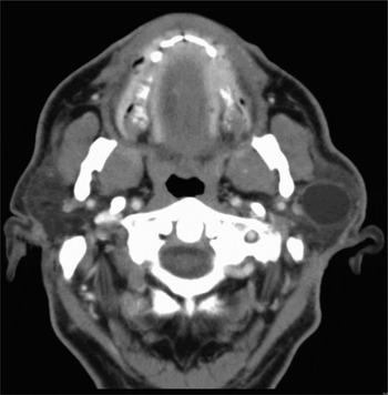
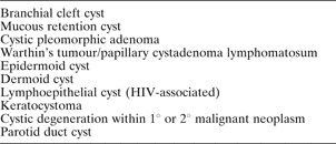
Pre-operative investigation included fine needle aspiration cytology (FNAC) and computed tomography (CT) scanning. Aspiration yielded thick, white material. Cytological examination identified numerous anucleate squames in a proteinaceous background. Computed tomography scans confirmed a 5 cm, cystic mass in the superficial aspect of the parotid gland (Figure 1). The differential diagnosis is summarised in Table I.
Axial computed tomography scan showing dermoid cyst in the left parotid gland.

Differential diagnosis of cystic lesions of the parotid gland

HIV = human immunodeficiency virus; 1° = primary; 2° = secondary
Surgery
A left, intra-parotid, cystic lesion was excised via a superficial lobectomy, with preservation of the facial nerve. The patient's post-operative course was uneventful, with a normally functioning facial nerve evident at day one.
Histopathology
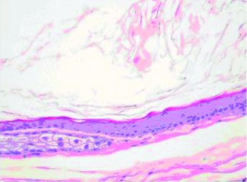
Histopathological examination of multiple transverse sections through the surgical specimen revealed parotid gland parenchyma comprising serous acini with intervening prominent fatty infiltration. There was a large cyst (55 × 26 × 14 mm) identified in the specimen. The lumen was filled with keratin and the lining comprised thin, stratified squamous, ortho-keratinised epithelium. The wall was composed of compressed, fibrous connective tissue and interspersed with sebaceous glands and focal collections of chronic inflammatory cells (Figure 2).
Photomicrograph of cyst lining, showing presence of sebaceous glands and intraluminal keratin debris (H&E; original magnification ×200).

Discussion
New and Erich reviewed 1485 cases of dermoid cysts, and highlighted the fact that 7 per cent of all such cysts occurred in the head and neck.Reference New and Erich1 The most common site was the orbit, accounting for 49.5 per cent. A further 23 per cent were found in the floor of the mouth, with the remainder occurring in various other sites.
Although several classifications of dermoid cysts have been described based on embryopathogenesis and microscopic findings, there is currently no universally accepted method.Reference New and Erich1, Reference Choi, Jin, Kim, Hong, Kim and Park2
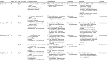
The occurrence of a dermoid cyst within the parotid gland is exceedingly rare. To our knowledge, there are only four previously published papers, documenting a total of six cases, in the English literature.Reference Choi, Jin, Kim, Hong, Kim and Park2–Reference Naujoks, Handschel, Braunstein, Emaetig, Depprich and Meyer5 The clinicopathological features of the reported cases in the English literature are summarised in Table II.
Summary of documented cases of intra-parotid dermoid cyst

Yrs = years; mths = months; M = male; NAD = no abnormality detected; MRI = magnetic resonance imaging; CT = computed tomography; FNAC = fine needle aspiration cytology; Pap = Papanicolau stain
New and Erich found no sex predilection for dermoid cysts. However, it is noteworthy that all those lesions which have been documented within the parotid gland have occurred in males (mean age 31 years).Reference New and Erich1 This was also the case in our patient.
Several authors have highlighted the difficulty in definitive pre-operative diagnosis of these lesions. This is largely due to the lesions' equivocal clinical and radiological findings. Definitive diagnosis in these cases has generally been retrospective, based on the macroscopic features of the excised specimen and histopathological examination.Reference Choi, Jin, Kim, Hong, Kim and Park2, Reference Moody, Avery and Harrison3, Reference Naujoks, Handschel, Braunstein, Emaetig, Depprich and Meyer5 Baschinsky et al. used FNAC, supplementing clinical and radiological assessments, as the diagnostic modality.Reference Baschinsky, Hameed and Keyhani-Rofagha4 Significantly, these authors highlighted the value of FNAC, and described pre-operative cytological findings, derived from FNAC samples, which were suggestive of dermoid cysts (see Table II). Cramer and colleagues also reported similar cytological features of a floor of mouth dermoid cyst, and recommended FNAC as the diagnostic procedure of first choice.Reference Cramer, Lampe and Downing6
• Dermoid cysts have only been rarely encountered in the parotid gland
• This case highlights the potential benefit of pre-operative FNAC
• a parotid aspirate displaying anucleate squames, in the absence of frankly dysplastic or malignant cells, in a radiologically benign lesion, is suggestive of a dermoid cyst
We disagree with Naujoks et al.,Reference Naujoks, Handschel, Braunstein, Emaetig, Depprich and Meyer5 who state that ‘investigations like FNAC may induce nerve violation and infection, and are irrelevant for the choice of operation on a process that appears to be benign on MRI’. Given the differential diagnosis for cystic pathology affecting the parotid gland (Table I), we would agree with Baschinsky and colleagues, and recommend FNAC as part of the diagnostic evaluation. In keeping with Baschinsky and colleagues' findings, our case highlights a finding of proteinaceous aspirate displaying ‘anucleate squames’ as being highly suggestive of parotid dermoid cyst.
When interpreting these cytological features, the integration of the clinical and CT findings is crucial. An important caveat is that, although no frankly dysplastic or malignant cells were identified in our case, the demonstration of anucleate squames would not exclude consideration of a well differentiated squamous cell carcinoma (SCC), either as an SCC that had undergone cystic degeneration, or as cystic degeneration of a metastatic (intra-parotid) lymph node.
Computed tomography and magnetic resonance imaging (MRI) scanning of parotid dermoid cysts usually demonstrate a well defined, low density mass consistent with an intra-parotid lipoma.Reference Moody, Avery and Harrison3, Reference Baschinsky, Hameed and Keyhani-Rofagha4 However, it has been suggested that a fat–fluid level appearing on an MRI or CT scan is a characteristic feature of a dermoid cyst.Reference Rosen, Wirtschafter, Rao and Wilcox7
Although dermoid cysts are generally regarded as benign entities, rare cases of malignant transformation of such cysts within the oral cavity have been reported.Reference Gold, Sheinkopf and Levy8
Treatment of dermoid cysts involves surgical excision. The standard approach for the surgical management of most parotid lesions is superficial lobectomy. Dermoid cysts are usually well encapsulated and this facilitates dissection. They may also contain yellowish-white, ‘cheesy’ contents, which may be observed during surgery.Reference Moody, Avery and Harrison3, Reference Naujoks, Handschel, Braunstein, Emaetig, Depprich and Meyer5 The cyst should be removed en bloc to minimise the risk of recurrence. To date, all documented cases of intra-parotid dermoid cysts appear to have been successfully treated with superficial parotidectomy. There has been no recurrence in our patient at the time of writing.
Conclusion
Dermoid cysts have only rarely been encountered in the parotid gland. This case highlights the potential benefit of pre-operative FNAC, and the fact that a parotid aspirate displaying anucleate squames, in the absence of frankly dysplastic or malignant cells and in a radiologically benign lesion, is suggestive of a dermoid cyst.