Introduction
Endoscopic sinus examination and paranasal space computed tomography (CT) enable the identification of nasal pathology and anomaly, most of which can be treated medically or surgically.
Turbinate anomalies resembling the middle turbinate comprise the bifid inferior turbinate, accessory middle turbinate and secondary middle turbinate. Bifid inferior turbinate has been defined as severe medial displacement and inferior rotation of the uncinate process.Reference Selcuk, Ozcan, Ozcan and Dere1 Accessory middle turbinate has been described as a medially and anteriorly folded uncinate process.Reference Lin, Lin, Su and Wang2 Secondary middle turbinate is a rare anatomical variation originating from the lateral wall of the middle meatus and usually projecting superomedially without obstruction of the ostiomeatal unit.Reference Lin, Lin, Su and Wang2
We present the first two published cases of a unilateral accessory middle turbinate with a polyp, which showed normal uncinate process development. One of these cases coexisted with a polyp from an inferomedially projecting, pneumatised, secondary middle turbinate.
Case reports
Case one
A 25-year-old woman presented at our clinic complaining of left nasal obstruction. The patient's history revealed that she had no other diseases and no previous nasal surgery.
Endoscopic examination revealed polypoid masses located medial to the inferior turbinate and anterior to the middle turbinate, together with a normal middle turbinate and uncinate process (Figure 1a).
(a) Endoscopic appearance of left nasal masses in case one: polypoid masses are located medial to the inferior turbinate (star), with a normal uncinate process (arrow) and middle turbinate (arrowhead). (b) Coronal and (c) axial paranasal space computed tomography (CT) scans show bifurcation of the left uncinate process with an obvious polyp (arrow). (d) Coronal paranasal space CT scan, using bone settings, shows a pneumatised left secondary middle turbinate with a polyp originating from the lateral wall of the ethmoidal bulla, just beneath the basal lamella, and projecting inferomedially (arrowhead).

Paranasal space CT scans revealed an inferomedially and anteriorly projecting bony lamella with attached polyp, originating from the uncinate process and attaching to the skull base superiorly (Figure 1b and 1c), together with a pneumatised bony structure with attached polyp projecting inferomedially from the left lateral wall of the ethmoidal bulla just beneath the basal lamella (Figure 1d).
A diagnosis of an accessory middle turbinate and secondary middle turbinate with polyps was established, based on the presence of the uncinate process.
The patient was treated successfully by endoscopic resection of the uncinate process, accessory middle turbinate and secondary middle turbinate.
Case two
A 39-year-old woman complaining of left nasal obstruction was referred to our clinic with a left nasal mass detected on magnetic resonance imaging brain scans. She denied any symptoms of chronic rhinitis or chronic sinusitis, and had no history of previous nasal surgery.
Upon endoscopic examination, a polypoid mass was detected in the left nasal cavity emerging anteriorly from the middle meatus.
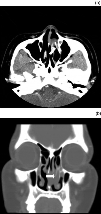
Paranasal space CT scans showed that the left uncinate process was fully developed, and a polyp was arising from a thick, inferomedially projecting, bony bifurcation from the uncinate process (Figure 2).
(a) Axial and (b) coronal paranasal space computed tomography scans in case two, showing a polyp originating from a thick, inferomedially projecting, bony lamella from the left uncinate process (arrow).

In addition, both endoscopic examination and paranasal space CT scanning revealed an accessory middle turbinate with a polyp.
The patient underwent endoscopic resection. Histopathological analysis confirmed the presence of a polyp.
Discussion
The uncinate process is normally a thin, sickle-shaped structure. Variations of uncinate process anatomy may obliterate the paranasal sinus drainage pathway but do not usually affect nasal airflow passage. If the uncinate process is bent medially and folded anteriorly to a greater than usual extent, it may give the impression that two middle turbinates are present, a condition referred to as accessory middle turbinate.Reference Lin, Lin, Su and Wang2 A bifid inferior turbinate could be confused with an accessory middle turbinate, but in fact a bifid inferior turbinate can be easily distinguished from an accessory middle turbinate on paranasal space CT scans because there is no uncinate process in a bifid inferior turbinate. It has been suggested that the upper part of the bifid inferior turbinate may represent a normal uncinate process,Reference Spear, Brietzke and Winslow3 or a severely medially displaced and inferiorly rotated uncinate process.Reference Selcuk, Ozcan, Ozcan and Dere1
In both the presented cases, the uncinate process showed normal development, reaching to the lamina papyracea or skull base, and the posterior free margin was markedly bent medially and anteriorly displaced, corresponding to the definition of an accessory middle turbinate. Interestingly, polyps originated from the anteromedially folding, bony part in both cases.
A review of the literature indicated that such a combination of pathology together with accessory middle turbinate had not previously been reported.
The secondary middle turbinate is an extremely rare nasal cavity variant that arises from the lateral wall of the ethmoidal bulla just beneath the basal lamella. An incidence of 0.8–14.3 per cent has been reported.Reference Khanobthamchai, Shankar, Hawke and Bingham4–Reference McLaughlin, Rehl and Lanza8 The secondary middle turbinate has been described as projecting medially and then turning superiorly, with no ostiomeatal unit obstruction.Reference Khanobthamchai, Shankar, Hawke and Bingham4–Reference Aksungur, Bicakci, Inal, Akgul, Binokay and Aydogan6 One case report described an inferomedially projecting, pneumatised, secondary middle turbinate.Reference Apaydin, Duce, Yildiz, Egilmez, Ozer and Talas9 In our first case, a left secondary middle turbinate showed pneumatisation and inferomedial projection, and a polyp originating from the secondary middle turbinate narrowed the ostiomeatal unit. A secondary middle turbinate might project inferomedially due to pneumatisation. There has been no previous report describing a polyp originating from a secondary middle turbinate, as in our first case.
• Accessory middle turbinate is an excessively medially and anteriorly folded uncinate process
• Secondary middle turbinate originates from the lateral middle meatus wall
• The first report of polyps originating from a unilateral accessory middle turbinate and secondary middle turbinate is presented
The complicated structure of the lateral nasal wall can be better understood by considering the embryological development of the turbinates. The inferior and middle turbinates have different embryological origins: the middle turbinate originates from the second ethmoturbinal, while the inferior turbinate develops from the maxilloturbinal. The uncinate process is regarded as the descending portion of the first ethmoturbinal.Reference McLaughlin, Rehl and Lanza8 A secondary middle turbinate might represent an additional turbinate originating embryologically from a section of the frontal ridge. (Normally, the frontal ridge evolves into the anterior ethmoidal cells.)Reference Aksungur, Bicakci, Inal, Akgul, Binokay and Aydogan6
Conclusion
A polyp extending from an accessory middle turbinate or secondary middle turbinate is an extremely rare anomaly. To our knowledge, this is the first report to present cases of polyps originating from an accessory middle turbinate and a secondary middle turbinate. Because the uncinate process is a landmark for endoscopic sinus surgery, pre-operative recognition of these anatomical variations is of particular importance in avoiding intra-operative complication.
Turbinate anomalies (including accessory middle turbinate and bifid inferior turbinate) are diagnosed endoscopically but their terminology can be confused. Therefore, we suggest that accessory middle turbinate and bifid inferior turbinate could be included amongst the recognised anatomical variations of the uncinate process.