Wolff–Parkinson–White syndrome is characterised by the presence of one or more accessory pathways that bypass normal atrioventricular nodal conduction.Reference Benson and Cohen 1 Several studies have reported left ventricular dysfunction that mimics dilated cardiomyopathy and/or septal aneurysmal dyskinesis in these patients.Reference Takeuchi, Tomita and Kasai 2 – Reference Dai, Guo and Li 6 The eccentric ventricular activation via the accessory pathway is thought to result in the asynchronous spread of ventricular depolarisation and significant dyssynchrony of the left ventricle.Reference Kwon, Bae and Kim 3 – Reference Ishizu, Seo and Igarashi 7 Although this phenomenon has been well-described using echocardiography,Reference Takeuchi, Tomita and Kasai 2 , Reference Tomaske, Janousek and Rázek 5 , Reference Dai, Guo and Li 6 electrophysiological mapping has been rarely demonstrated. Here, we present the case of a patient with Wolff–Parkinson–White syndrome with septal aneurysmal dyskinesis, who underwent three-dimensional mapping of the left ventricle before and after radiofrequency catheter ablation of accessory pathways. Abnormal activation wavefront arose from the aneurysmal septum and transmitted to the lateral wall prior to ablation, which normalised after successful radiofrequency catheter ablation.
Case report
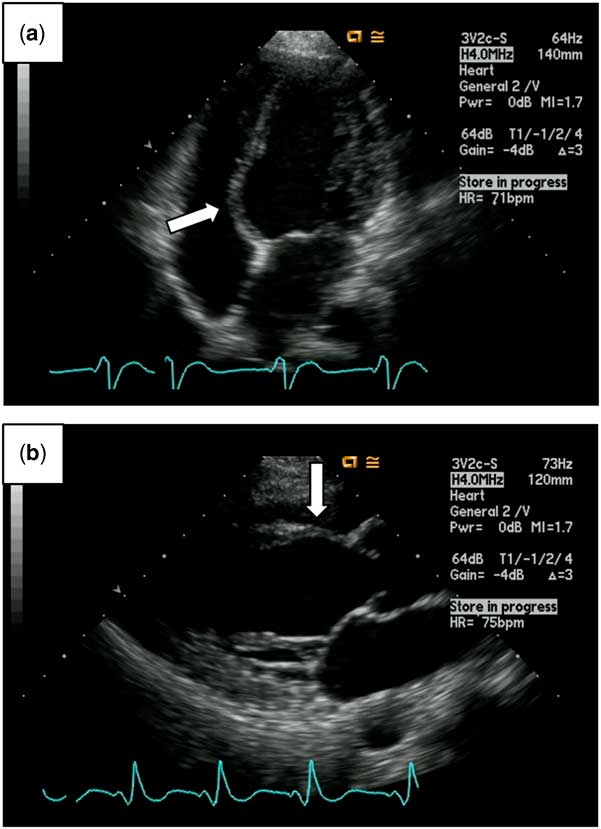
A 21-year-old man with Wolff–Parkinson–White syndrome and aneurysmal septal dyskinesis (Fig 1) underwent radiofrequency catheter ablation of the accessory pathways. He was referred to our hospital at 7 months of age because of a heart murmur. Delta waves and a short PR interval on 12-lead electrocardiography suggested Wolff–Parkinson–White syndrome. Echocardiography showed aneurysmal septal dyskinesis and left ventricular dysfunction mimicking dilated cardiomyopathy. He previously underwent medical therapy for several years, but these echocardiographic findings did not improve. Although he had remained asymptomatic, catheter ablation was pursued because of persistent left ventricular dysfunction that was suspected to be secondary to abnormal ventricular activation via the accessory pathway.
Two-dimensional echocardiography shows aneurysmal septal dyskinesis (white arrow) in a patient with Wolff–Parkinson–White syndrome. ( a ) Apical four-chamber view; ( b ) parasternal long-axis view.

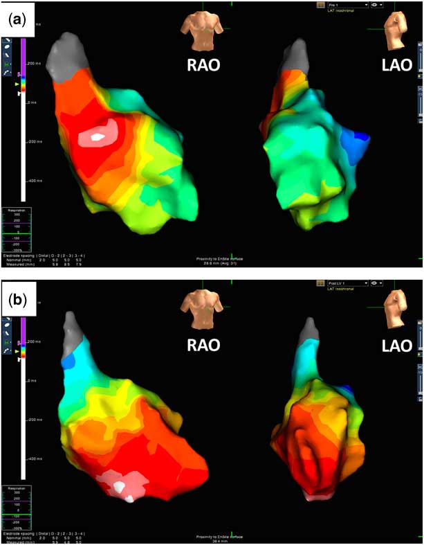
Written informed consent was obtained from the patient and his parents. An electrophysiological study was performed using standard pacing protocols.Reference Chen, Sun and Xu 8 , Reference Romero, Lupercio and Goodman-Meza 9 In brief, after performing local anaesthesia with 1% lidocaine, we obtained vascular access from the left femoral vein and right femoral artery. The entire procedure was performed without any fluoroscopy, and catheter navigation was guided by the Ensite NavX system (St. Jude Medical, Saint Paul, Minnesota, United States of America).Reference Chen, Sun and Xu 8 , Reference Romero, Lupercio and Goodman-Meza 9 Three-dimensional electroanatomic mapping of the left ventricle was also performed to evaluate the electrical wavefront propagation pattern. Geometrical reconstruction and an activation map of the left ventricle were acquired using a 7 F irrigated ablation catheter (FlexAbilityTM Ablation Catheter, St. Jude Medical) to avoid risk of clot formation. Electrophysiological examination revealed the right anterior and anterolateral locations of the broad accessory pathways (Supplementary Fig 1). The activation wavefront arose from the aneurysmal septum (Fig 2a), and was transmitted to the lateral wall. Radiofrequency ablation was targeted at the site of the earliest activation in the right free wall, with immediate loss of pre-excitation. Following ablation, the propagation of the left ventricle activation wavefront was normalised (Fig 2b).
Three-dimensional EnSite mappings of the left ventricle at sinus rhythm before ( a ) and after ( b ) radiofrequency catheter ablation. The earliest sites are shown in white. LAO=left anterior oblique; RAO=right anterior oblique.

Discussion
Wall motion abnormalities have been well described in patients with Wolff–Parkinson–White syndrome.Reference Takeuchi, Tomita and Kasai 2 – Reference Dai, Guo and Li 6 , Reference Ko 10 Studies regarding the relationship between global left ventricular dysfunction with ventricular pre-excitation and segmental wall motion abnormalities are limited.Reference Kwon, Bae and Kim 3 , Reference Dai, Guo and Li 6 , Reference Ishizu, Seo and Igarashi 7 We have demonstrated, for the first time, abnormal cardiac propagation of the left ventricle in a patient with Wolff–Parkinson–White syndrome who had septal aneurysmal dyskinesis and left ventricular dysfunction using three-dimensional voltage mappings.
Synchronous ventricular contraction and regional myocardial function crucially depends on timing; a delay in the electrical activation of a myocardial segment may contribute to cardiac-pump inefficiency.Reference Kwon, Bae and Kim 3 , Reference Dai, Guo and Li 6 , Reference Ko 10 Previous studies have hypothesised that ventricular pre-excitation can result in remodelling changes with segmental dyskinesia, leading to thinning, dyssynchrony, ventricular dilation, and, ultimately, ventricular dysfunction.Reference Dai, Guo and Li 6 The mechanism is thought to be similar to that of left bundle branch block or right ventricular apical pacing.Reference Kwon, Bae and Kim 3 , Reference Ko 10 The hypothesis of the accessory pathway-induced dyssynchrony has been verified indirectly using echocardiographic approaches; dyssynchronous wall motion or ventricular dilatation improved after the resolution of accessory pathways.Reference Takeuchi, Tomita and Kasai 2 , Reference Kwon, Bae and Kim 3 , Reference Tomaske, Janousek and Rázek 5 , Reference Dai, Guo and Li 6 Here, we have described, for the first time, the mechanism via three-dimensional electrophysiological mapping; the abnormal ventricular conduction via the accessory pathways improved after radiofrequency catheter ablation. Three-dimensional electroanatomic mappings of the left ventricle combined with radiofrequency catheter ablation to localise accessory pathways and determine abnormal propagation pattern might be useful in this population.Reference Ishizu, Seo and Igarashi 7
The occurrence of ventricular dysfunction in Wolff–Parkinson–White syndrome had been thought to depend on the location of the accessory pathway.Reference Kwon, Bae and Kim 3 – Reference Tomaske, Janousek and Rázek 5 , Reference Ko 10 The septal accessory pathways cause pre-excitation of a substantial part of the interventricular septum, inducing dyskinetic septal motion and a significant level of left ventricular dyssynchrony.Reference Kwon, Bae and Kim 3 , Reference Ko 10 Compared with septal accessory pathways, conduction through right or left free-wall pathways was considered to have less-detrimental effects on dyssynchronous wall motion and left ventricular function.Reference Kwon, Bae and Kim 3 Recently, Dai et alReference Dai, Guo and Li 6 reported left ventricular dysfunction and dilatation mimicking dilated cardiomyopathy event secondary to non-septal pathways. As just described in the present case, left ventricular voltage mappings combined with radiofrequency catheter ablation could certify the traditional hypothesis that dyssynchronous left ventricular motion caused by pre-excitation via accessory pathways may induce septal aneurysmal dyskinesis and left ventricular dysfunction.Reference Kwon, Bae and Kim 3 – Reference Tomaske, Janousek and Rázek 5 , Reference Ko 10 In addition, it might reveal the mechanisms of the electro-mechanical interaction in non-septal accessory pathways. Three-dimensional speckle-tracking echocardiography might be less invasive and a more useful approach in the future, although the tests have yet to be fully validated.Reference Ishizu, Seo and Igarashi 7
Left ventricular dysfunction in patients with Wolff–Parkinson–White syndrome may be underestimated or misdiagnosed in young patients.Reference Takeuchi, Tomita and Kasai 2 , Reference Kwon, Bae and Kim 3 , Reference Dai, Guo and Li 6 , Reference Ko 10 Radiofrequency catheter ablation in these patients results in mechanical resynchronisation, reverse remodelling, and improvements in left ventricular function.Reference Ko 10 Three-dimensional electroanatomic mappings combined with radiofrequency catheter ablation may be a useful approach to understand the mechanisms of electro-mechanical interaction and evaluate the treatment effects on patients with Wolff–Parkinson–White syndrome having ventricular dysfunction.
Acknowledgements
The authors thank Dr. Ryohei Matsuoka, Dr. Yusaku Nagatomo, Dr. Hiromitsu Shirouzu, Dr. Chiaki Iida, and Dr. Mamie Watanabe (all Department of Pediatrics, Japan Community Healthcare Organization, Kyushu Hospital) for assistance of radiofrequency catheter ablation and data collection.
Financial Support
This research received no specific grant from any funding agency, commercial or not-for-profit sectors.
Conflicts of Interest
None.
Supplementary Material
To view supplementary material for this article, please visit https://doi.org/10.1017/S1047951117001779