Isolated ventricular septal defects are common and are diagnosed in 1.5–3.5 per 1000 live births.Reference Hoffman and Kaplan1, Reference Hoffman and Rudolph2 They can be solitary or form part of a complex with other cardiac abnormalities. Of patients with surgically operated isolated ventricular septal defects, the majority are in a perimembranous position (80%), while the remainder (20%) are either muscular defects (inlet, apical, and outlet) or doubly committed and juxta-arterial defects.Reference Soto, Becker, Moulaert, Lie and Anderson3 Understanding the natural history of ventricular septal defects is complicated by variability in the size and position of the defect as well as in the occurrence of associated cardiac abnormalities. Many smaller defects will close spontaneously, and even if a small communication remains, there may be no implications for an individual's life expectancy or quality of life. Large defects will generally require surgical closure – indicated in infancy when infants fail to thrive or are at risk of pulmonary vascular disease. Management dilemmas centre on moderate-sized defects with normal pulmonary artery pressure or smaller defects with secondary complications such as aortic incompetence or endocarditis. In these cases, there is some debate as to when closure is indicated, and if so, by which route – surgical, hybrid, or transcatheter.
Muscular defects have been more amenable to transcatheter or hybrid closure even in infants as the defects are usually a sufficient distance from important structures including valves and conduction tissue. Closure of perimembranous defects, however, remains one of the most challenging procedures for the structural interventionalist. These defects lie in close proximity to the tricuspid valve and its sub-valve apparatus, the aortic valve and the conduction tissue. Defects are not infrequently complicated by aneurysmal fibrous tissue derived from the leaflets of the tricuspid valve.Reference Anderson and Wilcox4 It is therefore not surprising that most significant defects are closed surgically with excellent results.
The debate over whether to close or not to close significant perimembranous ventricular septal defects by transcatheter techniques centres on there being a higher rate of complications when compared with surgical alternatives. Surgical results are impressive and the complication rates extremely low.Reference Tucker, Pyles, Bass and Moller5, Reference Roos-Hesselink, Meijboom and Spitaels6 The chief complication under discussion is complete heart block, which can develop early or late after transcatheter closure.Reference Walsh, Bialkowski, Szkutnik, Pawelec-Wojtalik, Bobkowski and Walsh7–Reference Predescu, Chaturvedi, Friedberg, Benson, Ozawa and Lee13 It is impossible to place a device in a perimembranous ventricular septal defect without incurring the risk of atrioventricular block, given that the conduction axis penetrates through the postero-inferior margin of the defect.Reference Andersen, de Leval, Tsang, Elliott, Anderson and Cook14 However, what factors are most critical at governing this risk are poorly defined and not easily studied. The authors of most case series are in agreement that device size in relation to the size of the defect is likely to be a critical factor. Oversizing of devices is attractive as they are easier to deploy with higher success rates. However, choosing smaller devices and accepting lower success rates may be the key to avoiding heart block, along with the choice of device, defect position, and size of the patient. These issues constitute the central theme of this case series and analysis of the literature that follows. We therefore report on the immediate and medium-term follow-up results of using muscular amplatz ventricular septal defect and perimembranous amplatz ventricular septal defect devices to close perimembranous ventricular septal defects in children.
Materials and methods
The study is a retrospective case note review of all children referred for consideration of closure of a perimembranous ventricular septal defect between April, 2004 and February, 2010. Case notes were available for review on all patients who underwent procedures. The data collected retrospectively included demographics, surgical notes, intervention procedural data, and angiographic data. One of the authors (NW) was involved in all procedures, ensuring standardisation of technique. Each case was presented and approved for consideration of cardiac catheter closure to the institution's joint cardiology and surgery meeting before catheter evaluation was commenced. The indication for catheter closure of ventricular septal defect included signs of a significant left-to-right shunt with left ventricular volume load (increased left ventricular end-diastolic dimensions on the transthoracic echocardiogram indexed to body surface area, z score greater than 3 or calculated Qp:Qs greater than 2:1). Ventricular septal defect measurements were obtained from angiographic data, calibrated against a standard 5F pigtail catheter in a standard left anterior oblique projection. Measurements were confirmed independently by transoesophageal echocardiography. Success was defined as device closure of a perimembranous ventricular septal defect with resolution of left-to-right shunt and without significant complication. The lower weight limit before catheter closure was considered to be 10 kilograms. All patients have been followed up regularly. Adverse events were recorded, including procedural complications relating to vascular access (concerns over and management of distal limb perfusion, difficulties during or prolonged time taken while obtaining arterial access including resorting to an alternative vessel or cut down when not previously planned) and procedural catheter-related complications (haemodynamic instability or arrhythmia, device deployed in unsatisfactory position or embolisation post-deployment, haemolysis, stroke, and death).
Technique
All procedures were performed under general anaesthesia after obtaining informed written consent. Heparin (100 units per kilogram) was given after arterial access was obtained. In the majority of cases, initial access was obtained percutaneously with a 6 Fr Cook short paediatric sheath in the right femoral vein and a 5 Fr Cook sheath in the right femoral artery (Cook, Vandergrift, Pennsylvania, United States of America). All patients underwent left and right cardiac catheterisation under fluoroscopic guidance in the catheter laboratory including haemodynamic assessment of left-to-right shunt. Angiography was performed in single plane in standard projection (left anterior oblique). Transoesophageal echocardiography was an integral part of the procedure being used for assessment of both size and location of ventricular septal defect and then to guide device delivery in real time. This allowed immediate assessment of the effect of the device on the aortic and tricuspid valves as well as delivery of the left and right discs and their orientation relative to the interventricular septum. A brief attempt was made at crossing the ventricular septal defect directly from the right to the left ventricle, in order to avoid the need for an arteriovenous loop and to avoid the time taken in manipulating catheters in the left ventricle. In the main, however, the standard technique following haemodynamic assessment was to cross the ventricular septal defect from the left ventricle using a Judkins right 3.5 coronary catheter (5F, Cordis, New Jersey, United States of America) and then making an arteriovenous loop with a Terumo guide wire (0.035 inch, J, 260 centimetres, Terumo, Leuven, Belgium) snaring in the pulmonary artery or superior caval vein (either a 4F 10 millimetres or 6F 15 millimetres snare kit, EV3 Amplatz, Plymouth, Minnesota, United States of America). A sheath could then be delivered from the venous side to the proximal ascending aorta (Amplatzer TorqVue® delivery system, AGA Medical, Plymouth, Minnesota, United States of America). Generally, the sheath size chosen was 1 Fr greater than that suggested in the manufacturer's recommendations. The ventricular septal defect device was chosen depending on the size and position of the ventricular septal defect confirmed on two imaging modalities (angiography and transoesophageal echocardiography). The device was loaded according to the manufacturer's recommendations. The device was delivered to the end of the sheath such that an “olive” shaped configuration of the left ventricular disc protruded from the end of the delivery sheath while still in the ascending aorta. The side arm of the sheath was connected to a pressure manometer, such that as the sheath was withdrawn from the ascending aorta through the aortic valve to the left ventricle, the change from the arterial to the ventricular pressure waveform could be seen. This withdrawal of the sheath-device assembly was done slowly and with transoesophageal echocardiographic guidance. The left ventricular disc was then delivered, followed by a cautious withdrawal of the sheath to deliver the right ventricular disc. If the device pulled through, the procedure was repeated after further consideration of the size and type of device chosen and the technique and route for device delivery. Angiography with a 5 Fr pigtail catheter (Cordis, New Jersey, United States of America) and further transoesophageal echocardiography in multiple planes were used to confirm stable device position before release.
Literature complication analysis
All published series of transcatheter closure of ventricular septal defect were reviewed to identify those in which closure of perimembranous ventricular septal defect had been performed and results reported. Major procedural complication was defined as a need for early surgery because of new and significant aortic incompetence, device embolisation with failure of retrieval, or complete atrioventricular block necessitating pacemaker insertion. Arrhythmia not requiring treatment or intervention, including transient atrioventicular block, was not regarded as a major complication. Embolisation was only regarded as a complication if the device could not be retrieved and successfully redeployed. Aortic incompetence was regarded as a major complication, if it had newly arisen and required device removal and surgical ventricular septal defect closure. A total of 14 studies reporting the results of perimembranous ventricular septal defect closure by transcatheter techniques were evaluated.Reference Butera, Carminati and Chessa10–Reference Predescu, Chaturvedi, Friedberg, Benson, Ozawa and Lee13, Reference Bass, Kalra and Arora15–Reference Holzer, de Giovanni and Walsh24
Statistical analysis
Descriptive variables and statistical tests were performed in SPSS for Windows version 14 (Chicago, United States of America). The mean and median values and ranges are described. The two-tailed Student's t-test of unpaired samples was used to compare cases where complications occurred with overall case series means (significance level taken as p < 0.01).
Results
A total of 25 patients with a pre-procedural diagnosis of perimembranous ventricular septal defect considered likely to be suitable for transcatheter closure were assessed by cardiac catheterisation and transoesophageal echocardiography. These patients included two who were not considered suitable in view of a Qp:Qs less than 2:1 and the diagnostic procedure was terminated without attempt at ventricular septal defect closure. Transcatheter perimembranous ventricular septal defect closure was attempted on 25 occasions in 23 patients; successful device delivery failed in two of these. Successful closure of perimembranous ventricular septal defect was achieved in 21 of 23 cases (91.3%). In one of the 21 cases, the device embolised and a second device was inserted after successful retrieval of the first device; two procedures were therefore required. In a further two cases, transcatheter closure of ventricular septal defect was abandoned in favour of surgery as stable device position could not be achieved following two attempts in each case. In 20 cases, a device was inserted at the first procedure.
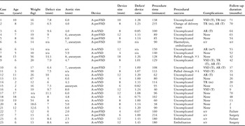
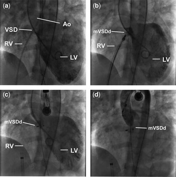
Procedural data are shown in Table 1. The median weight was 18.6 kilograms, ranging from 10 to 67 kilograms, at a median age of 8 years, with a range from 2 to 19 years. The mean ratio of occluder to ventricular septal defect size was 1.11, ranging from 0.75 to 1.33, with a median occluder size of 8 millimetres, ranging from 4 to 18 millimetres, and a median angiographically determined ventricular septal defect size of 7.85 millimetres, with a range from 4 to 14.3 millimetres. The median distance between the right coronary leaflet of the aortic valve and the superior edge of the ventricular septal defect measured on transoesophageal echocardiography from the left ventricle long axis view was 6 millimetres, with a range from 2 to 8 millimetres, see supplementary videos. In 14, an amplatz muscular ventricular septal defect device was inserted, with the remainder being perimembranous devices (7). In 19, the device was placed and was in a satisfactory position at the first attempt (Fig 1 and supplementary videos 1–3). In two, the device pulled through a total of seven times before an acceptable position was achieved (three times in case 8 and four times in case 11; Table 1). The device was upsized to a larger device in both of these procedures, in one case changing from an amplatz perimembranous ventricular septal defect to an amplatz muscular ventricular septal defect device (Fig 2).
Patient characteristics of the 23 cases in which pmVSD closure was attempted.

AR = aortic regurgitation; n/a = not applicable; n/r = not recorded; T = trivial; TR = tricuspid regurgitation; VSD = ventricular septal defect
m* refers to new onset AR regarded as a procedure related complication (m = mild)
Angiograms demonstrating placement of a muscular ventricular septal defect device (mVSDd) in an 18.6 kilogram 8-year-old child. (a) The left ventriculogram was obtained though a 5 Fr pigtail catheter from the right femoral artery. There is a 7-millimetre perimembranous ventricular septal defect (VSD). (b) An 8-millimetre mVSDd device has been placed after an arteriovenous loop was formed enabling delivery of the device withdrawing from the aorta to the left ventricle and finally to the right ventricle. (c) The device has been released. (d) There is no significant residual ventricular septal defect or new aortic regurgitation confirmed on the aortogram or on transoesophageal echocardiography (see supplementary transoesophageal echocardiography videos 1–3). RV = right ventricle; LV = left ventricle; Ao = Aorta.

Angiograms demonstrating placement of a perimembranous ventricular septal defect device (pmVSDd) in a 10 kilogram 3-year-old child, which repeatedly pulled through and was subsequently replaced with a muscular device (mVSDd). (a) The left ventriculogram was obtained though a 5 Fr pigtail catheter from the right femoral artery. There is a 7.5-millimetre perimembranous ventricular septal defect (VSD). (b) An “olive” of a 7-millimetre pmVSDd is protruding from the delivery sheath and the sheath is withdrawn and the device delivered across the septum (b′). (c) The pmVSDd pulled through and was removed after a second attempt. An 8-millimetre pmVSDd also pulled through. An 8-millimetre mVSDd was finally delivered successfully in its place. (d) There is no significant residual ventricular septal defect or new aortic regurgitation confirmed on the aortogram or on transoesophageal echocardiography. RV = right ventricle; LV = left ventricle; Ao = Aorta.

Percutaneous access was achieved in all cases. The median procedure time for the 21 procedures performed with fluoroscopic guidance was 100 minutes, ranging from 38 to 235 minutes, fluoroscopy time 25.5 minutes, ranging from 10.1 to 71.8 minutes, and radiation dose 1181 gray per square centimetre with a range from 184 to 3264 gray per square centimetre.
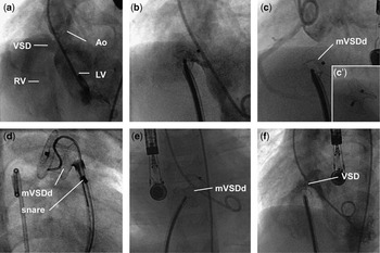
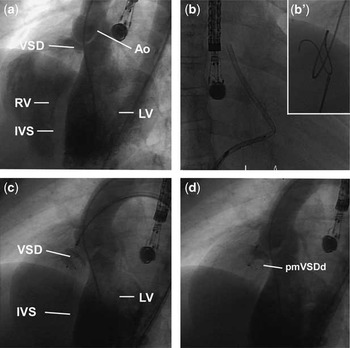
A total of 17 cases were straightforward with delivery of device at first attempt. In two cases, the device pulled through on three occasions in one case and four occasions in the other. In the first device, size was upsized from a 16- to 18-millimetre perimembranous ventricular septal defect device. In the second, upsizing from a 7- to 8-millimetre perimembranous ventricular septal defect device still failed and an 8-millimetre muscular ventricular septal defect device was placed successfully. Devices embolised on two occasions; the first was a perimembranous ventricular septal defect device, which was successfully retrieved from the aorta and replaced by a larger perimembranous ventricular septal defect device, which also pulled through (Fig 3). The case was discontinued in favour of surgery. In the second case, embolisation of a perimembranous ventricular septal defect device followed 3 days of haemolysis. There was a residual ventricular septal defect before device embolisation and blood transfusion was required. The device was retrieved from the pulmonary artery and a muscular ventricular septal defect device 2 millimetres larger than the first perimembranous ventricular septal defect device was successfully deployed. Failure to deliver a device through a persistently kinked delivery sheath occurred in one case, which necessitated changing the route of delivery from a retrograde venous to an antegrade aortic route (Fig 4). Both of the cases which failed had very little distance between the right coronary cusp and the ventricular septal defect measuring 2 and 2.5 millimetres, respectively (left ventricular outflow tract long axis view on transoesophageal echocardiography). In both cases, a larger device was tried but the device position was not satisfactory and the device pulled through.
Angiograms demonstrating failed placement of a muscular ventricular septal defect device (mVSDd) in a 13 kilogram 6-year-old child, which repeatedly pulled through and embolised. (a) The left ventriculogram was obtained though a 5 Fr pigtail catheter from the right femoral artery. There is an 8.4-millimetre perimembranous ventricular septal defect (VSD). (b) A 12-millimetre mVSDd has been placed across the ventricular septal defect. There is a significant residual defect both before and after release. The device sits obliquely across the defect. (c) The device was recaptured and redelivered with a more satisfactory position (c′). (d) The device embolises to the main pulmonary artery but is successfully retrieved. (e) A further attempt at delivery of the same size device is made several months later. (f) Despite the device appearing satisfactory, there is a significant residual defect suggesting that stable device position could not be achieved. The device was retrieved and the child referred for surgical closure. RV = right ventricle; LV = left ventricle; Ao = Aorta.

Angiograms demonstrating placement of a perimembranous ventricular septal defect device (pmVSDd) in a 21 kilogram 8-year-old child, which was unable to be delivered from a venous route necessitating delivery from an arterial route. (a) The left ventriculogram was obtained though a 5 Fr pigtail catheter from the right femoral artery. There is a 6-millimetre perimembranous ventricular septal defect (VSD). (b) Following the creation of an arteriovenous loop (b′), an 8-millimetre pmVSDd is unable to be advanced through a kinked delivery catheter. Despite using a catheter one size larger and a different delivery catheter, the catheter repeatedly kinks necessitating changing the route of device delivery. (c) An 8-millimetre pmVSDd device has been delivered across the defect. (d) There is no significant residual ventricular septal defect or new aortic regurgitation confirmed on the aortogram or on transoesophageal echocardiography. RV = right ventricle; LV = left ventricle; Ao = Aorta.

Transoesophageal echocardiography demonstrated a reduction in aortic regurgitation in one case with significant pre-procedure aortic regurgitation. In another case, mild aortic regurgitation was identified at the end of the procedure and was regarded as a complication. It was not considered significant enough to warrant device removal and surgical ventricular septal defect closure. In six cases, there was a trace of aortic regurgitation before procedure that was unchanged following device closure and subsequent follow-up. Tricuspid regurgitation increased in none but reduced in two cases after an on-table assessment. All had a persistent leak through the device on final angiography. There was no early or late mortality. None have developed heart block after an average follow-up of 42 months ranging from 2 to 71 months following device placement. Residual leak at 6-week follow-up occurred in five and persistent leak, although trivial, is still present in two.
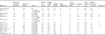
There were 14 published studies of perimembranous ventricular septal defect closure in 868 cases that were analysed (Table 2 and Supplementary Table 1). Since one study did not differentiate sufficiently between ventricular septal defect subtypes and it is possible that the data were presented in other later publications by the same authors, it was excluded. Of the remaining 13 studies, 618 procedures were successfully performed in 641 patients (96.4%). The median age at perimembranous ventricular septal defect closure was 10 years and weight 28 kilograms. The median Qp:Qs was 1.9. The median occluder size was 8 millimetres with an occluder/ventricular septal defect ratio of 1.26 and a median ventricular septal defect size of 7 millimetres. Major complications occurred in 18 (2.9%), 13 of which were complete atrioventricular block requiring insertion of a permanent pacemaker (2.1%). The median age of the patients who developed complications was 2.35 years and weight 8.65 kilograms. The median ventricular septal defect size was 10 millimetres. The patients who developed complications were significantly younger (p < 0.0001) and had larger ventricular septal defects (p < 0.01) than the overall patient cohort.
Details of 14 paper's describing closure of perimembranous VSDs and the complication rates observed

cAVB = complete atrioventricular block; PPM = permanent pacemaker; VSD = ventricular septal defect
*Paper analyses all VSD subtypes together with potential duplication of cases in **. This paper was therefore not included in the overall analysis
Discussion
Since the first surgical closure of ventricular septal defect in 1954, techniques have advanced such that ventricular septal defect closure is now associated with minimal morbidity and mortality.Reference Lillehei, Cohen, Warden, Ziegler and Varco25 Most historical surgical series report 30-day mortality of less than 1% with complete atrioventricular block in 1–2%, significant residual ventricular septal defect in 1–10%, and need for reoperation in 2%.Reference Roos-Hesselink, Meijboom and Spitaels6, Reference Andersen, de Leval, Tsang, Elliott, Anderson and Cook14, Reference Mavroudis, Ganzel, Cox and Polk26–Reference Backer, Winters and Zales30 An important caveat here is that younger children and larger defects than those routinely attempted by transcatheter techniques are often included in these series. Despite surgery being associated with patient discomfort, need for cardiac bypass, sternotomy and scar, the mortality from transcatheter ventricular septal defect closure will need to be approaching 0% and morbidity around 2–4% before it can generally be considered by many as an acceptable alternative, particularly given the implications of long-term pacing in children.
The natural history of small, untreated ventricular septal defects offers a further useful guide to assessing the need for ventricular septal defect closure and puts into context the risks associated with transcatheter and surgical techniques. Small perimembranous ventricular septal defects, defined as unequivocally restrictive and with normal left ventricular end-diastolic dimensions through childhood, are unlikely to close spontaneously in adult life (approximately 7–10% closure rates reportedReference Nadas31, Reference Corone, Doyon and Gaudeau32) and cannot be regarded as benign, the adult being at risk from a number of adverse events. Development of right ventricular outflow tract obstruction (3–7%Reference Nadas31, Reference Corone, Doyon and Gaudeau32), infective endocarditis (1.8–11%Reference Neumayer, Stone and Somerville33, Reference Gabriel, Heger and Innerhofer34), aortic regurgitation (2–7%Reference Hoffman and Rudolph35), and left ventricular dysfunction are all well described as significant risks associated with leaving small ventricular septal defects through adult life. A small defect with new aortic regurgitation or a persistent ventricular septal defect following an episode of infective endocarditis might be more widely considered as suitable for closure by many, although it would still be considered controversial to close such small ventricular septal defects. It is more difficult to define the natural history of larger ventricular septal defects with significant left ventricular volume loading because of the variability in size, location, and effect on left ventricular volume or aortic valve function. By extension, the risks are likely to be significantly greater than those for smaller defects outlined above.
Transcatheter closure of perimembranous ventricular septal defect was first reported two decades ago (1988) and techniques continue to advance rapidly.Reference Lock, Block, McKay, Baim and Keane36 The objective of this series was to assess persistent problems with the procedure, with particular emphasis on selection of cases likely to be associated with least morbidity. In this series, 14 defects were closed with a muscular ventricular septal defect device. This device is technically more straightforward to deliver and may have theoretical advantages in terms of risk of atrioventricular block, given the larger waist of the device (7 millimetres long). The amplatz muscular ventricular septal defect device has rims 4 millimetres larger than the waist. We found that the majority of perimembranous defects deemed suitable for transcatheter closure have sufficient distance from the aortic valve leaflets to safely allow use of a muscular device, and in many cases this has become our device of choice. In both cases, in which a stable device position could not be achieved, the distance between the leading edge of the ventricular septal defect and the right coronary cusp of the aortic valve (as measured on a transoesophageal echocardiography left ventricle long axis view) was insufficient to secure a device. It is possible that this distance is important as within this tissue runs the atrioventricular conduction axis. In addition to potentially providing for device stability during the continual motion of systole and diastole, it affords protection of the conduction axis within. Instability is one mechanism that may account for the relatively high incidence of atrioventricular block seen with transcatheter device closure, particularly in small hearts.
Two cases in our series failed, with a further case complicated by device embolisation with successful retrieval and redeployment. There were no other complications, and neither were there any occurrences of heart block. It is notable that published complications have occurred mostly in young children, with 90% occurring at less than 5 years of age and less than 15 kilograms. It seems prudent therefore, and is our practice, to delay closure of suitable perimembranous ventricular septal defects until the early school years, when the risk of complication is likely to be lower, without incurring significant delay before due consideration is given to cardiac surgical closure, should transcatheter closure fail. This work suggests that such a protocol should afford acceptable morbidity and be able to compete with surgical closure.
It is attractive to consider oversizing a device because of the associated ease of device delivery and lower risk of embolisation. Upsizing the device by one size, considering placing an alternative device type, or using an alternative delivery route (internal jugular vein or transarterial) constitute reasonable steps when undertaking technically challenging perimembranous ventricular septal defects that continue to seem amenable to transcatheter closure. We also favour delivering the device from the aorta, rather than from the left ventricle, upsizing the delivery catheter by one size to allow pressure monitoring guidance as the catheter and the partially protruding left ventricular disc are brought back to the left ventricle and the pressure waveform is seen to change. Simultaneous transoesophageal echocardiography, together with observing a change in the pressure waveform, allows for slow and controlled device delivery, while allowing for changing to delivery from the left ventricle, should this route fail, and avoiding prolonged manipulation of device and wire within the left ventricle. Given the difficulty with defining the exact morphology of ventricular septal defect with current imaging technology, the temptation to go beyond these steps and upsizing devices further should be avoided as complications are likely to be higher. In view of this, accepting and expecting failure rates in the region of 10–20% are likely to be associated with lower complication rates.
This study has some significant limitations related to its retrospective design and the lack of direct comparison with other therapeutic strategies, both conservative and surgical. Long-term follow-up is also an integral part of assessing the use of devices in children. Complications occurring in later life, including the late development of heart block, may be unknown entities for some time to come. This, like most interventions in paediatric cardiology, is an emerging field that will need to be kept under review: it is important that results continue to be published alongside ongoing debate as experience and technology develop.
We have shown that device closure of perimembranous ventricular septal defect is a safe alternative to surgery, capable of closing defects in a timely and safe manner. It may, however, commit children to long-term and unacceptable risks of heart block. We have not seen a heart block in our series to date, and it is possible that this may be a consequence of the technique of delivery, use of muscular devices, and a cautious sizing protocol. The indications for perimembranous ventricular septal defect device closure should be the same as those defined for surgical closure, but delaying until a child reaches 10–15 kilograms before proceeding to transcatheter closure seems prudent given the current devices and techniques. Applying caution when oversizing devices and accepting that some defects will be too morphologically complex to close will be vital, if morbidity is to be kept to a minimum. Given these caveats, we continue to advocate closure of perimembranous ventricular septal defect by transcatheter techniques as a safe alternative to surgical closure with acceptable morbidity.