Introduction
Among digenean species which have a redia stage in their life cycle, the pattern of two redial generations developing one after the other within the intermediate host is frequent. After their emergence from the sporocyst, the mother rediae constitute the first generation and produce the daughter rediae of the second generation. According to parasite species, the mother rediae produce either daughter rediae, or daughter rediae and cercariae, while daughter rediae form cercariae. In the case of Fasciolidae, for example, this two-generation pattern had been reported for Fascioloides magna (Swales, Reference Swales1935; Erhardová-Kotrlá, Reference Erhardová-Kotrlá1971) and Fasciolopsis buski (Lo, Reference Lo1967; Graczyk et al., Reference Graczyk, Alam, Gilman, Mondal and Ali2000).
In the case of the Fasciola genus, the existence of two redial generations is still open to question. Indeed, in the case of F. gigantica, up to five redial generations can develop within the snail host (Dinnik & Dinnik, Reference Dinnik and Dinnik1956, Reference Dinnik and Dinnik1964). In contrast, the life cycle of F. hepatica is more complex, as it comprises either a single redial generation (mother rediae), or two generations with the second generation (daughter rediae) only produced when snails are under adverse conditions, e.g. summer months (Thomas, Reference Thomas1883a, Reference Thomasb), winter (Leuckart, 1879–Reference Leuckart1886), cold (Kendall, Reference Kendall and Taylor1964, Reference Kendall1965) or drought (Styczynska-Jurewicz, Reference Styczynska-Jurewicz1965; Wilson & Draskau, Reference Wilson and Draskau1976). In spite of this contradiction (Andrews, Reference Andrews and Dalton1999), the concept of two redial generations in the life cycle of F. hepatica is still widely accepted by parasitologists.
In reality, the life cycle of F. hepatica is closely similar to that of F. gigantica, as up to four generations of F. hepatica rediae may succeed within the intermediate host. Studies have been carried out in our laboratory over the past 30 years, on serial sections of infected snails and then on live rediae using an image-processor. The results have demonstrated a more complex pattern of redial generations which is largely dependent on the behaviour of the first redia of the first generation. As many papers on the redial generations of F. hepatica have been published since 1978 (Rondelaud & Barthe, Reference Rondelaud and Barthe1978a, Reference Rondelaud and Barthe1982a, Reference Rondelaud and Bartheb, Reference Rondelaud and Barthe1987a; Augot et al., Reference Augot, Rondelaud, Dreyfuss, Cabaret, Bayssade-Dufour and Albaret1998, Reference Augot, Abrous, Rondelaud, Dreyfuss and Cabaret1999), it has been interesting to provide an update of these data to highlight the following three points: (1) the normal pattern of redial development and its variations; (2) the effect of some environmental or biotic factors on these redial generations; and (3) the consequences of such redial development patterns on cercarial productivity. Numerous data came from already published papers. They are completed with still unpublished findings.
Materials and methods
In our laboratory, eight French populations of Galba truncatula, living on the acid soils from the Limousin region (six populations) or in neighbouring sedimentary regions (two populations) were used for most experimental infections of snails with F. hepatica. All of them were known to be highly susceptible to F. hepatica infection, with greater than 60% prevalence when 4-mm high snails were exposed to single- or double-miracidium exposures with this digenean. Four other foreign populations (Spain, Morocco) of G. truncatula were also used to study variations in redial development when sympatric snails were exposed to an allopatric isolate of miracidia, or vice versa (Gasnier et al., Reference Gasnier, Rondelaud, Abrous, Boulard, Carreras, Diez-Banos and Cabaret2000; Goumghar et al., Reference Goumghar, Dreyfuss, Rondelaud, Benlemlih and Cabaret2001; Belfaiza et al., Reference Belfaiza, Moncef and Rondelaud2005). Most French isolates of F. hepatica eggs came from the slaughterhouse of Limoges and were collected from the gall-bladders of cattle. Specific isolates of eggs, originating from French lagomorphs, nutria and sheep, were also used for these studies. As for snails, Spanish and Moroccan isolates of F. hepatica eggs, collected from cattle, were also utilized in these experiments. Most trials were carried out by subjecting 4-mm high snails to single- or double-miracidium exposures with the digenean and by sacrificing several snails at weekly intervals. The others were made with 5-, 6- and 7-mm high snails or 3, 5, 10 or 20 miracidia of F. hepatica per snail, and by using the same protocol as above. Rediae present in every snail were counted, taking into account their physiological state, their generation and their contents.
The development of redial generations was also studied in 1211 G. truncatula naturally infected with F. hepatica and collected since 1993 from 168 cattle-breeding farms located in two French departments (Rondelaud et al., Reference Rondelaud, Vignoles and Dreyfuss2004). Snails were dissected under a stereomicroscope and counts of rediae were made under a microscope, according to their physiological state, their generation and their contents.
Results
Identification of redial generations
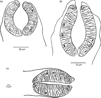
According to Thomas (Reference Thomas1883a, Reference Thomasb) and Kendall (Reference Kendall1965), mother rediae cannot be distinguished morphologically from daughter rediae. However, the redial generations can be identified according to the form and structure of their pharynx (Rondelaud & Barthe, Reference Rondelaud and Barthe1978a, Reference Rondelaud and Bartheb). Using serial sections of infected snails, these authors described three types of pharynxes (fig. 1). The first is that of first-generation rediae. The pharynx is ovoid in young rediae and progressively becomes spherical in older larvae after day 35 post-exposure (p.e.) at 20°C. Like the pharynx, the lumen is ovoid or spherical and its width grows proportionately with increasing length of the body. The second type comprises the first cohort of second-generation rediae (fig. 1b). The pharynx is smaller, ovoid and its lumen is clearly larger in the pre-intestinal zone than in the other zone. The third type is not distinct from a given redial generation, as it is observed in the second cohort of the second generation and in subsequent generations. The pharynx is small, ovoid and its long lumen is very narrow. Pharyngeal dimensions at day 49 p.e. are given in table 1. These values allow the first redia of the first generation to be distinguished from the other redial categories, even when this larva is old. When the rediae are alive, the determination of generations is not easy because of continuous movements of their pharynx under the microscope. If these larvae are fixed in 10% (v/v) formalin, i.e. 4% (w/v) formaldehyde, the pharynx walls are often distorted, thus making redial identification difficult.
Schematic drawings of pharynxes belonging to the first redial generation of Fasciola hepatica (a), the first cohort of the second generation (b), and the other cohorts and generations (c).

Pharyngeal dimensions of Fasciola hepatica rediae in relation to their generation and cohort at day 49 post-exposure in snails bred at 20°C (Augot et al., Reference Augot, Rondelaud, Dreyfuss, Cabaret, Bayssade-Dufour and Albaret1998, Reference Augot, Abrous, Rondelaud, Dreyfuss and Cabaret1999).

Developmental patterns of redial generations
The pattern depends on the behaviour of the first redia of the first generation after its exit from the sporocyst. If this larva remains alive throughout snail infection, at least up to day 49 p.e., the development is normal. In contrast, if the first redia dies during week 2 or week 3 of snail infection, the development is abnormal and two variants have been described (Rondelaud & Barthe, Reference Rondelaud and Barthe1982b; Belfaiza et al., Reference Belfaiza, Moncef and Rondelaud2005).
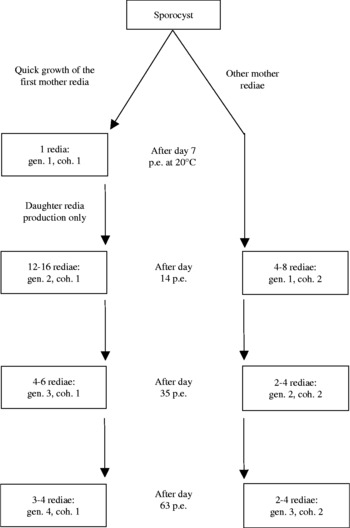
Figure 2 shows the four successive generations in the case of normal development. The numbers of live rediae for each generation and the dates of exit for the first rediae are also indicated. Two points are important. The first is the rapid emergence of the first mother redia from the sporocyst on day 7 p.e. (at 20°C), its rapid growth within the snail, and its production of only daughter redia. The second point is the time of this production. Indeed, the daughter rediae produced by the first mother redia become free during the same period as the other mother rediae which emerge later from the sporocyst. As a consequence, the free rediae, other than the first mother redia, observed between day 15 and day 35 p.e., are composed of two different cohorts, the first belonging to the first generation and the other to the second generation. A similar process also occurs for subsequent generations after day 35 and after day 63 p.e. The result of all this is a useful burden ranging from 28 to 43 live rediae at day 80 p.e. in snails originating from different populations of G. truncatula living on acid soils (the shell height of adults is often less than 8 mm). However, for the highest shell heights (up to 12 mm) of G. truncatula, the burden may rise up to a mean of 56 live rediae, essentially by a numerical increase of third- and fourth-generation rediae (Rondelaud & Barthe, Reference Rondelaud and Barthe1987a; Belfaiza et al., Reference Belfaiza, Rondelaud, Moncef and Dreyfuss2004).
Normal development of Fasciola hepatica redial generations in single-miracidium infections of Galba truncatula, with an indication of redial numbers for each redial cohort. In this case, the first redia of the first generation remained alive at least up to day 49 post-exposure (p.e.) at 20°C. Abbreviations: coh., cohort; gen., generation. From Belfaiza et al. (Reference Belfaiza, Rondelaud, Moncef and Dreyfuss2004), reprinted by permission of CABI Publishing and completed.

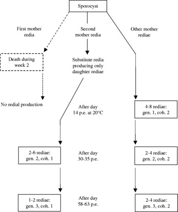
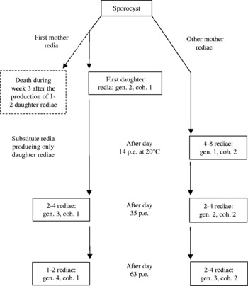
When the first redia of the first generation dies, daughter redia production is ensured by another free redia, either the second mother redia of the first generation (fig. 3), or the first daughter redia of the first cohort of the second generation (fig. 4). However, the number of daughter rediae produced by this substitute redia is lower (only 2–6) and occurs later over time (the first free rediae emerge after day 30 p.e. at 20°C). In single-miracidium infections of G. truncatula, the first developmental variant was seen in about 20% of infected snails, whereas the second variant was very scarce: less than 0.1% (Rondelaud & Barthe, Reference Rondelaud and Barthe1982b).
Abnormal development (first variant) of Fasciola hepatica redial generations in single-miracidium infections of Galba truncatula. The death of the first mother redia during week 2 post-exposure (p.e.) at 20°C induced the rapid growth of the second mother redia which produced only daughter rediae. Abbrevations: coh., cohort; gen., generation. From Belfaiza et al. (Reference Belfaiza, Rondelaud, Moncef and Dreyfuss2004), reprinted with permission of CABI Publishing and completed.

Abnormal development (second variant) of Fasciola hepatica redial generations in single-miracidium infections of Galba truncatula Abnormal development of redial generations (second variant) in single-miracidium infections of Galba truncatula with Fasciola hepatica. Before its death during week 3 post-exposure (p.e.) at 20°C, the first mother redia had produced 1 or 2 rediae and the first of these daughter rediae grew rapidly to produce only rediae. Abbreviations: coh., cohort; gen., generation.

A more surprising development of redial generations (Belfaiza et al., Reference Belfaiza, Moncef and Rondelaud2005) was noted in Moroccan G. truncatula infected by a sympatric isolate of F. hepatica miracidia. In parent snails, all free mother rediae produced second-generation rediae. However, in the F1 and F2 snails, daughter redia production was progressively ensured by the first mother redia. This abnormal development does not seem to be another variant, but rather the result of adaptation between two sympatric partners.
Factors influencing the developmental pattern of redial generations
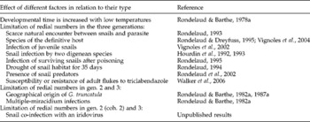
The factors are listed in table 2. Their effects on the development of the first three generations of F. hepatica were only studied up to day 49 p.e. at 20°C. Whatever the factor studied, the three generations were found in all snail groups. However, for some factors, variations in the numbers of free and live rediae were noted. Drought of the snail habitat for 35 days (Rondelaud, Reference Rondelaud1994), as well as seven biotic factors, have the effect of limiting redial numbers in the three generations. Among the biotic factors, the most important are the species of the definitive host from which eggs of F. hepatica are collected for snail infections (miracidia originating from eggs collected from cattle, sheep or nutria had a greater infectivity than those coming from lagomorphs: Rondelaud & Dreyfuss, Reference Rondelaud and Dreyfuss1995; Vignoles et al., Reference Vignoles, Ménard, Rondelaud, Agoulon and Dreyfuss2004), the susceptibility or the resistance of adult flukes to triclabendazole (Walker et al., Reference Walker, Hoey, Fletcher, Brennan, Fairweather and Trudgett2006), and the frequency of encounters between snails and the parasite in the field (Rondelaud, Reference Rondelaud1993). Limitation in redial numbers is different for the other three biotic factors. Indeed, the second and third generations are affected in the case of the geographical origin of snails (Rondelaud & Barthe, Reference Rondelaud and Barthe1982a, Reference Rondelaud and Barthe1987a) and the number of miracidia used per G. truncatula (Rondelaud & Barthe, Reference Rondelaud and Barthe1982a), whereas only the second cohort of the second generation and the third generation are affected if snails are already highly infected with an iridovirus at the time of miracidial exposure (unpublished data).
Impact of different environmental or biotic factors on the redial generations (gen.) of Fasciola hepatica.

Redial generations of F. hepatica in natural infections of snails
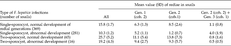
Table 3 shows the distribution of live rediae of F. hepatica found in a total of 731 G. truncatula collected from acid soils in June–July and in September–October over a 10-year period. Single-sporocyst infections are found in numerous snails, either with a normal development of redial generations (50.4%), or with an abnormal development (38.4%). Two-sporocyst infections are more scarce: 8.9% and 2.2%, respectively. The first three generations of rediae are present in all snails. In each type of F. hepatica infection (1- or 2-sporocyst), the numbers of first-generation rediae are close. The rediae belonging to the second cohort of the second generation and the third generation are few in numbers and this fact may be due to the characteristics of snail samples used for this study, i.e. snails harbouring cercariae-containing rediae. In spite of these fluctuations in the numbers of free rediae, the sequence of three redial generations developing one after the other was noted in all naturally infected snails, thus demonstrating the constancy of this process in all populations of G. truncatula when infected with F. hepatica.
Distribution of live rediae of Fasciola hepatica in 731 naturally infected Galba truncatula. From Rondelaud et al. (Reference Rondelaud, Vignoles and Dreyfuss2004), with kind permission of Springer Science+Business Media.
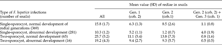
Abbreviations: coh., cohort; Gen., generation.
Consequences of redial development patterns on cercarial productivity
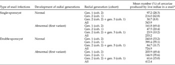
The existence of several developmental patterns for the rediae of F. hepatica and the lower numbers of rediae found during an abnormal development have consequences for cercarial productivity. Table 4 gives the results obtained at day 63 p.e. (at 20°C) in experimentally infected snails originating from acid soils. Snails showing a normal development of redial generations produce more cercariae than those with an abnormal development (a total of 343.2 cercariae up to day 63 p.e. instead of 233.2 in single-sporocyst infections, for example). Most cercariae in the case of a normal development are formed by the first cohort of the second generation, whereas they are produced by the second cohort of mother rediae when redial development is abnormal. In both cases, the roles of the second cohort of the second generation and that of the first cohort of the third generation in cercarial production are limited up to day 63 p.e.: less than 11% (single-sporocyst infections) or less than 16% (double-sporocyst infections). In long-surviving snails (at least up to day 102 at 20°C), the role of these two cohorts is also limited, as they produce 17% of the whole cercarial production in single-sporocyst infections and only 13.1% in double-sporocyst infections (Belfaiza et al., Reference Belfaiza, Rondelaud, Moncef and Dreyfuss2004).
Assessment of Fasciola hepatica cercarial productivity in the case of single- and double-sporocyst infections at day 63 post-exposure (p.e.) at 20°C. Single-sporocyst infection: from Augot & Rondelaud, Reference Augot and Rondelaud2001, with kind permission of Springer Science+Business Media; two-sporocyst infections, unpublished results.

* The following ratio: (highest mean number of germinal embryos in young rediae) − [(number of germinal embryos remaining in rediae at day 63 p.e.)+(number of morulae differentiated into daughter rediae)] is multiplied by the mean number of live and free rediae (in each cohort or each generation) to give the total number of cercariae.
Discussion
Four generations of F. hepatica rediae may develop one after the other within the intermediate host. This succession was verified in naturally infected snails as well as in those that were subjected to miracidia under laboratory conditions, thus suggesting that it is species dependent. As Thomas (Reference Thomas1883a, Reference Thomasb), Leuckart (1879–Reference Leuckart1886), Kendall (Reference Kendall and Taylor1964, Reference Kendall1965), Saint-Guillain (Reference Saint-Guillain1968), and Wilson & Draskau (Reference Wilson and Draskau1976) only described the presence of two redial generations, i.e. mother rediae and daughter rediae, in the life cycle of F. hepatica, an explanation must be given. In their reports, Thomas (Reference Thomas1883a, Reference Thomasb) and Leuckart (1879–Reference Leuckart1886) did not describe the rapid growth of the first mother redia and the production of only daughter redia, so that the mother rediae described by these authors might correspond to the group constituted by the rediae of the first generation and those belonging to the first cohort of the second generation. In the same way, the daughter rediae reported by Thomas (Reference Thomas1883a, Reference Thomasb) might correspond to rediae which became free after day 35 p.e., i.e. the second cohort of the second generation and the first cohort of the third generation. This interpretation might also partly explain the variations that several authors (Thomas, Reference Thomas1883a, Reference Thomasb; Leuckart, 1879–Reference Leuckart1886; Kendall, Reference Kendall1965; Styczynska-Jurewicz, Reference Styczynska-Jurewicz1965; Wilson & Draskau, Reference Wilson and Draskau1976) have noted in daughter redia production when infected snails were under adverse conditions. In contrast, the studies performed in our laboratory do not confirm the succession of three or more cohorts into each redial generation of F. hepatica, as reported by Ginetsinkaya (Reference Ginetsinkaya1988) in his review.
The role of the first redia of the first generation in daughter redia production had already been reported in the life cycle of Fasciola gigantica (Rakotondravao et al., Reference Rakotondravao, Moukrim, Hourdin and Rondelaud1992) and in that of Echinostoma paraensei (Sapp et al., Reference Sapp, Meyer and Loker1998). As demonstrated in table 4, this redia had a great importance in the larval development of F. hepatica, as the daughter rediae produced (first cohort of the second generation) led to the development of most cercariae in the case of a normal development. However, its role in daughter redia production might change for the following two reasons. The first argument was proposed by Rondelaud & Barthe (Reference Rondelaud and Barthe1987b) in single-miracidium infections of Austropeplea tomentosa and of Pseudosuccinea columella. In snails showing a rapid increase of their shell height during the experiment (up to 12.6 mm for A. tomentosa and 15.6 mm for the other lymnaeid), the formation of rediae belonging to the first cohort of the second generation (23–26 rediae) was ensured by two mother rediae, i.e. the first two rediae that emerged from the sporocyst. The second reason is given in the report by Belfaiza et al. (Reference Belfaiza, Moncef and Rondelaud2005). According to these authors, each of mother rediae, after its exit from the sporocyst, formed several rediae of the second generation (the two cohorts cannot be identified) when Moroccan G. truncatula were exposed to miracidia of a sympatric isolate of F. hepatica. Under these conditions, one may wonder about the reasons of such changes for these mother rediae and two, perhaps complementary, hypotheses may be proposed. The first is to admit that regulation of the redial population would be ensured by the host snail, and particularly by its size, as there is a relationship between the shell height of snails and the quantity of free rediae found within them (Zischke, Reference Zischke1967). However, evolution in the role of these mother rediae over time by adapting to a new snail host or to an unusual snail cannot be excluded.
As demonstrated in figs 2 and 3 and table 4, the rediae that become free after day 35 p.e. or day 63 p.e. are few in numbers. Even if these larvae are important for maintaining the redial population, their role as cercarial producers seems limited. Indeed, in single-sporocyst infections, with a normal development of rediae, the second cohort of the second generation and the first cohort of the third generation, for example, were able to form only 8.9–10.2% of the cercarial population at day 63 p.e. (table 4), or a total of 19.5% between days 102 and 118 p.e. at 20°C (Belfaiza et al., Reference Belfaiza, Rondelaud, Moncef and Dreyfuss2004). In our opinion, these rediae must be considered as substitute larvae and their development would only occur to replace old rediae of previous generations. Two points may support this hypothesis. First, most rediae belonging to the second cohort of the second generation and the first cohort of the third generation grow slowly between day 35 and day 63 p.e. Second, the number of full-grown rediae during the above period is always close to that of empty larvae from previous generations (unpublished results). As most of their cercariae become free within the snail's haemocoel after day 63 p.e. at 20°C, one may wonder about the participation of these rediae in cercarial shedding after day 20 of the patent period (generally corresponding to day 69 p.e. at 20°C), because the number of cercariae produced was low and discontinuous at that time (Audousset et al., Reference Audousset, Rondelaud, Dreyfuss and Vareille-Morel1989).