INTRODUCTION
Over a century ago, Lauterborn (Reference Lauterborn1895) discovered an unusual freshwater testate amoeba near Ludwigshafen, Germany, which he formally described as the new genus Paulinella with Paulinella chromatophora Laut. as the type species. The test (shell) of this organism consists of about ten rows of endogenously produced siliceous plates or scales that are elongated and rectangular in shape with rounded corners. These scales are also curved to enable their placement in five columns around the circumference of the test. There are usually three specialized scales surrounding the pseudostomal aperture through which, in living cells, the filose pseudopodia extend. The species name (chromatophora) was selected by Lauterborn to draw attention to the presence of usually two plastid-like inclusions (cyanelles) in the Paulinella cell. Significantly, Lauterborn did not include the presence of the ‘chromatophores’ that he found in the German specimens in his description of the new genus Paulinella, but instead relegated this feature to the type species. This was an insightful decision, because two additional species, P. ovalis (Wulff) (Johnson et al., Reference Johnson, Hargraves and Sieburth1988) and P. indentata (Hannah et al., Reference Hannah, Rogerson and Anderson1996) have since been thoroughly studied in cultures with no evidence of the P. chromatophora-like cyanelles in either of them.
Soon after the German discovery, P. chromatophora was discovered in America (Kepner, Reference Kepner1905). It is now known worldwide (Melkonian & Mollenhaurer, 2005), mainly from the sediments of freshwater lakes and ponds, but also from brackish marine environments (Pankow, Reference Pankow1982). More recently, three additional marine species have been discovered: P. ovalis, P. indentata (see above) and P. intermedia (Vørs, Reference Vørs1993). This paper reports the discovery of six new species of Paulinella from marine beach sands and nearshore benthic sediments of Canada's west coast (Pacific Ocean) and thereby more than doubles the number of known species of this unusual genus. Descriptions were based on detailed study of the tests using both light and electron microscopy and include new data on both P. indentata and P. chromatophora included here as ‘controls’ on methodolgy, since both of these species were available in samples collected from the same marine locations (for P. indentata) and from freshwater locations in Ontario Canada (for P. chromatophora).
MATERIALS AND METHODS
Sample collection sites were near Skidegate, Haida Gwaii (Queen Charlotte Islands), located in the Pacific Ocean approximately 360 km north-west of Vancouver Island, British Columbia, Canada at locations specified in Nicholls (Reference Nicholls2007) for collections made in June 2006 and in Nicholls (in press) for collections made in July 2007. Additional collections using similar methods were made at a more southerly location in Pacific Ocean coastal waters (southern Vancouver Island) in July 2008. Samples were collected at low tide from 30–40 cm deep holes dug in the beach sand a few metres from the edge of the sea, and from surface detrital material at the bottom of small shallow tide pools. Samples were shipped by airmail in 500 ml polycarbonate bottles and examined immediately on arrival at the laboratory in Ontario, when salinity was also measured with a refractometer (model FG-211, Sun Instruments Corp., Torrance CA) with automatic temperature compensation. Salinity of all samples collected from Haida Gwaii in 2006 and 2007 and from the Esquimalt, Vancouver Island site in 2008 indicated essentially no freshwater dilution effects (S = 31–32 ppt).
Subsamples were examined for initial detection of Paulinella specimens with an inverted microscope using either dark-ground illumination at 200× magnification or phase contrast at 160× magnification. Measurements of test dimensions were made with an inverted microscope using open preparations (no coverglass) using 40× dry or 100× oil-immersion objectives and 15× eyepieces, one of which was equipped with a precalibrated reticule.
Descriptive statistics for the test dimensions (mean, median, standard deviation, minimum, maximum and coefficient of variation) were performed in CoStat (CoHort Software, 1995). For electron microscopy and for permanent slide preparations (type specimens held by the Canadian Museum of Nature (see below)), sub-samples were alternately washed with distilled water and concentrated by sedimentation several times to remove sea salts. Specimens were then isolated with a single hair brush and a micropipette, were air dried on No. 1 cover glasses, and cemented to glass slides with Canada balsam for permanent mounts. For scanning electron microscopy (SEM), specimens were transferred to a fragment of cover glass or polycarbonate plastic with a micropipette, dried at room temperature, glued to a SEM stub, and coated with gold before examination and image acquisition. For transmission EM, washed specimens were transferred to a TEM copper location grid previously coated with a film of Butvar-B98 (J.B. EM Services, Dorval, Quebec, Canada).
Descriptions of scale arrangement on Paulinella tests includes the term ‘non-overlapping’, defined here for Paulinella as meaning arranged in parallel columns (but the sides and ends of individual scales may overlap slightly those of adjacent scales). In contrast, ‘overlapping’ or ‘imbricated’ scales in the Paulinella context means the scales are not arranged in parallel columns but the rows are offset so that the ends of individual scales of a given row are positioned about the middle of scales in the rows immediately anterior and posterior to the given row forming a brickwork pattern over multiple rows of scales.
RESULTS
1Note: alternative classifications exist in the modern protist literature (one higher level classification could include ‘Rhizaria’ and ‘Cercozoa’ in place of Rhizopoda and Filosea). Alternative classification systems are, however, in a state of flux (Adl et al., Reference Adl, Leander, Simpson, Archibald, Anderson, Bass, Bowser, Brugerolle, Farmer, Karpov, Kolisko, Lane, Lodge, Mann, Meisterfeld, Mendoza, Moestrup, Mozley-Standridge, Smirnov and Spiegel2007). The classification used here is that presented in Corliss (Reference Corliss1994) because it uses a familiar and stable system of hierarchical names that will be familiar to students of testate amoebae.
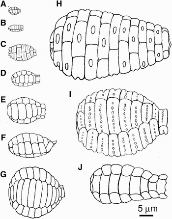
Diagrammatic representation of the 10 known Paulinella species, emphasizing overall differences in test sizes and shapes. (A) P. ovalis (Wulff) Johnson, Hargraves & Sieburth; (B) P. intermedia Vørs; (C) P. indentata Hannah, Rogerson & Anderson; (D) P. carsoni sp. nov.; (E) P. agassizi sp. nov.; (F) P. suzukii sp. nov.; (G) P. chromatophora Lauterborn; (H) P. gigantica sp. nov.; (I) P. multipora sp. nov.; (J) P. lauterborni sp. nov.

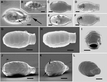
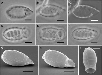
Paulinella carsoni sp. nov. (A) living cell (small arrow) with a filopod (large arrow) emerging from its test; (B) LM image of an empty test showing scale covering; (C–F) LM images of four different specimens showing variability in test shape; (G–L) SEM images; (G, H, J) tests in lateral views (anterior collar surrounding the pseudostomal aperture at right); (I, L) apical view of pseudostomal aperture; (K) lateral posterior view showing swollen distal ridges on posterior scales (arrows). Scale bars = 2 μm.

DIAGNOSIS
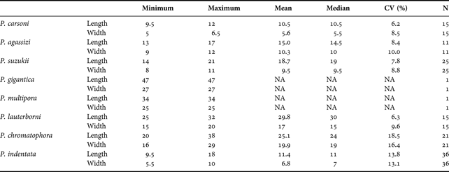
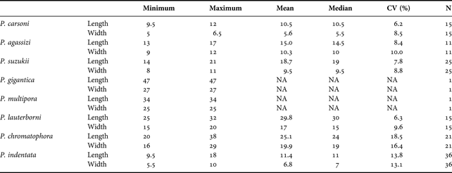
Tests elongate, sub-cylindrical, 9.5–12 μm long and 5–6.5 μm wide (Table 1). Five columns and 5–7 rows of non-overlapping, siliceous, oblong scales. Collar prominent, consisting of three wide scales with slightly concave exteriors bent to form a short column surrounding the elliptical pseudostomal aperture, about 1.5 × 3 μm. Scales of the test body with slightly thickened lateral rims.
Descriptive statistics for tests (shells) of the eight species of Paulinella studied for this paper. CV, coefficient of variation; N, number of tests measured; NA, not applicable. All measurements in μm and represent the longest and widest parts of the tests.
ETYMOLOGY
The discovery and investigation of this species in 2006–2007 coincided with the 100th anniversary of the birth of Rachel Carson (1907–1964). The specific epithet (carsoni) is intended to commemorate the contributions of this noted American marine biologist and award-winning author.
TYPE SPECIMEN
The type specimen mounted in Canada balsam on a glass slide, is retained at the Canadian Museum of Nature (Invertebrate Zoology Division), Ottawa, Ontario, Canada; Catalogue No. CMNI 2009-0001.
TYPE LOCALITY
Pacific Ocean beach near Gillatt (Grassy) Island, Haida Gwaii (Queen Charlotte Islands, British Columbia, Canada (53.241°N, 131.898°W)).
MATERIAL FROM TYPE LOCALITY
Retained by the author in sample No. V-2032, collected 27 June 2006.
Paulinella agassizi sp. nov. (A–D) LM images of tests showing variation in shape (A–C) and configuration of scale pattern (D); (E–I) SEM images of tests showing scalloped anterior edge of collar scales (E, F), apical view of pseudostomal aperture (G), scale pattern over the posterior of a test (H), and pattern of small pores on scales (I). Scale bars = 5 μm.

DIAGNOSIS
Tests 13–17 μm long and 9–12 μm wide (Table 1); tests vase-shaped, widest slightly posterior to the middle of the test. Siliceous scales on test oblong with rounded corners and organized in 5–6 rows of five non-overlapping columns; some specimens reveal a small area of overlapping (brickwork pattern) scales, most often aborally as a result of the misalignment of a single row of scales. Collar surrounding the pseudostomal aperture comprised three specialized scales with scalloped margins. Pseudostomal aperture elliptical, approximately 50% of test width.
ETYMOLOGY
The discovery and investigation of this species in 2006–2007 coincided with the 200th anniversary of the birth of Louis Agassiz (1807–1873). The specific epithet (agassizi) is intended to commemorate the contributions of this noted Swiss-American zoologist, naturalist and teacher.
TYPE SPECIMEN
The type specimen mounted in Canada balsam on a glass slide, is retained at the Canadian Museum of Nature (Invertebrate Zoology Division), Ottawa, Ontario, Canada; Catalogue No. CMNI 2009-0002.
TYPE LOCALITY
Pacific Ocean beach near Gillatt (Grassy) Island, Haida Gwaii (Queen Charlotte Islands, British Columbia, Canada (53.241°N, 131.898°W)).
MATERIAL FROM TYPE LOCALITY
Retained by the author in sample No. V-2044, collected 27 June 2006.
Paulinella suzukii sp. nov. (A–F) LM images of tests in lateral views (A–C), in ventral views (D–E), and dorso-lateral view (F). Note that B and C represent different focal planes of the same specimen; (G–I) SEM images of a test in lateral view (G), latero-ventral view (H), and ventral view (I). Scale bars = 5 μm.

DIAGNOSIS
Tests 14–21 μm long and 8–11 μm wide (Table 1) with a wide and prominent anterior concave hood-like collar scale partially covering the pseudostomal aperture with an apical–ventral opening at an angle of about 45° to the long axis of the test. Two additional collar scales surround the aperture overlapping the main scales of the test body at their ends. These two scales are of similar morphology as the scales of the main test body (elongate with rounded ends and parallel sides), but unlike the main test body scales which are oriented at right angles to the long axis of the test, they follow the 45° angle of the pseudostomal aperture. Body scales in 5–6 rows of five parallel columns (non-overlapping).
ETYMOLOGY
The specific epithet (suzukii) pays tribute to the well-known Canadian geneticist, nature advocate and science educator, David Suzuki.
TYPE SPECIMEN
The type specimen mounted in Canada balsam on a glass slide, is retained at the Canadian Museum of Nature (Invertebrate Zoology Division), Ottawa, Ontario, Canada; Catalogue No. CMNI 2009-0003.
TYPE LOCALITY
Pacific Ocean beach near Gillatt (Grassy) Island, Haida Gwaii (Queen Charlotte Islands, British Columbia, Canada (53.241°N, 131.898°W)).
MATERIAL FROM TYPE LOCALITY
Retained by the author in sample No. V-2044, collected 27 June 2006.
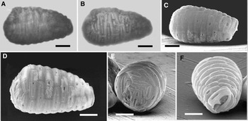
Paulinella gigantica sp. nov. (A–B) LM images of a test, focused on the edge (A), and focused on the scales covering the test (B); (C–F) SEM images of lateral (C, D), posterior (E) and anterior (F) views of a test. Scale bars = 10 μm.

DIAGNOSIS
Test 47 μm long and 27 μm wide, sub-cylindrical with a rounded aboral terminus and tapering anteriorly to a prominent terminal pseudostomal aperture. Three pseudostomal collar scales, one of which is broadly domed with a simple scalloped anterior edge with one median anteriorly-directed peak. Exterior convex surfaces of collar scales distinctively ornamented with a centrally located elongated pit exposing the hollow interior of the scale. Scales of the test body with similar pits, but of a less elongated shape (some nearly circular), except on posterior body scales where pits are greatly elongated. Scales of the test body overlapping (imbricated) over about 10–12 rows.
ETYMOLOGY
The specific epithet (gigantica) refers to the large size of this species (the largest of all 10 known Paulinella species).
TYPE SPECIMEN
The type specimen mounted in Canada balsam on a glass slide, is retained at the Canadian Museum of Nature (Invertebrate Zoology Division), Ottawa, Ontario, Canada; Catalogue No. CMNI 2009-0004.
TYPE LOCALITY
Pacific Ocean beach near Gillatt (Grassy) Island, Haida Gwaii (Queen Charlotte Islands, British Columbia, Canada (53.241°N, 131.898°W)).
MATERIAL FROM TYPE LOCALITY
Retained by the author in sample No. V-2032, collected 27 June, 2006.
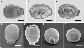
Paulinella multipora sp. nov. (A–C) LM images of a test focused on the margin (A) and on the scale covering (B, C); (D–G) SEM images of a test in lateral views (D, E), apical view, showing collar scales surrounding the pseudostomal aperture (F), and posterior view (G). Scale bars = 10 μm.

DIAGNOSIS
Test globular, 34 μm long and 25 μm wide, covered with 5 columns of non-overlapping scales in 7–8 rows. Scales with 5–10 pores or depressions in a single row located in a median depression through the length of the scale. Pores of variable degree of development and definition approximately 0.3–1 μm in diameter. Collar scales three, surrounding the elliptical aperture, each with single row of well-defined pores.
ETYMOLOGY
The specific epithet (multipora) denotes the single row of conspicuous pores in the middle of the scales surrounding the pseudostomal aperture and test body.
TYPE SPECIMEN
The type specimen mounted on a fragment of a microscope cover glass and coated with gold (used here for SEM examination), is mounted on a glass slide and protected under a raised cover glass and is retained at the Canadian Museum of Nature (Invertebrate Zoology Division), Ottawa, Ontario, Canada; Catalogue No. CMNI 2009-0005.
TYPE LOCALITY
Pacific Ocean beach near Gillatt (Grassy) Island, Haida Gwaii (Queen Charlotte Islands, British Columbia, Canada (53.241°N, 131.898°W)).
MATERIAL FROM TYPE LOCALITY
Retained by the author in sample No. V-2071, collected 13 July 2007.
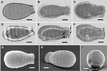
Paulinella lauterborni sp. nov. (A–F) LM images of tests showing variation in shape (A–C), scalloped edges of collar scales (E, arrows), and scattered pores on scales (D–F). Note that D–F are the same specimens as A–C, respectively; (G–I) SEM images of a test in lateral view (G), latero-aboral view (H) and apical view (I). Scale bars = 5 μm.

DIAGNOSIS
Tests widest in the posterior one-third and tapering anteriorly to a moderate constriction approaching the region of the collar. Tests 25–32 μm long and 15–20 μm wide (Table 1). Test comprised five columns and 8–9 rows of non-overlapping siliceous, curved, rectangular scales with rounded corners. Scales with 4–5 rows of small pores, about 0.4 μm in diameter (about 7–10 pores per row). Collar formed from three specialized scales with scalloped margins and imparting a slight flare to the anterior region of the test.
ETYMOLOGY
The specific epithet (lauterborni) pays tribute to Robert Lauterborn (1869–1952), German ecologist/protistologist and discoverer of the genus Paulinella.
TYPE SPECIMEN
The type specimen mounted in Canada balsam on a glass slide, is retained at the Canadian Museum of Nature (Invertebrate Zoology Division), Ottawa, Ontario, Canada; Catalogue No. CMNI 2009-0006.
TYPE LOCALITY
Pacific Ocean beach at Esquimalt, south shore of Vancouver Island, British Columbia, Canada (48.41°N, 123.48°W).
MATERIAL FROM TYPE LOCALITY
Retained by the author in sample No. V-2103, collected 22 July 2008.
OTHER PAULINELLA SPECIES
Two other previously known species (P. chromatophora and P. indentata) were relatively common in my collections and data from these were obtained using identical methods as for the new species described above in order to provide comparative material and to maximize the comprehensiveness of this treatment of the genus. Only P. ovalis and P. intermedia are missing from this treatment, as I have not yet found them.
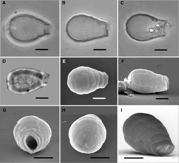
Paulinella chromatophora Lauterborn Reference Lauterborn1895. (A–D) LM images; (A) living specimen with sausage-like cyanelles (arrows); (B–D) tests, focused on the edge (B) and on the scales (C–D) showing pores; (E–I) SEM images of test in lateral view (E), apical view (F), showing structure of the collar scales (G–H) and pores on scales covering test body (I, arrows). Scale bars A–F, I = 5 μm; G–H, = 2 μm.

Tests from freshwater locations in Ontario (both hardwater and softwater sites with specific conductance >200 μm cm−1 and <50 μm cm−1, respectively) revealed a wide range in size: 20–38 μm long × 16–29 μm wide (Table 1). Test morphology agreed well with Lauterborn's (1895) original description of this species. Scales are organized in parallel series of five columns in 7–11 rows (mean and median = 9; N = 21). SEM revealed scales ornamented with scattered small shallow pits or depressions that appear superficial in SEM (Figure 8I), but which are dramatically evident with critical light microscopy (oil immersion phase contrast; Figure 8C, 8D). The external curved surface of collar scales possesses two raised ridges running parallel to the long axis of the scale and forming a rectangular central area (Figure 8G, H). In some specimens, there is a scale-like structure oriented on its edge blocking the pseudostomal aperture which may function as a specialized stopper or plug during transition to a resting cyst stage in the life cycle of this species (Figure 8G).
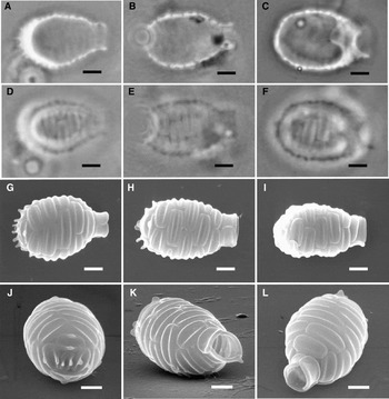
Paulinella indentata Hannah, Rogerson & Anderson Reference Hannah, Rogerson and Anderson1996. (A–F) LM images of tests, focused on the edges (A–C) and on the interlocking scales (D–F). Note that D–F are the same specimens as A–C, respectively; (G–L) SEM images of tests in lateral views (G–I) showing shape variation, posterior scales with marginal protuberances (J), latero-apical views showing arrangement of collar scales (K–L). Scale bars = 2 μm.

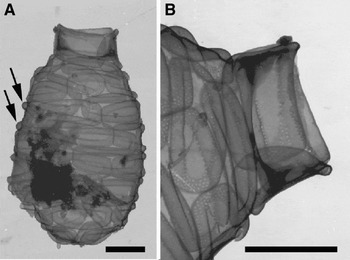
Paulinella indentata Hannah, Rogerson & Anderson Reference Hannah, Rogerson and Anderson1996. TEM images of scale structure. (A) Scale covering over a whole test showing swollen marginal rim of a scale (arrows); (B) detail of collar scales and anterior test scales showing small pores confined to the marginal rim and the edges of the central indented zone. Scale bars = 2 μm.

Tests from the Canadian Pacific Ocean locations (Haida Gwaii) were 9.5–18 μm long and 5.5–10 μm wide (Table 1). While the posterior scales of this species are most distinctive with their marginal labiate processes (Figure 9G, 9H, 9J), these processes were poorly developed in a few specimens (Figure 9I). Identification of such specimens could still be made with light microscopy, however, knowing that the marginal swelling on the scales of P. indentata imparted a corrugated appearance to the margins of tests of this species, while in P. carsoni (with which it overlapped in size in the Canadian material) revealed only flat scale margins with the light microscope. More importantly, P. indentata has the overlapping pattern of scale arrangement while P. carsoni has parallel columns of scales. While pores as described by Hannah et al. (Reference Hannah, Rogerson and Anderson1996) were not evident on the scales of Canadian specimens examined with SEM, pores of about 0.05 μm diameter occupying the same scale locations as those observed in the Scottish type material (the marginal rims and along the edge of the central depression) were observed in a specimen observed with TEM (Figure 10B).
DISCUSSION
Each of the new species described here has highly distinctive test morphology that cannot be confused with other species of this genus. Evidently, light microscopy is adequate for detection and identification of these species, even among the small members of the genus. For example, C. carsoni can be distinguished from C. indentata with high magnification LM by the lack of the prominent marginal bulges on the scales, which in P. indentata are readily apparent in optical cross-section along the lateral margins of the P. indentata test. The papillae-like protuberances on posterior scales of P. indentata are also usually visible with high magnification phase contrast microscopy.
Given the highly distinctive morphology of both Paulinella multipora and P. gigantica, there can be no question of their status as distinct species; but more specimens of each need to be found in order to describe the range in morphology of each. Like P. intermedia Vørs (Reference Vørs1993), both were described on the basis of a single specimen. Thus far, many hours of searching have failed to produce more specimens of either species.
More serious questions remain about P. intermedia owing to its description only by TEM, thus precluding important information that should be possible to obtain from tilted views of additional specimens using SEM. Does it really have ‘3–4 columns of scales’? Certainly it is difficult to determine the answer to this question from the single TEM image (Vørs' figure 1). Five columns of scales in those species with non-overlapping scale patterns would appear to be typical for the genus based on the new information presented here, so a species with only three or four columns would represent to some degree an ‘outlier’ within the genus. It is also difficult to conclude from Vørs' figure 1 that the scales in P. intermedia are arranged in parallel columns. Answers to these questions about P. intermedia are important because they may either strengthen or weaken the distinction between P. intermedia and P. carsoni. There can be no questioning the five parallel rows of scales in P. carsoni. Other, perhaps less significant, differences include a test with a relatively narrower but taller collar; a test that is about 2× larger with a more rounded aboral end than that presently known for P. intermedia.
With the discovery of the new species described here, certain characteristics common to all species of Paulinella are now apparent. The genus can be divided into two groups based on the organization of scales covering the test. Most common is the series of five parallel columns of non-overlapping scales found in P. ovalis, P. intermedia (?), P. carsoni, P. agassizi, P. suzukii, P. chromatophora, P. multipora and P. lauterborni. Only P. indentata and P. gigantica possess the overlapping (interlocking brickwork) pattern of scales. All species have a series of usually three specialized collar scales surrounding the pseudostomal aperture. Pores organized in rows or in less organized scattered distributions are common on scales. Electron microscope revelations of such pores on P. chromatophora scales were reported by Kies (Reference Kies1974; his figure 22), but the images presented here appear to be the first clear demonstration by LM of this feature.
With a mean test width and length of 6.8 and 11.4 μm, respectively, the Canadian P. indentata was considerably smaller than those reported by Hannah et al. (Reference Hannah, Rogerson and Anderson1996) from the type locality (the Firth of Clyde, Scotland; mean test length and width of 9.8 and 15.8 μm, respectively). Only two of the 36 Canadian specimens measured were within the range of Scottish test widths (8.3–10.6 μm), and 11 of the 36 were within the range of the test lengths (11.7–17 μm) reported from Scotland (Hannah et al., Reference Hannah, Rogerson and Anderson1996). It is possible that the Canadian test dimensions may reflect those found in natural habitats, while the larger Scottish specimens perhaps resulted from the artificial laboratory cultures from which specimens for measurement were taken (Hannah et al., Reference Hannah, Rogerson and Anderson1996). Beyond the test size differences, there appear to be no other major morphological differences between specimens representing the two widely separated populations.
A similar wide range in size was found among Ontario (Canada) freshwater populations of P. chromatophora, (by far the highest % coefficient of variation among all species investigated), and is consistent with data from other locations as summarized by Vørs (Reference Vørs1993; test lengths = 17–35 μm; test widths = 15–20 μm; rows of scales = 9–12). Johnson et al. (Reference Johnson, Hargraves and Sieburth1988) suggested that the variability in number of rows of scales in this species might be related to geography, with the North American populations having the lower number (9) and European populations with 11–12 rows. The data presented here for Ontario do not support this hypothesis, however. In 21 specimens measured, from eight different populations, the number of rows of scales ranged from 7 to 11 (mean = 9.1; median = 9).
The significance of the species name (chromatophora) was pointed out by Lauterborn as signifying a unique phenomenon among colourless protists and suggested the presence of some form of symbiotic relationship between the host testate amoeba and a blue-green alga-like symbiont. This insightful observation has held through to the present and indeed was the basis for the endosymbiont hypothesis of the late 20th Century explaining the rise of plastid-bearing organisms. Marin et al. (Reference Marin, Nowack and Melkonian2005) demonstrated the uniqueness of the photosynthetic organelle in P. chromatophora and concluded that the evolution of photosynthetic organelles from blue-green algae was not likely a single event, and that in the case of P. chromatophora, it probably happened more recently than is believed for other extant products of evolutionary symbioses (e.g. the Glaucoplantae). Still, the evolutionary development of P. chromatophora must be ancient and very well advanced because P. chromatophora in wild material and long-term cultures has never been observed to engage in phagotrophic nutrition (Marin et al., Reference Marin, Nowack and Melkonian2005; Melkonian & Mollenhauer, Reference Melkonian and Mollenhauer2005) as do P. ovalis (Johnson et al., Reference Johnson, Hargraves and Sieburth1988) and P. indentata (Hannah et al., Reference Hannah, Rogerson and Anderson1996), instead depending solely on photosynthesis attributable to its included cyanelles. Furthermore, P. chromatophora cyanelles mechanically extruded in the laboratory are not able to exist independently of the ‘host’ Paulinella cell (Marin et al., Reference Marin, Nowack and Melkonian2005); the two likely originally independent entities are now apparently fully mutually dependent with the cyanelle functioning as an organelle within the Paulinella cell.
It is significant that no other Paulinella species other than P. chromatophora has been observed with cyanelle-like inclusions, although only living specimens of P. ovalis, P. indentata and P. carsoni have been observed. This is not likely a consequence of the unrelatedness of the species, because morphologically there is strong evidence that all ten species likely shared a common ancestor. The fact that only the cyanelle-bearing species, P. chromatophora, has been found in freshwater and the remaining nine species appear to be exclusively marine, suggests that the evolution of photosynthesis in Paulinella might have been in some way associated with its freshwater tolerance. Among the features common to each of the species, whether freshwater or marine, are: (1) test with a number of rows of endogenously-produced siliceous scales organized either in five parallel columns (but see discussion of P. intermedia, above) or in alternating ‘brick-work’ interlocking pattern (per P. indentata and P. gigantica); (2) test with usually three specialized collar scales surrounding the anterior oval pseudostomal aperture. Typically the collar scales consist of two longer curved scales touching or slightly overlapping at one end and bridged at their opposite end by the third shorter, overlapping scale; and (3) a series of pores or small shallow pits ornamenting the scales on the test.
There are, of course, some minor deviations from the typical Paulinella morphological model described above. For example, the collar scales of P. suzukii are asymmetrical in shape and orientation and characterized by the presence of a single large hood-like scale that partially covers the pseudostomal aperture modifying the normally anteriorly-directed pseudostome of other Paulinella species into an aperture with an evident ventral component (i.e. the opening is directed in about equal parts ventrally and anteriorly). Scales on the body of the Paulinella tests appear to be more often hollow than not, but there is wide variability among the species. For example, in P. indentata, only the two tube-like swollen margins of the scale are hollow (revealed by Hannah et al. (Reference Hannah, Rogerson and Anderson1996) and in some of my material—SEM images of broken scales not included here). In P. gigantica, a large area of the scale is hollow and is open to the exterior environment through large circular or elliptical pores in the distal layer of the scale. In general, however, based on the test morphology of the 10 known species, the genus is well-defined.
ACKNOWLEDGEMENTS
I thank Doug Burles (Gwaii Haanas National Park Reserve and Haida Heritage Site) and Dr David Evans for their meticulous attention to sample collection, and site documentation for the Haida Gwaii and south Vancouver Island collections, respectively. Drs N. Yan and I. Coe facilitated my use of the electron microscopes at York University, Toronto. Karen Rethoret, Biology Department, York University, provided technical assistance with the SEM and TEM.