INTRODUCTION
The jacks and pompanos are tropical and subtropical species belonging to the family Carangidae that have been exploited worldwide as food and game fishes. The Florida pompano (Trachinotus carolinus) is a species common to the southeastern Atlantic and Gulf of Mexico coasts of the United States and it has been demonstrated to be an important candidate for aquaculture with high commercial value (Riley et al. Reference Riley, Weirich and Cerino2009).
In fishes, several parasitoses have been recognized to be important both in the wild and under fish farming conditions (Lom and Dyková, Reference Lom, Dyková, Lom and Dyková1992; Lom, Reference Lom2002). Among these parasites are the microsporidians (Phylum Microsporidia), which are obligate intracellular eukaryotic parasites with minute dimensions, unusual characteristics, and which are phylogenetically related to fungi (Lom and Nilsen, Reference Lom and Nilsen2003). At the present time, there are more than 156 species grouped in 18 genera found in marine and freshwater fishes from different geographical areas (Lom, Reference Lom2002; Azevedo and Matos, Reference Azevedo and Matos2003; Baquero et al. Reference Baquero, Rubio, Moura, Pieniazek and Jordana2005; Casal et al. Reference Casal, Matos, Teles-Grilo and Azevedo2008; Diamant et al. Reference Diamant, Goren, Yoke, Galil, Klopman, Huchon, Szitenberg and Karhan2010).
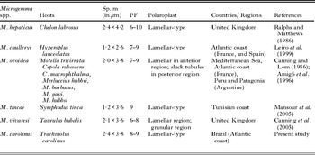
Among Microsporidia, the genus Microgemma is a small group containing 5 species, all of them parasitizing the liver of marine fishes: M. hepaticus, type species, was found in Chelon labrosus caught in the United Kingdom (Ralphs and Matthews, Reference Ralphs and Matthews1986); M. caulleryi in Hyperoplus lanceolatus on the French and Spanish Atlantic coast (Leiro et al. Reference Leiro, Paramá, Ortega, Santamarina and Sanmartin1999); M. ovoidea in different hosts from the Mediterranean Sea, Peru and Patagonia (Canning and Lom, Reference Canning and Lom1986; Amigó et al. Reference Amigó, Salvadó, Gracia and Vivarès1996); M. tinca in Symphodus tinca captured in Tunisia (Mansour et al. Reference Mansour, Prensier, Jemaa, Hassine, Metenier, Vivarès and Cornillot2005) and M. vivaresi parasitizing Taurulus bubalis in the United Kingdom (Canning and Curry, Reference Canning and Curry2005; Canning et al. Reference Canning, Feist, Longshaw, Okamura, Anderson, Tse and Curry2005).
Microsporidiosis has been found relatively rarely among the Brazilian aquatic fauna when compared to the other geographical areas and especially given the extensive ichthyofauna diversity in Brazil, with about 8000 described species just in the Amazonian region (Cellere et al. Reference Cellere, Cordeiro and Adriano2002). Two new genera and species, Amazonspora hassar in the gills of the species Hassar orestis (Azevedo and Matos, Reference Azevedo and Matos2003) and Potaspora morhaphis in the coelomic cavity of Potamorhaphis guianensis (Casal et al. Reference Casal, Matos, Teles-Grilo and Azevedo2008) from Amazonian fauna have recently been described. Four other microsporidian species from the same region have also been described: Loma myrophis in the subepithelial gut tissue of Myrophis platyrhynchus (Azevedo and Matos, Reference Azevedo and Matos2002); L. psittaca in the intestinal mucosa of Colomesus psittacus (Casal et al. Reference Casal, Matos, Teles-Grilo and Azevedo2009); Kabatana rondoni in the skeletal muscle of the abdominal cavity of Gymnorhamphichthys rondoni (Casal et al. Reference Casal, Matos, Teles-Grilo and Azevedo2010); and Microsporidium brevirostris in the skeletal muscle adjacent to the abdominal cavity of the teleost fish Brachyhypopomus brevirostris (family Hipopomidae) (Matos and Azevedo, Reference Matos and Azevedo2004).
In this paper, we described a new microsporidian species through morphological and ultrastructural observations. Phylogenetic relationships to other fish infecting microsporidian species, based on molecular data of SSU rRNA gene were also inferred.
MATERIALS AND METHODS
Fish, location of infection and prevalence
Thirty specimens of the marine fish Trachinotus carolinus Linnaeus, 1766 (Teleostei, Carangidae) (Florida pompano; Brazilian common name ‘pampo’), were collected between June and August 2010 on the Atlantic coast of the ‘Barra da Lagoa’ Beach (27°34′S/48°25′W), near the city of Florianópolis, Santa Catarina State, Brazil. The specimens were anaesthetized by MS 222 and later measured (12–25 cm in length). The parasitosis was detected by the presence of xenomas located only in the liver which contained spores identified as being of a microsporidian species.
Electron microscopy
For transmission electron microscopy (TEM), small fragments of the infected liver were fixed in 3% glutaraldehyde in 0·2 M sodium cacodylate buffer (pH 7·4) for 12 h at 4 °C, washed overnight in the same buffer at 4 °C and post-fixed in 2% OsO4 buffered in the same solution for 3 h at the same temperature. After dehydration in an ascending ethanol series and propylene oxide, the fragments were embedded in Epon. The semithin sections were stained with methylene blue-Azure II for light microscopy. The ultrathin sections were contrasted with both aqueous uranyl acetate and lead citrate and observed with a JEOL 100CXII TEM operated at 60 kV.
DNA isolation and PCR amplification
The xenomas were dissected from the fish, following homogenization, in order to isolate the spores and these were then stored in 80% ethanol at 4 °C. The genomic DNA of about 5 × 10⁶ spores was extracted using a GenElute™ Mammalian Genomic DNA Miniprep Kit (Sigma) following the manufacturer's instructions for animal tissue, except for the incubation time. The DNA was stored in 50 μl of TE buffer at −20 °C until used. The majority of the region coding for the small subunit (SSU) rRNA gene was amplified by PCR using the primers V1f (5′CACCAGGTTGATTCTGCC3′) and 1492r (5′GGTTACCTTGTTACGACTT3′) (Vossbrinck et al. Reference Vossbrinck, Baker, Didier, Debrunner-Vossbrinck and Shadduck1993; Nilsen, Reference Nilsen2000). PCR was carried out in 50 μl reactions using 10 pmol of each primer, 10 nmol of each dNTP, 2 mM of MgCl2, 5 μl 10X Taq polymerase buffer, 1·25 units Taq DNA polymerase (Invitrogen products), and 3 μl of the genomic DNA. The reactions were run on a Hybaid PxE Thermocycler (Thermo Electron Corporation, Milford, MA, USA). The amplification program consisted of 94 °C denaturation for 5 min, followed by 35 cycles of 94 °C for 1 min, 50 °C for 1 min and 72 °C for 2 min. A final elongation step was performed at 72 °C for 10 min. Then, 5 μl aliquots of the PCR products were electrophoresed through a 1% agarose 1X Tris-acetate-EDTA buffer (TAE) gel stained with ethidium bromide.
DNA cloning and sequencing
PCR products for the SSU gene with an approximate size of 1400 bp were obtained by excising the band. Before cloning, it was purified with NucleoSpin Extract II (Macherey-Nagel). Two samples of DNA from different extractions of DNA genomic were cloned into a pGEM-T Easy Vector System II (Promega). The JM109 competent cells, high efficiency (Promega) were transformed and then 3 positive clones were selected using the blue-white colour screening method. The minipreps were carried out with a NucleoSpin Plasmid (Macherey-Nagel), the inserts cloned were confirmed by digestion with the restriction enzyme EcoRI (Promega) and were then sequenced in both directions with the universal sequencing primers T7 forward/SP6. Sequencing was done using BigDye Terminator v1.1 of Applied Biosytems Kit and the sequence reactions were run on an ABI3700 DNA Analyzer (Perkin-Elmer, Applied Biosystems).
Distance and phylogenetic analysis
The various forward and reverse sequence segments were aligned and corrected manually with Clustal W (Thompson et al. Reference Thompson, Higgins and Gilson1994) using MEGA 5 software (Tamura et al. Reference Tamura, Peterson, Peterson, Stecher, Nei and Kumar2011), and ambiguous bases were clarified using corresponding ABI chromatograms. To evaluate the relationship of Microgemma carolinus n. sp. to other microsporidians, a similarity search was performed using the BLAST program. We used 21 rDNA sequences belonging to the microsporidian species that parasitize fish species. The sequence and NCBI Accession number data obtained from GenBank are as follows: Kabatana takedai (AF356222); Kabatana newberryi (EF202572); Kabatana rondoni (FJ843105); Kabatana seriolae (AJ295322); Kabatana sp. (EU682928); Microgemma caulleryi (AY033054); Microgemma tincae (AY651319); Microgemma vivaresi (AJ252952); Microsporidium milevae (EF990668); Microsporidium GHB1 (AJ295324); Microsporidium sp. RSB1 (AJ295323); Microsporidium sp. SH (EU871680); Potaspora morhaphis (EU534408); Spraguea americana (AF056014); Spraguea americana (AY465876); Spraguea lophii (AF104086); Spraguea lophii (AF033197); Spraguea lophii (AF056013); Spraguea gastrophysus (GQ868443); Spraguea sp. (AB623034); Tetramicra brevifilum (AF364303). Nucleospora salmonis (U78176) was used as outgroup.
Alignment using Clustal W in MEGA 5 software with an opening gap penalty of 10 and a gap extension penalty of 4 for both pairwise and multiple alignments was performed. Subsequent phylogenetic and molecular evolutionary analyses were also conducted in MEGA 5, being the distance estimation for the most similar microsporidian carried out using the Kimura-2 parameters model distance matrix for transitions and transversions.
For the phylogenetic tree reconstructions, we included all 21 sequences selected, as well as the outgroup sequence. The maximum parsimony (MP) analysis was performed using the close neighbour interchange heuristic option with a search factor of 2 and a random initial trees addition of 2000 replicates. The Neighbor-Joining (NJ) method was performed using Kimura-2 parameters as a substitution model (Saitou and Nei, Reference Saitou and Nei1987) while for the Maximum Likelihood (ML) method the GTR substitution model was used (Guindon and Gascuel, Reference Guindon and Gascuel2003). Clade support was assessed with bootstrapping of 100 replicates.
RESULTS
Morphological description of the parasite
Light microscopy
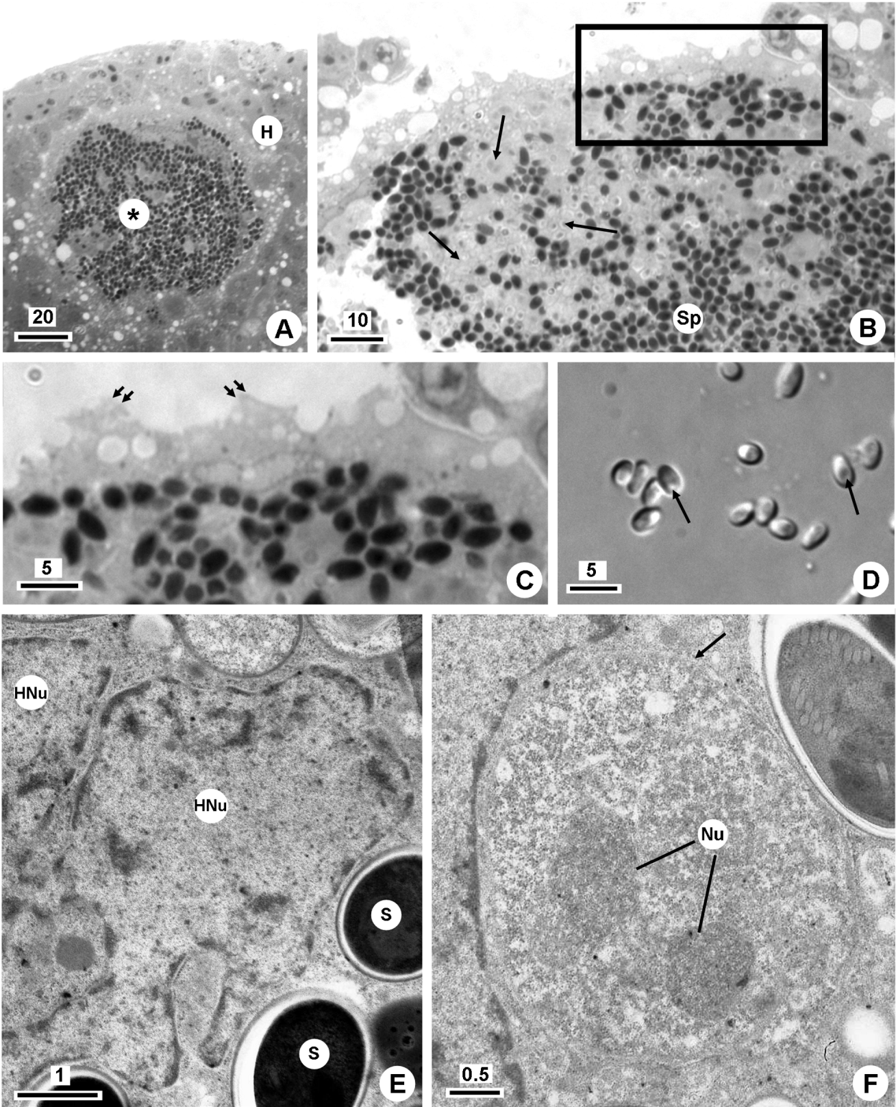
Several xenomas (up to ∼1500 μm in diameter) were observed distributed randomly in the liver parenchyma (Fig. 1A). They contained different developmental stages, with mainly mature spores in the central region and merogonic and sporogonic stages at the periphery of the xenoma (Fig. 1B). Near the plasmalemma the cytoplasm presented some vacuolization and, parallel to it. several elongated ramifications of the hypertrophic nucleus were observed. The surface of the xenoma had a very irregular outline as a consequence of the differentiation of numerous small projections (Fig. 1C). Encapsulations by fibroblasts and/or collagen layers were not observed.
(A–F) Light and transmission electron micrographs of Microgemma carolinus n. sp. infecting the liver of the marine teleost fish Trachinotus carolinus (Scale bars in μm). (A) Semi-thin section of a xenoma (*), containing numerous spores, located in the liver parenchyma. Host (H). (B) Semi-thin section of the periphery of a xenoma showing different developmental stages (arrows) and mature spores (Sp). The boxed area is enlarged in (C). (C) Detail of the xenoma membrane with some small projections (double arrows). (D) Fresh spores released from the xenoma observed in DIC, showing the pyriform to ellipsoidal shape and their prominent posterior vacuole (arrows). (E) Ultrathin section of a xenoma showing an irregular and hypertrophic nucleus (HNu) and some spores (S) in direct contact with the host-cell cytoplasm. (F) Ultrathin section of a merogonial plasmodium showing 2 nuclei (Nu) and a cisterna of the RER surrounding it.

Based on the morphological aspects of the spores (Fig. 1D) and the developmental stages the parasite described in the present paper was identified as belonging to the phylum Microsporidia.
Ultrastructural observations
The developmental stages including the meront, sporont and sporoblast cells are described. The merogonial plasmodium of round to ovoid shape was characterized as a multinucleate cell with unpaired nuclei. The cytoplasm, containing numerous free ribosomes, and the plasmalemma, was in close contact with a cisterna of the rough endoplasmic reticulum (RER) of the host cell (Fig. 1F). Near to the meronts several ramifications of the host nucleus, presenting masses of peripheral heterochromatin, compared to the nuclei of the different developmental stages of the parasite, were distinguished (Fig. 1E and F).
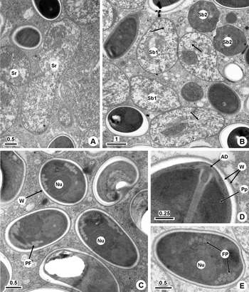
Gradually the membrane of the merogonic plasmodium acquired a densification by the deposition of amorphous material and then this cell transformed into a sporogonic plasmodium (Fig. 2A). The cytoplasm of these cells (i.e. sporoblast mother cells) had more density than that of the meront cells, giving rive to sporoblasts through a process of plasmotomy. These cells exhibited irregular contours, gradually differentiating into numerous membranes, usually as arrays of flattened sacs or as small vesicles. The sporoplasm became dense, and the endospore (the internal portion of the wall) became more evident as the typical organelles of the spores were formed (Fig. 2B).
(A–E) Transmission electron micrographs of Microgemma carolinus n. sp. infecting the liver of the marine teleost fish Trachinotus carolinus (Scale bars in μm). (A) Ultrathin section of the sporonts (Sr). (B) Some sporoblasts in different developmental stages. Younger sporoblasts (Sb1) in differentiation showing numerous cisternae of the Golgi (arrows) and older sporoblasts (Sb2). (C) Spores sectioned at different planes showing the wall (W), polar filament (F) and the nucleus (Nu). (D) Detail of the anterior region of a spore showing the wall (W) composed of 2 distinct layers (exospore and endospore), anchoring disc (AD) and polaroplast (P). (E) Ultrastructural detail of the polar filament coils (PF) and the nucleus at a central position of the spore (Nu).

Systematic position
Phylum Microsporidia Balbiani, 1882; Class Marinosporidia Vossbrinck and Debrunner-Vossbrinck, Reference Vossbrinck and Debrunner-Vossbrinck2005; Family Tetramicridae Matthews and Matthews, Reference Matthews and Matthews1980; Group IV according to the classification proposed by Lom and Nilsen (Reference Lom and Nilsen2003); Genus Microgemma Ralphs and Matthews, Reference Ralphs and Matthews1986.
Description of the species
Name:Microgemma carolinus n. sp.
Type host:Trachinotus carolinus (Teleostei, Carangidae) with 12–25 cm in length on average.
Type locality: Atlantic coast of the ‘Barra da Lagoa’ Beach (27°34′S/48°25′W), near the city of Florianópolis, Santa Catarina State, Brazil.
Site of infection: Xenomas located in the liver.
Prevalence of infection: 30% (9/30) (6/19 females, 3/11 males).
Type specimens: One slide containing mature free spores of the hapantotype was deposited in the International Protozoan Type Slide Collection at the ‘Instituto Nacional de Pesquisa da Amazônia’ (INPA), situated in the city of Manaus (Amazonia State), Brazil, under the ‘INPA’ no. 002/12.
Etymology: the epithet name ‘carolinus’ derives from the species epithet of the host species (Trachinotus carolinus).
Description of the spore: Numerous mature spores, slightly pyriform to ellipsoidal with rounded ends and measuring 3·8 ± 0·4 × 2·4 ± 0·4 μm (n = 50), were observed (Fig. 1D). The spores were uninucleate and the nucleus (∼0·9 μm in diameter) occupied a central position between the apical polaroplast and the basal vacuole (Fig. 2C and E). The polaroplast was lamellate and bipartite, with the elements of distal position somewhat dilated (Fig. 2D). The isofilar polar filament, measuring ∼0·1 μm (n = 25) in diameter, exhibited an angle of tilt of about 45° (n = 10) and posteriorly consisted of packed single or double layer coils with 8–9 turns (Fig. 2E). The posterior vacuole occupied about one third of the total spore length (Fig. 1D). The spore wall, measuring 85 ± 12 nm (n=50), exhibited 2 layers: an electron-dense exospore of ∼30 nm in width and an electron-lucent endospore of ∼55 nm in width (Fig. 2D, Table 1).
Comparative measurements from Microgemma spp. infecting the liver of marine fishes

Molecular and phylogenetic analysis
An almost complete SSU rDNA gene sequence (incomplete at the 3′ end) of Microgemma carolinus n. sp. was obtained through amplification with the primer pair V1f/1492r. A genetic sequence of 1295 bp in length, with a GC content of 49·4%, after removal of the forward and reverse primers was deposited in GenBank (Accession number JQ085991). A Blast search for similar sequences to the 16S rDNA gene confirmed relationships to other microsporidia that parasitize fishes, with the best score being that for the Microgemma vivaresi (AJ252952).
Previously all sequences that have a fish as host were aligned and the most parsimonious tree showed that Microgemma carolinus n. sp. was positioned in the group IV following the classification of Lom and Nilsen (Reference Lom and Nilsen2003). For a second alignment 21 sequences of the group IV were selected. These sequences belonged to the Kabatana, Microgemma, Potaspora, Spraguea, Tetramicra genera and also included 4 Microsporidium species. Nucleospora salmonis (U78176) was chosen as an outgroup sequence, since this is also a microsporidian that parasitizes fish. After the trimming of the 3′ end SSU rDNA, and the exclusion of the sites which could only be aligned ambiguously, the alignment resulted in 1304 bp.
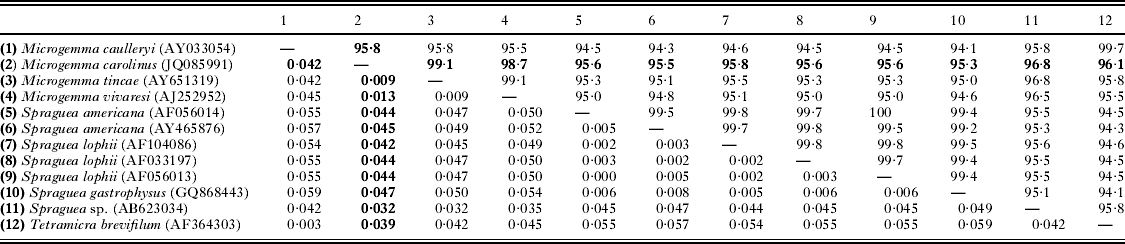
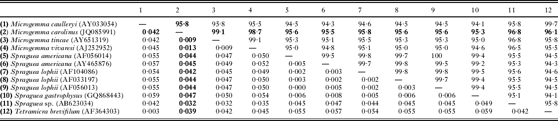
Based on pairwise comparisons among the SSU rRNA gene, the minimum genetic distance (Kimura 2-parameter) was observed with Microgemma tincae (AY651319) and Microgemma vivaresi (AJ252952), 0·9% and 1·3%, respectively. For all other microsporidia, including Microgemma caulleryi (AY033054), the percentage was higher than 3·2% (Table 2).
Comparison of some SSU rDNA sequences: percentage of identity (top diagonal) and pairwise distance (bottom diagonal) obtained by Kimura-2 parameter analysis.
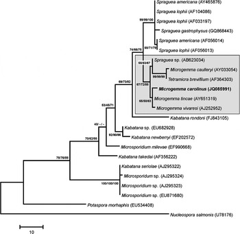
In all phylogenetic analyses (MP, ML, NJ), there was strong evidence for one monophyletic clade, with a bootstrap of 67% (MP), 72% (ML) and 88% (NJ), that included all Microgemma species, another one that also belongs to the family Tetramicridae, i.e. Tetramicra brevifilum and a species classified as Spraguea sp. (Fig. 3).
Maximum parsimony tree of the SSU rDNA sequence of Microgemma carolinus sp. nov. and other selected microsporidian species. The numbers on the branches are bootstrap confidence levels on 100 replicates corresponding to (MP/ML/NJ) trees. GenBank Accession numbers are in parentheses after the species names and the scale is given under the tree. Microgemma carolinus n. sp. is placed within group IV (Lom and Nilsen, Reference Lom and Nilsen2003) that includes the sequences of the genera Kabatana, Microgemma, Potaspora, Spraguea, Tetramicra.

Discussion
The characterization of the parasitosis in freshwater and marine fishes with high commercial value (in this group are included the Trachinotus spp.) is very important in aquaculture. From the observations described above it can be seen that there are a number of morphological characteristics of the parasite presented here that correspond to those recognized in the genera Microgemma Ralphs and Matthews, Reference Ralphs and Matthews1986 and Tetramicra Matthews and Matthews, Reference Matthews and Matthews1980, both included in the family Tetramicridae (Lom and Dyková, Reference Lom, Dyková, Lom and Dyková1992; Lom and Nilsen, Reference Lom and Nilsen2003). These correspondences include the ultrastructural organization of the xenoma wall; aspects of the developmental stages, such as multinucleate meronts dividing by plasmotomy; and, sporogonial plasmodial development in direct contact with the host cell cytoplasm during all stages and producing numerous sporoblasts by budding.
Investigating more closely the comparison with the only species of the genus Tetramicra, some differences were found: such as the locality of the infection, which in T. brevifilum is in the connective tissues throughout the body musculature; the surface of the xenoma has several anastomosed microvillous processes containing ribosomes and microtubules; and finally, the tetrasporoblastic development produces spores with a conspicuous electron-dense inclusion in the posterior vacuole (Matthews and Matthews, Reference Matthews and Matthews1980).
Concerning, the Microgemma species, there are 5 species recognized: M. hepaticus (Ralphs and Matthews, Reference Ralphs and Matthews1986); M. caulleryi (formerly named Glugea caulleryi) (Leiro et al. Reference Leiro, Paramá, Ortega, Santamarina and Sanmartin1999); M. ovoidea (formerly named Microsporidium ovoideum) (Amigó et al. Reference Amigó, Salvadó, Gracia and Vivarès1996); M. tincae (Mansour et al. Reference Mansour, Prensier, Jemaa, Hassine, Metenier, Vivarès and Cornillot2005); M. vivaresi (Canning et al. Reference Canning, Feist, Longshaw, Okamura, Anderson, Tse and Curry2005). All of these share a propensity to infect the parenchyma of the liver of marine fishes (Table 1). In Microsporidia a correlation in the locality of the infection and the identity of the parasite at the level of the genus is frequently observed. For example, the Pleistophora (Canning and Nicholas, Reference Canning and Nicholas1980) and Kabatana (Lom et al. Reference Lom, Dyková and Tonguthai1999) species adopt the muscles as the locality of infection while the Spraguea (Freeman et al. Reference Freeman, Yokoyama and Ogawa2004) and Loma (Casal et al. Reference Casal, Matos, Teles-Grilo and Azevedo2009) species parasitize the central nervous system and the gills, respectively. The Microgemma like other Microsporidia genera is represented by few species. This lack is easily explained because, in general, different stages of development of the life cycle of the parasite are necessary to enable classification at the level of family, genus and species.
When comparing the morphological and ultrastrutural aspects of the Microgemma species several similarities with the species presented here can be observed. These include the organization of the xenoma, which is not encapsulated. The xenoma is delimited by a single membrane, with an extremely irregular outline, containing many small projections similar to microvilli (Canning and Curry, Reference Canning and Curry2005). The xenoma also exhibits a stratified organization with a vacuolated peripheral region free of parasites and rich in mitochondria, as well as a network formed by a hypertrophic nucleus (Ralphs and Matthews, Reference Ralphs and Matthews1986; Amigó et al. Reference Amigó, Salvadó and Gracia1998; Canning and Curry, Reference Canning and Curry2005). A cisterna of the RER surrounding the merogonic stages is another common characteristic (Canning et al. Reference Canning, Feist, Longshaw, Okamura, Anderson, Tse and Curry2005; Mansour et al. Reference Mansour, Prensier, Jemaa, Hassine, Metenier, Vivarès and Cornillot2005). On the other hand, there are some minor ultrastructural differences, such as the presence of membranous structures in the cytoplasm of the meront cells exhibited in Glugea caulleryi, a species transferred to the Microgemma genus (Leiro et al. Reference Leiro, Paramá, Ortega, Santamarina and Sanmartin1999); or the absence of the formation of sporoblast mother cells as is verified in M. ovoidea (Amigó et al. Reference Amigó, Salvadó, Gracia and Vivarès1996) and M. vivaresi (Canning et al. Reference Canning, Feist, Longshaw, Okamura, Anderson, Tse and Curry2005). Comparing the morphology of the spore of the species described here only minor differences were found, however, except in relation to M. tincae (Mansour et al. Reference Mansour, Prensier, Jemaa, Hassine, Metenier, Vivarès and Cornillot2005) which has a very elongated shape.
Regarding the host species there are some reports of microsporidians infecting fish belonging to the family Carangidae but there have been no previous studies using LM and TEM observations in the Brazilian aquatic fauna. The species Kabatana (Microsporidium) seriolae has been described in the muscle tissues of the yellowtail species Seriola quinqueradiata, in which it is the causative agent of ‘Beko’ diseases (Egusa, Reference Egusa1982; Lom et al. Reference Lom, Dyková and Tonguthai1999). In the Indian Ocean, Pleistophora carangoidi was found in Carangoides malabaricus (Narasimhamurti and Sonabai, Reference Narasimhamurti and Sonabai1977) and several microsporidia have also been reported in the liver of fish caught in Senegalese waters. Microsporidium chloroscombri was found in Chloroscombrus chrysurus (Toguebaye et al. Reference Toguebaye, Marchand and Faye1989). Microsporidium spp. were also observed in Caranx crysos, C. senegallus, Selene dorsalis and Trachurus trachurus (Faye, Reference Faye1992; Lom, Reference Lom2002). In New Zealand a microsporidian was found in the pericardial cavity and nervous trunks of Trachurus declivis (Lom, Reference Lom2002; Faye, Reference Faye1992).
In tandem with the above ultrastructural and morphological comparisons, phylogenetic analysis further confirmed a strong relationship with the Microgemma genus and especially the species, M. caulleryi (AY033054), and, even more closely, M. tincae (AY651319) and M. vivaresi (AJ252952). These species, and another one also belonging to the same family Tetramicridae, Tetramicra brevifilum (AF364303), formed a clade supported by 67% (MP), 72% (ML), 88% (NJ) bootstrap. Recently an rRNA gene sequence (AB623034) belonging to a microsporidian classified as Spraguea sp., a parasite of the greater amberjack, Seriola dumerili, from Japan was deposited, with the report that it is clustered with those species (Miwa et al. Reference Miwa, Kamaishi, Hirae, Murase and Nishioka2011). This event is unexpected because it is known that Spraguea species formed a monophyletic clade and all of them have lophiid fishes as hosts (Freeman et al. Reference Freeman, Yokoyama and Ogawa2004).
Analysing the pairwise distance between Microgemma species, the smallest genetic distance was observed between M. carolinus and M. tincae (0·9%), similar to M. vivaresi and M. tincae species. Each of these species (M. vivaresi and M. tincae) are parasites described autonomously in the same year, although it seems clear when comparing their ultrastructural aspects that they do indeed represented different species (Canning et al. Reference Canning, Feist, Longshaw, Okamura, Anderson, Tse and Curry2005; Mansour et al. Reference Mansour, Prensier, Jemaa, Hassine, Metenier, Vivarès and Cornillot2005). In all phylogenetic trees, therefore, these 3 species form a distinct cluster supported by 65% (MP), 58% (ML), 63% (NJ) bootstrap.
Regarding M. caulleryi, curiously, the SSU rRNA gene sequence (Cheney et al. Reference Cheney, Lafranchi-Tristem and Canning2000) presents more phylogenetic affinities with T. brevifilum than with other Microgemma species, with a genetic distance of only 0·3%. As previously suggested by Mansour et al. (Reference Mansour, Prensier, Jemaa, Hassine, Metenier, Vivarès and Cornillot2005) the sequencing of the SSU rRNA gene of the type species, M. hepaticus (Ralphs and Matthews, Reference Ralphs and Matthews1986) and M. ovoidea (Amigó et al. Reference Amigó, Salvadó, Gracia and Vivarès1996) could help to clarify the overall taxonomy of this clade in relation to the family Tetramicridae, which comprises 2 genera.
Taken together, the molecular phylogenetic analysis of the SSU rRNA gene sequence of the species observed in this study, in addition to ultrastructural data and host specificity, support the conclusion that this parasite is a new microsporidian species, which is herein named Microgemma carolinus n. sp.
ACKNOWLEDGMENTS
This work has been partly supported by the Eng°. A. Almeida Foundation (Porto, Portugal), ‘CNPq’, ‘CAPES’ (Brazil) and the Visiting Professor Programme of King Saud University (Riyadh, Saudi Arabia). We would like to thank the iconographic work of João and Joana Carvalheiro and the proof-reading assistance of Karl Woodgett. This work complies with the current laws of the countries in which it was performed.