Cyanotic CHD is a heart defect present at birth that results in low blood oxygen levels. There are certain cardiac defects where patients may not have apparent clinical cyanosis because of increased pulmonary blood flow, although they are traditionally classified into cyanotic heart disease with increased blood flow owing to the presence of admixture physiology and developed evident cyansosis with the beginning of pulmonary hypertension. Overall, cyanotic heart defects account for ~25% of all CHDs.Reference Mitchell, Korones and Berendes 1 There are few prospective studies that have demonstrated the different spectrum of cyanotic CHD in neonatal age; however, no previous study has tried to find out the relative spectrum of congenital cyanotic heart disease in overall children of age 0–18 years.Reference Li, Long and Liu 2 This prospective observational study was undertaken to describe the frequency of various forms of congenital cyanotic heart disease diagnosed by echocardiography in children. This study has demonstrated the relative proportion of cases with congenital cyanotic heart disease evaluated in a tertiary cardiac care centre.
Methods
Study period
This was a prospective observational study conducted in a tertiary cardiac care centre in South India over a period of 3 months − June, 2013 to August, 2013.
Inclusion criteria
All patients under 18 years of age who visited paediatric cardiac clinic within this study period of our hospital with clinically suspected congenital cyanotic heart disease were provisionally included in this study. They underwent thorough echocardiographic evaluation − 2D echocardiography and Doppler echocardiography − in our hospital by experts in paediatric echocardiography. After echocardiographic evaluation, those who had definite diagnosis of congenital cyanotic heart disease were finally included in this study. We had also included those patients who had palliative shunt operation or corrective surgery for congenital cyanotic heart disease and came for follow-up during our study period. Those patients who were not clinically cyanosed, although echocardiography revealed cardiac anomaly suggestive of congenital cyanotic heart disease with increased pulmonary blood flow or admixture lesion, were included in this study – such as acyanotic tetralogy of Fallot; total anomalous pulmonary venous drainage without obstruction and so on.
Exclusion criteria
We had excluded those patients who had normal cardia with central or peripheral cyanosis, for example, pulmonary arterio-venous fistula, congestive heart failure, methemoglobinemia and so on. We had also excluded patients with primary or idiopathic pulmonary hypertension and acquired cyanosis from Eisenmenger’s syndrome.
Classification of disease
After final inclusion in this study, patients were classified according to the cardiac defects and age at presentation. We had arbitrarily divided the children into four groups, namely, neonatal period (0–1 month), infancy (1–12 months), preschool age (>1–6 years), and school age/adolescence (>6–18 years).
Investigations
All patients were subjected to investigations such as oxygen saturation, chest X-ray, electrocardiogram, and echocardiography. Few patients underwent cardiac catheterisation before corrective surgery for cardiac malformation. All patients with pulmonary atresia with ventricular septal defect, double outlet right ventricle, ventriculo-arterial discordance, single ventricle, tricuspid atresia, and few cases with tetralogy of Fallot, where branch pulmonary arteries could not be visualised well in echocardiography, underwent cardiac catheterisation. Sometimes, cardiac catheterisation was performed to rule out pulmonary arterio-venous fistula.
Ethics
We have obtained clearance from our institutional ethical committee before beginning the study. Written consent was taken from the attendants of these children.
Statistics
Data were entered in MS excel. Data were analysed by software SPSS version 16. Although we have considered the use of χ2 or Fisher test statistics to compare cyanotic CHD distribution between age groups and cyanotic CHD distribution according to pulmonary blood flow characteristics, it was not possible because of small sample size.
Results
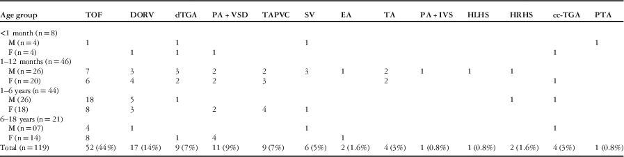
Within the study period, 136 children aged 0–18 years presented with cyanosis, and of them 119 children were diagnosed of having cyanotic CHD through clinical and echocardiographic evaluation. Of the 17 excluded patients, five had Eisenmenger’s syndrome, five patients were diagnosed to have left ventricular failure due to dilated cardiomyopathy, four patients had primary pulmonary hypertension, one patient had congestive heart failure due to neonatal Coarctation, and another two patients had pulmonary arterio-venous fistula. Of the study patients, 68 were male and 60 were female. The various diagnoses of cyanotic CHD in each age group are presented in Table 1.
Distribution of various cyanotic CHD.

DORV=double outlet right ventricle; dTGA=complete transposition of great arteries; HLHS=hypoplastic left heart syndrome; HRHS=hypoplastic right heart syndrome; IVS=intact ventricular septum; L-TGA=congenitally corrected transposition of great arteries; PA=pulmonary atresia; PTA=persistent truncus arteriosus; SV=single ventricle; TA=tricuspid atresia; TAPVC=total anomalous pulmonary venous connection; TOF=tetralogy of Fallot; VSD=ventricular septal defect.
Neonatal period (0–1 month)
We only had eight patients − four male and four female − in this age group. Among the male babies, tetralogy of Fallot, persistent truncus arteriosus, single ventricle with pulmonary valvular stenosis, and transposition of the great arteries were seen in one case each. Among the female babies, double outlet right ventricle with pulmonary valvular stenosis, pulmonary atresia with ventricular septal defect with the major aortopulmonary collateral arteries with increased pulmonary blood flow, transposition of great vessels with large ostium secundum atrial septal defect, increased pulmonary blood flow, congenitally corrected transposition of great vessels − the presence of both atrio-ventricular and ventriculo-arterial discordance − with ventricular septal defect, and pulmonary valvular stenosis were seen in one case each.
Infantile period (1–12 months)
A total of 46 babies − 26 male and 20 female − were presented within this period. Of them, seven male and six female babies had tetralogy of Fallot, which was the most common congenital cyanotic heart disease in this age group. The next common lesion was double outlet right ventricle, which was seen in seven cases. Of the babies, five had transposition of great vessels and another five had total anomalous pulmonary venous connection. Of all the babies mentioned, four had pulmonary atresia with ventricular septal defect and tricuspid atresia was also observed in another four babies; three male babies had single ventricle, and one male baby had Ebstein’s anomaly with decreased pulmonary blood flow and cyanosis. Pulmonary atresia with intact ventricular septum, hypoplastic right-heart syndrome, and hypoplastic left-heart syndrome was observed in one case each among the male babies, and one female baby had cyanosis due to congenitally corrected transposition of great vessels with ventricular septal defect and pulmonary valvular stenosis.
Preschool age group (>1–6 years)
A total of 44 children − 26 male and 18 female − presented in this age group. Of them, 26 – 18 male and 8 female – children had tetralogy of Fallot and its variants such as absent pulmonary valve (one male child), absent left pulmonary artery (two male children), associated Gerbode’s defect (one male child), and so on. Of the children, eight – five male and three female – had double outlet right ventricle, and four of them had total anomalous pulmonary venous connection. Pulmonary atresia with ventricular septal defect was observed in two children. Among the male children, each had transposition of great vessels, hypoplastic right-heart syndrome, and congenitally corrected transposition of the great vessel with ventricular septal defect, and pulmonary valvular stenosis, and another female child had single ventricle with both right ventricular infundibular and pulmonary valvular stenosis.
School age/adolescent (>6–18 years)
A total of 21 patients – 7 male and 14 female – had presented in this age group. Of them, 12 had – four male and eight female – tetralogy of Fallot, which is the most common congenital cyanotic heart disease. Of the eight girls, four had pulmonary atresia with ventricular septal defect and decreased pulmonary blood flow. Among this age group of patients, each patient had double outlet right ventricle, Ebstein’s anomaly, transposition of great vessels with ventricular septal defect and pulmonary valvular stenosis, congenitally corrected transposition of great vessel with ventricular septal defect, and pulmonary valvular stenosis.
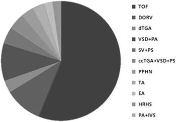
Distribution of cases according to the diagnosis of congenital cyanotic heart disease with decreased pulmonary blood flow (Fig 1)
Bar diagram showed the distribution of cases according to the diagnosis of cyanotic CHD with decreased pulmonary blood flow. ccTGA=congenitally corrected transposition of great arteries; DORV=double outlet right ventricle; dTGA=complete transposition of great arteries; EA=Ebstein anomaly; HRHS=hypoplastic right heart syndrome; IVS=intact ventricular septum; PA=pulmonary atresia; PPHN=persistent pulmonary hypertension in newborn; PS=pulmonary stenosis; SV=single ventricle; TA=tricuspid atresia; TOF=tetralogy of Fallot; VSD=ventricular septal defect.

Of the total number of our study patients, 87 (73%) had cyanotic CHD with decreased pulmonary blood flow. Tetralogy of Fallot (60%) was the most common cause of cyanotic CHD with decreased pulmonary blood flow. Of the 52 cases with tetralogy of Fallot in this study, two had acyanotic tetralogy of Fallot, six (12%) had associated atrial septal defect, four (8%) cases had right-sided aortic arch, two patients had tetralogy of Fallot with an absent pulmonary valve, (4%) and another three patients (6%) had an absent left pulmonary artery. Of the patients, nine (10%) with double outlet right ventricle had decreased pulmonary blood flow, and of these cases, two had situs inversus with dextrocardia and two had right-sided aortic arch. Of the patients with transposition of great vessels, three (3%) had decreased pulmonary blood flow owing to the presence of pulmonary valvular stenosis. Of the patients with pulmonary atresia with ventricular septal defect, nine (10%) had decreased pulmonary blood flow and four had right-sided aortic arch (44%). In all, four patients with single ventricle (4%), four patients with congenitally corrected transposition of great vessels and ventricular septal defect with pulmonary valvular stenosis (4%), three patients with tricuspid atresia (3%), two patients with Ebstein’s anomaly (2%), two patients with hypoplastic right heart syndrome (2%), and one patient with pulmonary atresia with intact ventricular septum (1%) had decreased pulmonary blood flow. All single ventricle patients in our study had left ventricular morphology and were associated with pulmonic or sub pulmonic stenosis. Of the four cases with congenitally corrected transposition of great vessel and ventricular septal defect with pulmonary valvular stenosis, three had either dextrocardia or mesocardia (75%).
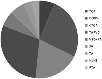
Distribution of cases according to the diagnosis of congenital cyanotic heart disease with increased pulmonary blood flow (Fig 2)
Bar diagram showed the distribution of cases according to the diagnosis of cyanotic CHD with increased pulmonary blood flow. DORV=double outlet right ventricle; dTGA=complete transposition of great arteries; HLHS=hypoplastic left heart syndrome; PA=pulmonary atresia; PTA=persistent truncus arteriosus; SV=single ventricle; TA=tricuspid atresia; TAPVC=total anomalous pulmonary venous connection; TOF=tetralogy of Fallot; VSD=ventricular septal defect.

Only 32 (27%) of our study patients with cyanotic CHD had increased pulmonary blood flow. Of these cases, two patients with tetralogy of Fallot (6%) had no persistent cyanosis, although they had history of cyanosis while crying. We had included these cases as they had similar cardiac malformations as what is seen in classic tetralogy of Fallot and needed similar intracardiac correction; of the 32 patients, eight patients with double outlet right ventricle (25%), six patients with transposition of great vessels (19%), nine patients with total anomalous pulmonary venous connection (28%), two patients with pulmonary atresia with ventricular septal defect (6%), two patients with single ventricle (6%), one patient with tricuspid atresia (3%), and one case each with hypoplastic left heart syndrome (3%) and persistent truncus arteriosus (3%) had increased pulmonary blood flow. Of the patients with pulmonary atresia with ventricular septal defect and single ventricle, two patients each had cardiac catheterisation demonstrating the presence of a large patent duct and other aortopulmonary collaterals. The patients with tricuspid atresia had ventriculo-arterial discordance with increased pulmonary blood flow. Of the eight cases with double outlet right ventricle, one patient had Taussig–Bing anomaly (12%), and of the six cases with transposition of great vessels two patients (33%) had intact ventricular septum. Of the nine cases with total anomalous pulmonary venous connection, five cases had supracardiac type (55%), two cases had cardiac type (22%), and another two cases had mixed-type − both supracardiac and cardiac − total anomalous pulmonary venous connection (22%). Although non-obstructive total anomalous pulmonary venous connection is classically presented with features of heart failure and cyanosis developed if they had pulmonary hypertension, and as all of these nine patients presented in their later infancy and early childhood, they had presented with minimal cyanosis − oxygen saturation ranges from 85 to 90% − and mild-to-moderate pulmonary hypertension at the time of presentation.
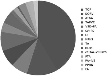
Figure 3 demonstrated the overall proportion of congenital cyanotic heart disease in our study population. Tetralogy of Fallot and its variants were the most common type (44%) of cyanotic CHD in our patients followed by double outlet right ventricle (14%), transposition of great vessels (7%), total anomalous pulmonary venous connection (7%), pulmonary atresia with ventricular septal defect (9%), single ventricle with pulmonary stenosis (5%), Ebstein’s anomaly (1.6%), hypoplastic right-heart syndrome (1.6%), tricuspid atresia (3%), hypoplastic left-heart syndrome (0.8%), congenitally corrected transposition of great vessel with ventricular septal defect with pulmonary valvular stenosis (3%), persistent truncus arteriosus (0.8%), and finally pulmonary atresia with intact ventricular septal defect (0.8%).
Bar diagram demonstrated overall prevalence of cyanotic CHD in our study population. ccTGA=congenitally corrected transposition of great arteries; DORV=double outlet right ventricle; dTGA=complete transposition of great arteries; EA=ebstein anomaly; ES = eisenmenger's syndrome; HLHS=hypoplastic left heart syndrome; HRHS=hypoplastic right heart syndrome; IVS=intact ventricular septum; PA=pulmonary atresia; PPHN=persistent pulmonary hypertension in newborn; PTA=persistent truncus arteriosus; PS=pulmonary stenosis; SV=single ventricle; TA=tricuspid atresia; TAPVC=total anomalous pulmonary venous connection; TOF=tetralogy of Fallot; VSD=ventricular septal defect.

Discussion
Prevalence of CHD ranges from 3.7 to 17.5/1000 live births. Cyanotic heart defects account for ~25% of all CHDs.Reference Ferencz, Rubin and McCarter 3 , Reference Bolisetty, Daftary, Ewald, Knight and Wheaton 4 All cyanotic CHDs fall in the category of severe CHD.Reference Hoffman and Kaplan 5 They could be divided into two groups depending on the pulmonary blood flow. Patients who have congenital cyanotic heart disease with decreased pulmonary blood flow are called as having tetralogy of Fallot physiology and those who have congenital cyanotic heart disease with increased pulmonary blood flow are called as having transposition of great arteries physiology.Reference Humayun and Atiq 6 Echocardiography is the initial diagnostic test of choice to diagnose CHDs, and here in this study we have used it for diagnostic confirmation of congenital cyanotic heart disease.Reference Sadoh, Uzodimma and Daniels 7 – Reference Akhtar and Ahmed 9 Of our study patients, 73% had tetralogy of Fallot physiology, whereas 27% patients had transposition of great arteries physiology. Presentation and management are different among these groups of patients.Reference Shah, Singh, Pandey, Kalakheti and Bhandari 10
Within the last 3 months, 136 children presented with intermittent or persistent peripheral or central cyanosis. Of them, 119 patients who had confirmed cyanotic CHD through echocardiographic evaluation were included in this study. Similar to previous studies, we also found that tetralogy of Fallot and its variant were the most common type, with proportion of 44% among all congenital cyanotic heart disease, and when we considered in the group of patients with congenital cyanotic heart disease and decreased pulmonary blood flow, tetralogy of Fallot comprised 60%.Reference Waldman and Wernly 11 Tetralogy of Fallot with absent pulmonary valve was seen in 4% of patients with tetralogy of Fallot. Double outlet right ventricle was the next common congenital cyanotic heart disease with prevalence of 14%, and among them, 9/17 patients had tetralogy of Fallot physiology and rest (8/17) had transposition of great arteries physiology. Pulmonary atresia with ventricular septal defect was seen in 9% of cases and among them 44% had right-sided aortic arch and this finding was also similar to the previous studies. Both transposition of great vessels and total anomalous pulmonary venous connection were seen in 7% of our study patients. However, in our study, total anomalous pulmonary venous connection was the most common cause of congenital cyanotic heart disease with increased pulmonary blood flow followed by double outlet right ventricle rather than transposition of great vessels, and this finding was not similar with the previous studies.Reference Grifka 12 Although we had come across all forms of congenital cyanotic heart diseases, few congenital cyanotic heart diseases, which occurred right after birth such as hypoplastic left heart syndrome, transposition of great vessels, persistent truncus arteriosus, tricuspid atresia, pulmonary atresia with intact ventricular septum and so on, were seen less frequently in this study.
Limitations of the study
We have several limitations in this study. First, as we had conducted this study in a tertiary cardiac care centre, we did not get true epidemiological spectrum of different congenital cyanotic heart diseases in the community. Second, as we did not have obstetrics and neonatal department in our hospital, congenital cyanotic heart disease, which occurred right after birth and which needed to be addressed immediately after birth such as transposition of great vessels, hypoplastic left heart syndrome and so on, were seen less frequently, and this could modify the data presented in this study. As a previous study showed that 35–40% neonates with cyanotic CHD expired during their hospital stay, epidemiological data of cyanotic CHD will be different from our data.Reference Humayun and Atiq 6
As our centre is the largest tertiary care centre in our country, and it is the only governmental tertiary-level cardiac care hospital in the state where it is situated, and it also caters population from other part of our country, we are used to get good numbers of congenital cyanotic heart disease to assess the proportion of different congenital cyanotic heart disease in the community. We need to prioritise the foetal and early neonatal diagnosis of cyanotic CHD through foetal echocardiography, neonatal oxygen saturation testing, neonatal echocardiography, and colour Doppler screening as majority of the neonates with congenital cyanotic heart disease showed survival with appropriate management.Reference Li, Long and Liu 2 , Reference Humayun and Atiq 6 , Reference Reich, Miller and Brogdon 13 Although we had conducted this study over a short period of time, we observed that this trend was similar in all these 3 months.
Conclusion
Although this study did not provide any novel data, this study was relevant in presenting spectrum of different congenital cyanotic heart diseases in Indian children (0–18 years) so that we can assess the burden created by them; at the same time, we must be familiar with the echocardiographic diagnosis and management of all complex congenital cyanotic heart disease. In this study, tetralogy of Fallot was the most common diagnosis in patients with congenital cyanotic heart disease and decreased pulmonary blood flow, whereas total anomalous pulmonary venous connection was the most common diagnosis in patients with congenital cyanotic heart disease and increased pulmonary blood flow.
Acknowledgements
S.P. planned the study, designed the protocol, collected and analysed the data, and drafted the manuscript. U.M.K., J.M., and A.S. were involved in the diagnosis of the patients. J.M., R.K.S., and M.C.N. corrected the manuscript. All authors approved the final version of this manuscript. We acknowledge the patients and their parents who have given their full consent for the publications of the images and data.
Financial Support
This research received no specific grant from any funding agency, commercial, or not-for-profit sectors.
Conflicts of Interest
None.
Ethical Standards
Ethical clearance was received from the Institutional Ethical Committee before starting this study and an appropriate standard was maintained throughout the study.