

INTRODUCTION
The family Sertulariidae is probably the largest within Hydrozoa, with about 500 species according to Bouillon et al. (Reference Bouillon, Gravili, Pagès, Gili and Boero2006). It is well known for its colony structure and hydrotheca morphology as taxonomic classification is mainly based, from the very beginning, on skeletal characters of the perisarc (i.e. Naumov, Reference Naumov1960; Millard, Reference Millard1975). Besides, the scarcity of data on the coenosarc is striking, except for the presence of a caecum, considered of great taxonomic importance from Broch (Reference Broch1918), and of a mantle, though not systematically investigated by the author. Hydranth morphology is still largely unknown because it has to be studied on living colonies. Indeed, the ability of hydranths to withdraw inside the hydrotheca for protection, which is evident upon fixation of the animal for preservation, prevents observation. Differences between genera and species are then unrecognized (personal observations). Two rare sertulariid hydranth features however are already described, both of them nematocyst-bearing organs. The first is a tongue-shaped structure called ligula by Thornely (Reference Thornely and Herdman1904) for the species he named Sertularia ligulata. The second was described by Billard (Reference Billard1925) as a single abcauline ‘racket-shaped’ mass of cnidocysts in Salacia tetracythara Lamouroux, Reference Lamouroux1816 and Salacia hexodon (Busk, Reference Busk and MacGillivray1852). Billard, who observed only preserved and contracted hydranths, did not give a special name to this structure and said that it could be a modified tentacle situated within the circle of tentacles. Bouillon et al. (Reference Bouillon, Gravili, Pagès, Gili and Boero2006), in their glossary, define the term ligula as ‘an extensible outgrowth armed with cnidocysts from the base of the adcauline side of hydranths in some Salacia and Sertularia (a nematophore?)’. This mixes the two features into a single one and misses the difference in location—abcauline for the organ described by Billard for the Salacia species, adcauline for the ligula. In contrast to Cornelius (Reference Cornelius, Barnes and Crothers1995), for whom ligula and nematophores are separated, they also question the appropriateness of calling it a nematophore, a term used within the thecates for specialized zooids present in several families but not in the Sertulariidae (Gravier-Bonnet, Reference Gravier-Bonnet2004).
Salacia tetracythara was originally described from Australia and has been reported several times for other locations in tropical and subtropical regions of the Indo-Pacific province (see Rees & Vervoort, Reference Rees and Vervoort1987 for references). Further, the species was reported at Fiji (Gibbons & Ryland, Reference Gibbons and Ryland1989), Sagami Bay, Japan (Hirohito, 1995), Australia (Watson, Reference Watson2000) and Kei Islands, Indonesia (Schuchert, Reference Schuchert2003). However the original description of the hydranth by Billard (Reference Billard1925), from specimens collected in Indonesia (and not from Madagascar as stated by Rees & Vervoort, Reference Rees and Vervoort1987), was never completed or revised, probably because all the authors focused descriptions on skeleton (perisarc).
The present work is the first carried out on living animals of S. tetracythara, a necessary condition for observing soft tissues (coenosarc) and hydranth morphology and behaviour, as already recommended for the study of hydroids (Cornelius, Reference Cornelius1998; Gravier-Bonnet & Bonnet, Reference Gravier-Bonnet and Bonnet2000; Gravier-Bonnet, Reference Gravier-Bonnet2008, this volume), and illustrated by earlier work on sertulariids (Nutting, Reference Nutting1904). The results bring new data on hydranth morphology (presence of a mantle and of a cluster of nematocysts), and corroborate and complete Billard's description of the modified tentacles.
MATERIALS AND METHODS
Materials studied are from several islands of the south-western Indian Ocean belonging to the Comoros (Mohéli, Mayotte) and Mascarene (La Réunion, Rodrigues) archipelagos (Gravier-Bonnet & Bourmaud, unpublished results). Living colonies were sampled by SCUBA diving mainly on coral reefs and on nearby rocky shores from 5 to 30 m. Collected manually, they were put underwater into plastic bags or jars with a large volume of water to prevent soft tissue damage, and then transported inside isothermic containers. The study of living colonies with a stereomicroscope (Leica M420 or M3Z) was undertaken as soon as possible. Skeletal characteristics of each colony allowed unambiguous identification according to existing descriptions in the literature. Additional laboratory studies and measurements were carried out in La Réunion with a compound microscope equipped with Nomarski interference (Nikon Eclipse 80i). Microphotographs were taken with digital cameras (Nikon Coolpix 4500 and DS-Fi1). Video sequences were registered with a Sony Camescope. Specimens were then fixed in 4% formaldehyde in seawater.
SYSTEMATICS
Salacia tetracythara Lamouroux, Reference Lamouroux1816: 214, pl. 6, figures 3a, B–C; Deslongchamps, Reference Deslongchamps1824(*); Bedot, Reference Bedot1901: 462, 499; Stechow, Reference Stechow1922: 150; Stechow, Reference Stechow1923: 214; Billard, Reference Billard1909a: 1064; Billard, Reference Billard1924: 54–55, 66; Billard, Reference Billard1925: 202–204, figure 47, pl. 8, figures 27–28; Dawydoff, Reference Dawydoff1952: 55 (***); Pennycuik, Reference Pennycuik1959: 194; Mammen, Reference Mammen1965: 54, figure 87; Rédier, Reference Rédier1966: 81; Van Praët, Reference Van Praët1979: 891–892, figure 61; Rees & Vervoort, Reference Rees and Vervoort1987: 103–104, figure 6d; Gibbons & Ryland, Reference Gibbons and Ryland1989: 414–415, figure 31; Hirohito, 1995: 183, figure 60a–c; Watson, Reference Watson2000: 23, figure 18A–F; Schuchert, 2005: 181–182, figure 37 Gravier-Bonnet, Reference Gravier-Bonnet, Payri and Richer de Forges2006: 122.
Serialaria tetracythera: Schweigger, Reference Schweigger1820: 426 (**); Bedot, Reference Bedot1901: 462.
Salacia tetracyttara: Lamouroux, Reference Lamouroux1821: 15, pl. 67, figures 7 & 9 (***); Bale, Reference Bale1884: 116–119.
Thuiaria fenestrata Bale, Reference Bale1884: 116–119, pl. 7, figure 7, pl. 9, figure 14; Bale, Reference Bale1888: 773; Kirkpatrick, Reference Kirkpatrick1890: 604; Bale, Reference Bale1894: 103, pl. 4, figure 2; Nutting, Reference Nutting1905: 934, 950; Hartlaub, Reference Hartlaub1905: 617; Bartlett, Reference Bartlett1907: 42, 64, figure; Thornely, Reference Thornely1916: 149.
Calyptothuiaria opposita von Campenhausen, Reference von Campenhausen1896: 312–313, pl. 15, figure 7.
Thuiaria tetracythara: Billard, Reference Billard1909a: 1064; Billard, Reference Billard1909b: 319–320; Bedot, Reference Bedot1925: 450; Leloup, Reference Leloup1937: 5, 44.
Articles not at my disposal, references from: (*) Bale, Reference Bale1884; (**) Bedot, Reference Bedot1901; (***) Rees & Vervoort, Reference Rees and Vervoort1987.
REMARKS ON TAXONOMY
1—the term ‘tetracythara’ was choosen by Lamouroux in reference to a tetramerous structure that was not confirmed further. In the original description—in French and in Latin ‘cellulis terebitus, elongatis, quaternatim coalescentibus; oribus annulatis, quasi verticillatis; ovariis ovoïdeis truncatis’—and on the drawings given by Lamouroux (Reference Lamouroux1816, figure 3a, B, pl. 6), there are four tubes (one not drawn, hidden by the others) ending in identical cups (supposed to be the orifice of the hydrothecae) at the same level, verticillate. This is far from following descriptions of the species that are all near that of the synonym Thuiaria fenestrata Bale (Reference Bale1884). Bale, discussing his new species, cited the commentaries in the Dictionnaire des Zoophytes of Lamouroux et al. (Reference Lamouroux, Bory de St Vincent and Deslongchamps1824) by Deslongchamps on ‘the true structure of Salacia tetracyttara’ after he had studied the collection of Lamouroux. Deslongchamps was the first to remark on the absence of tetramerous verticillate structure in the specimen described by Lamouroux in 1816. Further, Billard (Reference Billard1909b, p. 320; 1925, p. 203), examining the type of Lamouroux, supposed that the presence of triangular spaces below the hydrothecae could have induced an error by Lamouroux who interpreted the specimen as having four hydrothecae in a profile view. Bale (Reference Bale1884, p. 119, footnote) remarked: ‘As the genus Salacia seems to have been founded on characters which had no existence in the type species, Lamouroux’ name has of course no claim to acceptance'. For a while, the species was then cited either as Thuiaria tetracythara or Thuiaria fenestrata, until Billard (Reference Billard1924) re-established the genus Salacia after having re-examined specimens of tetracythara when taking into account new generic characters proposed for sertulariids by Levinsen (Reference Levinsen1913, operculum) and Broch (Reference Broch1918, caecum). He also proposed Calyptothuiaria opposita Von Campenhausen, Reference von Campenhausen1896 as a synonym of S. tetracythara. Then, and until now, all other authors have agreed with Billard.
2—the Sertularia crisioides Lamouroux identified by Busk (Reference Busk and MacGillivray1852: 389) is given by Bale (Reference Bale1884: 117–118) as a synonym of T. fenestrata from a suggestion of Busk himself, and further as a synonym of S. tetracythara. This synonymy seems doubtful because Busk (Reference Busk and MacGillivray1852: 387) classified the species in a group with alternate cells (hydrothecae).
3—Pennycuik (Reference Pennycuik1959) described from Queensland, Australia, a variety differing in hydrotheca length. New findings of fertile specimens, from Australia (Watson, Reference Watson2000) and Indonesia (Schuchert, Reference Schuchert2003), cover the gap she noticed between her material and the typical form.
4—Hirohito (1995) doubtfully assigned his material from Japan to Salacia tetracythara, remarking that it differed slightly from specimens he examined from Australia, Indonesia and the Indian Ocean, which resembled rather that of Billard, with long hydrothecae and racket-shape nematocyst groups. Conversely, specimens from Japan lack nematocyst-bearing organs. They also lack the characteristic triangular ‘window’ beneath the hydrotheca floor and the slight zigzag shape of the stem. Moreover, figures given by Hirohito (1995) show obviously shorter hydrothecae (figure 60a–b) and differing in length/diameter ratio from those from the Indian Ocean (figure 60c). With such differences, specimens from Japan might be assigned to a different species to be described when fertile colonies are found.
MATERIAL EXAMINED
La Réunion—Cap La Houssaye: 21 February 2005, 10–15 m; 16 March 2005, 12–15 m; 1 April 2005, 10 m; 4 August 2005, 10–15 m; 13 January 2006, 10–15 m; 13 April 2007, 12 m. Boucan: 6 June 2001, 12–14 m (Trois Grottes); 23 March 2005, 15–20 m (Tour); 4 July 2005, 30 m (Grandes Gorgones); 27 April 2005, 15–20 m (Tour). Saint-Leu: 17 February 2005, 10 m (Langoustière); 28 October 2006, 30 m (Saliba).
Rodrigues—Le Chaland Sud: 28 November 2006, ROD16, 15 m; Le Chaland Nord: 29 November 2006, ROD32, 25 m; Cotton Bay: 12 June 2006, ROD104, 15–20 m.
Mayotte—Passe en S: 25 October 2006, MAY92, 40–55 m and MAY102, 20–26 m; Saziley: 26 October 2006, MAY142, 12–15 m; 27 October 2006, MAY192, 5–8 m.
Mohéli—Ouallah Un: 18 October 2006, MOH33, 2–6 m; M'Chaco: 21 October 2006, MOH79, 12–18 m.
RESULTS
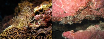
Colonies erect and rigid, in one plane, light brown, up to 10 cm, settled on organic hard substrates on the outer slope of coral reefs and on basaltic cliffs, usually covered with diatoms, microscopic algae and particulate matter, this giving them a characteristic mucky appearance but not affecting the behaviour of the hydranth which remained able to extend and retract inside the hydrotheca (Figure 1). Stem rigid, mostly unbranched or with few branches, often polysiphonic at the base, nodes usually not visible, sub-opposite hydrothecae in two lateral rows with no contact between the rows, three hydrothecae between two successive hydrocladia on one side, hydrocladial apophyses not well demarcated from the hydrocladium (Figure 2A).
Salacia tetracythara colonies (in situ, about 4–5 cm high): on the left long hydranths emerging from the ‘mucky’ skeleton are visible (underwater photographs by D. Caron at La Réunion on the left and C. Bourmaud at Rodrigues on the right).

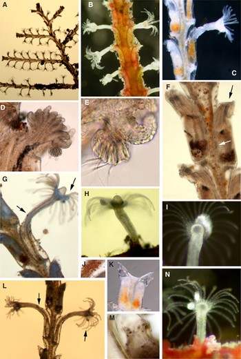
Hydrocladia straight and rigid, strictly in one plane, alternate, of maximal length at median part of stem (then diminishing slightly and regularly from middle towards base and top) (Figures 1 & 2A), nodes almost not visible and usually detected only by a weak diameter constriction of hydrocladia between two successive pairs of hydrothecae, opposite or sub-opposite hydrothecae settled in two lateral rows with no contact, members of one row in contact or not, sometimes slightly overlapping (Figure 2B, C, F, G, L). Coenosarc on the growing tips usually bright orange.
Salacia tetracythara trophosome: (A–C) parts of colony and hydrocladiae with hydranths contracted and in extension (1 cm); (D, E) one modified tentacle on abcauline side of a semi-contracted hydranth, with racket-shaped nematocyst-bearing organ (E: close-up view); (F) hydranths retracted into hydrothecae (0.5 cm); (G, H) profile view of hydranths in extension showing a single modified tentacle and adcauline nematocysts cluster (G): the bilateral symmetric shape in (G) is at rest, the circular one in (H) is during release of waste products; (I) big nematocyst-bearing organ covering the base of several tentacles at a time; (J) bright orange cells in the mantle of hydrocladia coenosarc; (K) profile view of hydrothecae with smooth orifice; (L) a pair of hydranths with columns obviously in two parts, the basal digestive part being swollen by gastrovascular flux on left; (M) mantle tightened under the hydrotheca orifice during hydranth contraction (tips of tentacles on base), with coloured cells; (N) the pair of modified tentacles on a hydranth viewed from underneath. On (F, G) and (L), the arrows indicate the location of adcauline nematocysts clusters and modified tentacles (microphotographs by N. Gravier-Bonnet).

Hydrotheca tubular, almost entirely adnate, adnate adcauline wall straight except distally, ending by a short free adcauline wall slightly curved, abcauline wall straight curving distally, with one small intrathecal abcauline thickening just under the aperture corresponding to operculum hinge, hydrothecal opening untoothed and approximately oval, almost parallel to main axis (Figure 2C, F, G, K, L). Operculum of one valve being an extension of the abcauline wall, bending just underneath the aperture, closing the hydrotheca in an intrathecal location in front of the orifice; pushed out by the hydranth while extending; during extension, remaining in contact with the column and becoming almost invisible; never opening further whereas the column itself remains in contact with the skeleton at the orifice of the hydrotheca, and thus the hydrotheca always remaining closed, even during the extension of the hydranth (Figure 2C, D, G, L).
Mantle a thin layer of ectoderm lining the entire inner side of the skeleton of the colony, including the hydrotheca; at many points continuous with the ectoderm surrounding the coenosarcal tubes inside stems, branches and hydrocladia; also linked to the ectoderm of the hydranth inside the hydrotheca; entirely enclosing hydranths when they are retracted, because wrapped around them (Figure 2M); mainly transparent (Figure 2K) but, in many areas of the stems and hydrocladia, it includes glandular cells with large and deep orange secretive granules (Figure 2J).
Hydranth with no caecum, due to its attachement to the skeleton on hydrotheca floor only (Figure 2G, L); column very extensible, length of extended hydranth two times that of the hydrotheca (1 mm), differentiated into proximal digestive and distal parts differing for both endoderm and ectoderm, bearing a small refringent nematocyst cluster in the middle of the adcauline side, located just above the hydrothecal aperture on extended hydranths (Figure 2F, G); large conical rounded hypostome (Figure 2G, H) surrounded by a crown of 22 to 28 tentacles, two of which are modified by large nematocyst-bearing organs on abcauline side (Figure 2 N); tentacles of regular diameter, rounded at tip, with very small nematocysts dispersed and not prominent; marked bilateral symmetry in extension, obvious during tentacle folding and enhanced by abcauline nematocyst organs and adcauline cluster (Figure 2A, B, G, I, L, N). Column, tentacles and hypostome uncoloured except for white circle at tentacular level (visible in blue-grey on Figure 2G); stomach often exhibiting a deep orange coloration arising from pigmented glandular cells and turnover of waste products (Figure 2C, K).
Nematocyst-bearing organs on ‘modified tentacles’ as one pair per hydranth, both on abcauline side, looking singular in lateral view especially on contracted hydranths, the first hiding the second, with about 2–3 tentacles in between (Figure 2G, H, L, N); racket-shaped, strongly refringent, formed of a bundle of large microbasic mastigophores (Figure 2D, E, N); linked to the base and on the external side of a single tentacle when small (Figure 2D), but the larger ones attached to several tentacles (Figure 2I); much larger than a tentacle: about 40/55 µm on a contracted hydranth; usually with no autonomy but moving during tentacle activity and changing shape slightly, except for a large one, almost rounded in shape and flat, that was seen unfolding very slowly while, consequently, elongating.
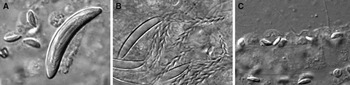
Cnidome: microbasic mastigophores of two kinds very different in shape, size and location (Figure 3A–C). Tentacles: small, pip-shaped, undischarged capsules 5.1–5.8/1.6–2.1 µm; tubule everting longitudinally, shaft a bit longer than capsule (Figure 3C). Nematocyst-bearing organs and nematocysts clusters: large, banana-shaped, capsules 26–28/4.3–4.7 µm (undischarged), 23–26/3.2–4.3 µm (discharged); tubule everting with an angle from 120 to 90°, shaft as long as the capsule, 26/1 µm, enlarging slightly from base to tip and equipped with big spines (Figure 3A, B).
Salacia tetracythara cnidome. (A–C) Microbasic mastigophores nematocysts of two types: (A) undischarged large and small; (B) large discharged; (C) small undischarged and discharged on tentacles (microphotographs taken with compound microscope on more or less squashed preparations by N. Gravier-Bonnet).

DISCUSSION
The discovery of the presence of Salacia tetracythara in the Mascarene (La Réunion and Rodrigues) and Comoros (Mohéli and Mayotte) archipelagos extends the distribution of this tropical Indo-Pacific species to the south-west of the Indian Ocean.
Morphological characters of the trophosome of the specimens studied (colonies, hydrocladiae and hydrothecae) agree with those already described and figured (Bale, Reference Bale1884; Billard, Reference Billard1925; Gibbons & Ryland, Reference Gibbons and Ryland1989; Watson, Reference Watson2000; Schuchert, Reference Schuchert2003). According to the authors, the hydrotheca orifice of S. tetracythara is either said to be toothed (i.e. ‘two obscure lateral and one large pointed adcauline’ for Gibbons & Ryland, Reference Gibbons and Ryland1989) or smooth (Billard, Reference Billard1925; Hirohito, 1995) as in this study. I agree with Billard (Reference Billard1924, Reference Billard1925) who explained clearly that these teeth, previously checked by Bale (Reference Bale1884) for T. fenestrata, are not true teeth but angles made up by the skeleton plates of the hydrotheca wall at the orifice, due to its peculiar non-circular shape (Billard, Reference Billard1925, figure 47P). But it is possible that these angles may be more marked in some specimens (Watson, Reference Watson2000, figure 18D).
This study observes and describes in S. tetracythara for the first time: (i) a mantle lining the whole colony and the hydrothecae; and (ii) an adcauline nematocyst cluster on the hydranth column. According to the literature at my disposal, there is no other such cluster described for a sertulariid. The mantle is noted in several but not all sertulariid species, sometimes under the name ‘ectoderm lining’, and also in thyroscyphids (Millard, Reference Millard1975; see Bouillon et al., Reference Bouillon, Gravili, Pagès, Gili and Boero2006 for a definition). Nutting (Reference Nutting1904) recalled that Hartlaub (Reference Hartlaub1901) suggested that the hydrotheca was lined by an epithelial membrane in Sertularella species, but he did not succeed in finding it. As presence of the mantle seems not to be a constant feature within a single genus, according to genera diagnosis in use presently, it has never been used for classification. However, it is probably an important character to take in consideration to separate groups of species, maybe at generic level. What is the function of the mantle, and what functional difference its presence or absence implies for an individual is unknown.
Present results on S. tetracythara hydranths confirm the presence in the species of abcauline modified tentacles as interpreted by Billard (Reference Billard1925). These results pinpoint their location and content (microbasic mastigophores), and reveal the presence of two per hydranth instead of one, these tentacles imparting strong bilateral symmetry to the hydranth. Identical to all others in shape, size, and structure, the modification they exhibit is a basal connection to a nematocyst-bearing organ. The width and length of one organ are variable, probably related to the number of large nematocysts inside. In consequence, when it reaches a large size, it is connected not to a single tentacle, very thin at the base, but to several at once. There are then several modified tentacles per hydranth and two large nematocyst-bearing organs suspected to grow enough to join in a single big one (Figure 2I). The behaviour of modified tentacles appears to be unaffected by the presence of nematocyst-bearing organs. Observed by light microscopy, these organs look like clusters of nematocysts, but they probably are true organs. Indeed, on video sequences read at low-speed, a large one was recorded slowly unfolding, and then reaching about twice its former length, as if it had been enrolled on itself when at rest. This obscure behaviour was checked once only though hydranths were studied several times. It has to be confirmed by additional observations.
Among the family Sertulariidae, the single tentacle with large nematocysts noted for Salacia hexodon (Busk, Reference Busk and MacGillivray1852) by Billard (Reference Billard1925) and Schuchert (Reference Schuchert2003), but not by Watson (Reference Watson2000), looks like the modified tentacles of S. tetracythara for its shape and location. Future studies of living specimens should confirm if it is really single or paired as in S. tetracythara. Another peculiar feature concerning tentacles is a smaller structure—perhaps composed of nematocysts—that was described for Sertularia exerta Allman, Reference Allman1888, as a little cushion-like prominence at the base of every tentacle (Allman, Reference Allman1888). Within the Haleciidae, modified tentacles characterizing the genus Nemalecium, the nematodactyls, are twin and not symmetrically disposed within the tentacle circle as those of S. tetracythara, but large nematocysts are distributed along their whole length (Bouillon, Reference Bouillon1986) instead of being gathered separately at the base. They are strictly defined as modified tentacles since the presence of big nematocysts of a special type (pseudostenoteles) goes with a change of their shape, size and behaviour.
The ligula, as described for Sertularia loculosa Busk, Reference Busk and MacGillivray1852, differs from the modified tentacles of S. tetracythara: (i) it does not belong to the hydranth but originates from the mantle; (ii) it has an adcauline position; (iii) there is only one per hydrothecal unit; and (iv) is very extensible (Migotto, Reference Migotto1996). Consequently, they have not to be confused, as in the definition cited in the introduction (Bouillon et al., Reference Bouillon, Gravili, Pagès, Gili and Boero2006). ‘Nematocyst batteries’ is a vivid term used by Millard (Reference Millard1975) to describe nematocyst-bearing organs in the sertulariids (which at that time included thyroscyphids). This general term is useful in gathering together different sorts of nematocyst groupings, and authors still use it nowadays (Bouillon et al., Reference Bouillon, Gravili, Pagès, Gili and Boero2006). Millard (Reference Millard1975, figure 81C) represented the annular fold (a special shape of the mantle) of the genus Thyroscyphus as linked to the hydranth column at its base and equipped with two nematocyst batteries. Recently Schuchert (Reference Schuchert2003) noted a pad containing a dense cluster of large nematocysts on the mantle of Thyroscyphus torresi (Busk, Reference Busk and MacGillivray1852). The single extensible ligula of Sertularia ligulata Thornely, Reference Thornely and Herdman1904, has such a location. It was doubtfully termed a dactylozoid by Billard (Reference Billard1925) and a nematophore by Bouillon et al. (Reference Bouillon, Gravili, Pagès, Gili and Boero2006). Different terms used by authors for the same part highlight problems of terminology encountered in the specialized hydrozoan vocabulary and illustrate the need for more research on the anatomy and function of structures that have been most often described from fixed material.
In a literal sense, the term nematophore (i.e. bearing nematocysts) could be used for the ligula and for the different nematocyst-bearing organs. But in thecates, nematophores are independent of hydranths and of hydrothecae. They are modified polyps, like the dactylozoids of athecates, whereas ligulae and nematocyst-bearing organs are specialized organs adjunct to the polyp and its extension (the mantle). In a recent revision, the author demonstrated that the so-called nematophores are an artificial grouping to be separated into three types differing in structure and function (Gravier-Bonnet, Reference Gravier-Bonnet2004). Though probably evolved separately, they all share a protective/defensive function, as does the adcauline nematocyst cluster of S. tetracythara, whereas some nematophores have additional functions that require further investigation.
Nematocyst batteries are rare amongst the sertulariids, but the morphology of hydranths is until now poorly documented. Those described as linked to the tentacles of S. tetracythara are unusual, being intermediate between a simple cluster and a true organ with its own characteristic shape and behaviour, such as a ligula or a nematophore. Additional histological and SEM studies might throw light on their structure and on their relation to hydranth and tentacles.
The study of peculiar features of the coenosarc, including hydranths, has to be generalized for the Sertulariidae. It brings to the hydroids new data that add more complexity to this family, hitherto well known for homogeneity. For example, a lot of the sertulariids species are provided with hydranths exhibiting a strong bilateral symmetry in extension (personal observation), a character that could be interesting for use in classification as well as the presence of a mantle. Coenosarcal features are used in other families at different levels of classification, and the caecum is already used in the sertulariids for genera, whereas Calder (Reference Calder1991) suggested that ‘the systematic importance of the character should be carefully reassessed’. More data from more species are necessary for the level of use of nematocysts batteries, modified tentacles and ligula. But, to be consistent, as for example the presence of a nematodactyle in a hydranth of a Haleciidae induces its classification in the genus Nemalecium instead of Halecium, then the presence of a ligula in a Sertularia must lead to the creation of a new genus. For the genus Salacia, as tetracythara is the type species, the presence of modified tentacles has to be included in the diagnosis, while other species not provided with this character might be moved to a new genus.
ACKNOWLEDGEMENTS
Many thanks to Chloé Bourmaud and David Caron for the collection of living specimens and in situ photographs, and to the referees for their remarks and suggestions, and also to Elaine Robson and the referees for having improved the English in the manuscript. This work was supported by funds from the ‘Conseil Régional de la Réunion’ and the ‘Direction de l'Agriculture et de la Forêt de Mayotte’, with the collaboration of ‘Lagonia’ and ‘Ocean'obs’ (Mayotte), the association ‘Vie Océane’ (Mohéli) and SHOALS of Rodrigues.