Introduction
The jugular bulb, a vital structure responsible for draining both intracranial and extracranial compartments, is positioned posterolaterally within the pars vascularis of the jugular foramen. At the skull base, the bulb continues as the internal jugular vein, receiving tributaries from several veins, including the middle and superior thyroid, lingual, facial, pharyngeal, and inferior petrosal sinus.Reference Singh and Das1 Its complex anatomical relationships, bordered anteriorly by the internal carotid artery and lower cranial nerves and posteriorly by the sigmoid sinus and occipital bone, pose unique challenges in lateral skull base surgery.Reference Giotta Lucifero, Baldoncini, Bruno, Tartaglia, Ambrosi and Marseglia2
A high-riding jugular bulb, characterised by an extension into the hypotympanum or above the tympanic annulus, complicates surgical interventions due to its proximity to critical structures such as the internal auditory canal and the posterior semicircular canal.Reference Manjila, Bazil, Kay, Udayasankar and Semaan3 Despite definitions and subclassifications by researchers (e.g. Park et al.,Reference Park, Shen, Loberg and Westhofen4 Sasindran et al.Reference Sasindran, Joseph, Abraham and Hiremath5), a consensus on precise high-riding jugular bulb criteria remains elusive. High-resolution computed tomography (CT) venography of temporal bone imaging is the preferred diagnostic tool for accurate classification.Reference Park, Shen, Loberg and Westhofen4, Reference Sasindran, Joseph, Abraham and Hiremath5
The Manjila et al. classification system proposes a practical framework based on local skull base landmarks and the presence or absence of dehiscences.Reference Manjila, Bazil, Kay, Udayasankar and Semaan3 This system’s applicability spans various pathologies, from vestibular and jugular foramen schwannomas to cochlear implants, aiding pre-operative planning and optimising surgical outcomes. Comprehensive classification of the jugular bulb, validated through a meticulous review of literature and imaging studies, holds promise for enhancing the precision and safety of lateral skull base surgery, a crucial concern for neuro-otologists. This retrospective analysis aims to validate the Manjila et al.Reference Manjila, Bazil, Kay, Udayasankar and Semaan3 classification system and its intra-operative implications.
Materials and methods
Study setting
This single-centre retrospective observational study aimed to validate the Manjila et al.Reference Manjila, Bazil, Kay, Udayasankar and Semaan3 classification of the high jugular bulb in the context of otology and lateral skull base surgery, conducted at a tertiary care centre from 2022 to 2023, adhering to STROBE (strengthening the reporting of observational studies in epidemiology) guidelines. The study included patients who underwent lateral skull base or otological surgery between January 2022 and December 2023, with pre-operative high-resolution CT and/or magnetic resonance imaging (MRI) scans of the temporal bone and detailed intra-operative records documenting the jugular bulb’s position. Patients with previous skull base surgeries, congenital anomalies affecting the jugular bulb, or incomplete imaging data were excluded.
Patient identification was facilitated through the hospital’s electronic medical records system using surgical codes and radiological reports. Pre-operative data included demographics, clinical indications for surgery, and imaging data where the Manjila et al.Reference Manjila, Bazil, Kay, Udayasankar and Semaan3 classification was applied as follows: Type 1: no jugular bulb; Type 2: below inferior margin of posterior semicircular canal; Type 3: between inferior margin of posterior semicircular canal and inferior margin of internal auditory canal; Type 4: above inferior margin of internal auditory canal; Type 5: combination of dehiscence, which was further subtyped (a and b) based on middle-ear dehiscence. A type equal to or above 3 is known as a high-riding jugular bulb.
To ensure inter-rater reliability, this classification was independently applied to pre-operative imaging by a neuroradiologist and a neuro-otologist. Intra-operative data included the position of the jugular bulb as noted in surgical reports, with procedures categorised into middle-ear surgeries with minimal bone drilling, mastoid explorations, translabyrinthine approaches for cerebellopontine angle lesions, cochlear implantation, temporal bone malignancy resections, and surgeries involving the jugular bulb. Complications related to the jugular bulb during surgery, such as venous injury or cranial nerve deficits, were noted. Post-operative data reviewed included outcomes such as complications, hearing preservation, and facial nerve function, with meticulous documentation of any adverse events related to the surgical procedure.
Imaging protocol
CT venography was performed using a multi-detector CT scanner, Siemens SOMATOM Definition Flash Dual Source Dual Energy 128 × 2 slices CT scanner (Erlangen, Germany). A non-ionic iodinated contrast medium (iohexol 350 mgI/mL) was administered intravenously, with the dosage calculated based on patient body weight (1.5 mL/kg). The scan range extended from the skull base to the vertex to encompass the entire cerebral venous system. Acquisition parameters included a slice thickness of 0.5 mm for high-resolution images and a reconstruction interval of 0.5 mm to optimise spatial resolution. The imaging protocol utilised 120 kVp and automatic mA modulation to ensure consistent image quality while minimising radiation dose. The scan time was approximately 60–70 seconds post-contrast administration to optimally capture the venous phase.
For post-processing and image analysis, raw data were reconstructed using a high-resolution bone algorithm to enhance the visibility of osseous landmarks and venous structures. Multiplanar reformation was performed in axial, coronal, and sagittal planes to provide comprehensive views of the jugular bulb in relation to adjacent anatomical structures. Additionally, 3D volume-rendered images were created to visualise the jugular bulb and its spatial orientation within the venous system.
Outcome measures
Primary outcome
Concordance between pre-operative imaging classification of the jugular bulb using the Manjila et al.Reference Manjila, Bazil, Kay, Udayasankar and Semaan3 classification and intra-operative findings was the primary outcome.
Secondary outcomes
The incidence of surgical complications related to the jugular bulb, such as venous bleeding and cranial nerve palsy, were the secondary outcomes. Alterations in the surgical corridor, changes in patient positioning, or employment of additional surgical techniques to manage the high-riding jugular bulb were documented. The study also recorded the surgeon’s assessment of the difficulty of the procedure in the presence of a high-riding jugular bulb, using a standardised scale (e.g. a Likert scale from 1 to 5) to quantify perceived difficulty. Additionally, functional outcomes, such as hearing preservation and facial nerve function, and their correlation with jugular bulb classification were recorded.
Statistical methods
Cohen’s kappa coefficient was used to assess the agreement between the pre-operative imaging classifications by the neuroradiologist and neuro-otologist and the intra-operative findings. Descriptive statistics were utilised to summarise demographic and clinical data. Pearsons’s correlation was used to evaluate the relationship between jugular bulb classification and surgical or functional outcomes.
Results
Demographic and clinical features of the cohort
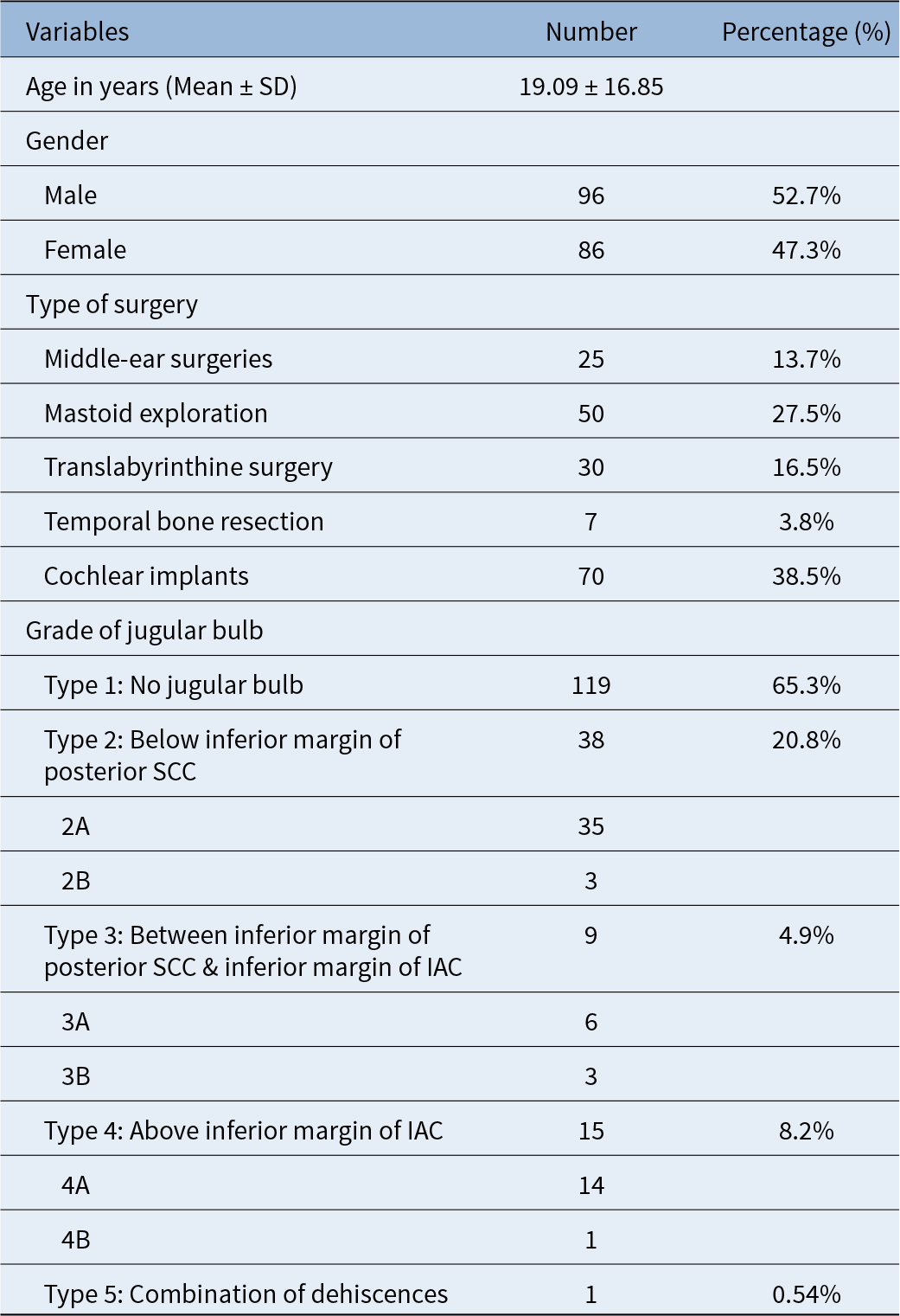
The study gathered data from 182 patients, comprising 96 males (52.7 per cent) and 86 females (47.3 per cent). The mean age of the patients was 19.09 ± 16.85 years, with a median age of 13.5 years (range 4–30 years). The relatively younger age group may be attributed to most patients being paediatric patients undergoing cochlear implantation. Table 1 shows clinico-demographic details of the cohort.
Clinico-demographic features; SCC = semicircular canal; IAC = internal auditory canal

Incidence and inter-observer agreement of high-riding jugular bulbs
Table 1 describes the distribution of various types of jugular bulbs. The grades of the jugular bulb in the study were categorised as follows: Type 1, indicating no jugular bulb, was observed in 119 patients (64.4 per cent). Type 2, where the jugular bulb is below the inferior margin of the posterior semicircular canal, was seen in 38 patients (15.3 per cent), and was further subdivided into 2A (35 patients) and 2B (3 patients). Type 3, with the jugular bulb between the inferior margin of the posterior semicircular canal and the inferior margin of the internal auditory canal, was noted in nine patients (4.9 per cent), including 3A (6 patients) and 3B (3 patients). Type 4, where the jugular bulb is above the inferior margin of the internal auditory canal, was found in 15 patients (8.2 per cent), and was subdivided into 4A (14 patients) and 4B (1 patient). Finally, Type 5, representing a combination of dehiscences, was present in one patient (0.5 per cent).
There was substantial concordance between the pre-operative imaging classification of the jugular bulb using the Manjila classification and intra-operative findings, with a Cohen’s kappa coefficient of 0.78 (95 per cent confidence interval (CI) = 0.72–0.84) between the neuro-otologist and neuroradiologist. This indicates a strong agreement between their assessments. The agreement between two neuro-otologists was also strong, with a Cohen’s kappa coefficient of 0.82 (95 per cent CI: 0.76–0.88).
The contributory value of individual grades of classification
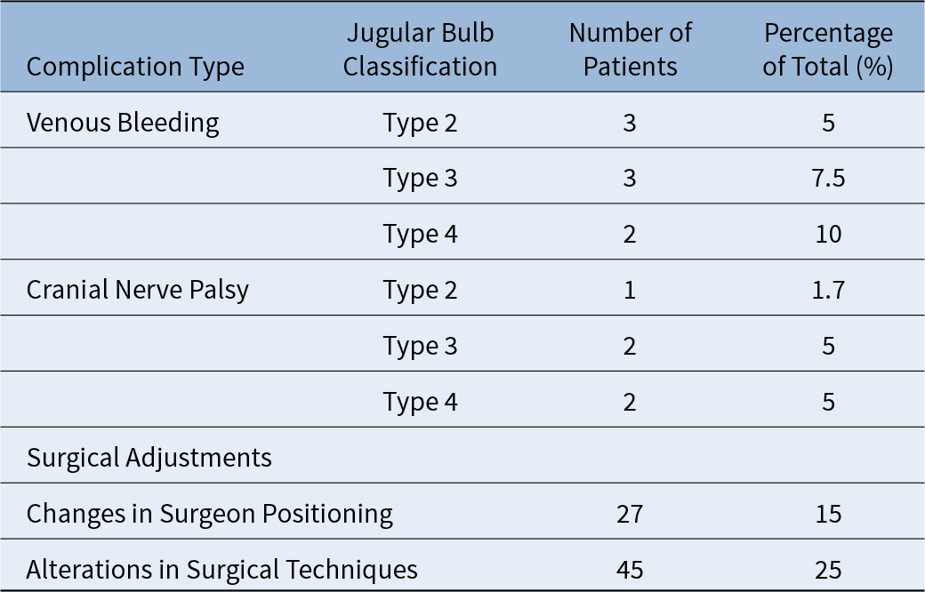
Table 2 details the complications and surgical implications of the jugular bulb. The complications were documented on the left, right, and bilateral sides, with most patients experiencing complications on the right side (118 patients, 64.8 per cent), followed by the left side (58 patients, 31.9 per cent), and bilateral complications in six patients (3.3 per cent). The incidence of surgical complications related to the jugular bulb was noted in 15 per cent of the cases across various grades of high-riding jugular bulbs. Specifically, venous bleeding was the most common complication, occurring in 10 patients (5.5 per cent). The incidence of venous bleeding varied with the classification of the jugular bulb, observed in three patients (5.0 per cent) with Type 2, three patients (7.5 per cent) with Type 3, and two patients (10 per cent) with Type 4. Cranial nerve palsy was observed in five patients (2.7 per cent), distributed across different grades of jugular bulb: one patient (1.7 per cent) with Type 2, two patients (5.0 per cent) with Type 3, and two patients (5.0 per cent) with Type 4. However, only one patient had a cranial nerve deficit (facial nerve) directly attributed to a high-riding jugular bulb. These findings underscore the increased risk of complications in patients with higher grades of jugular bulb, emphasising the importance of meticulous pre-operative planning and intra-operative management.
Intra-operative complications and adjustment

Alterations in the surgical corridor and surgical techniques were required in 25 per cent of the cases, and changes in surgeon positioning were required in 15 per cent of the cases, to manage the high-riding jugular bulb. The surgeon’s assessment of the procedural difficulty in the presence of a high-riding jugular bulb averaged 3.8 on a Likert scale (from 1 to 5), indicating a perceived high difficulty.
Functional outcomes revealed that hearing preservation was achieved in 85 per cent of the patients, while facial nerve function was preserved in 98.6 per cent. There was no correlation between the jugular bulb classification and functional outcomes, according to the Pearson correlation (r = 0.45, r = 0.067, respectively), with reduced complication rates observed in patients with lower grades of the Manjila et al.Reference Manjila, Bazil, Kay, Udayasankar and Semaan3 classification.
Discussion
The Manjila et al.Reference Manjila, Bazil, Kay, Udayasankar and Semaan3 classification, a comprehensive staging for jugular bulb, said to have utility in both otological and lateral skull base procedures. In our attempt to validate this staging system, we found a significant incidence (13.7 per cent) of high-riding jugular bulbs. Strong concordance between pre-operative imaging and intra-operative findings was observed, with Cohen’s kappa coefficients of 0.78 and 0.82 for the neuro-otologist and neuroradiologist and between two neuro-otologists, respectively. Complications occurred in 15 per cent of cases, with venous bleeding being the most common. Higher Manjila et al.Reference Manjila, Bazil, Kay, Udayasankar and Semaan3 grades correlated with increased complication rates. Alterations in the surgical approach were necessary in a significant number of cases, and the procedure’s difficulty was rated high. Functional outcomes showed 85 per cent hearing preservation and 90 per cent facial nerve function, with no significant correlation between jugular bulb classification and functional outcomes, but reduced complication rates in lower Manjila et al.Reference Manjila, Bazil, Kay, Udayasankar and Semaan3 classification grades.
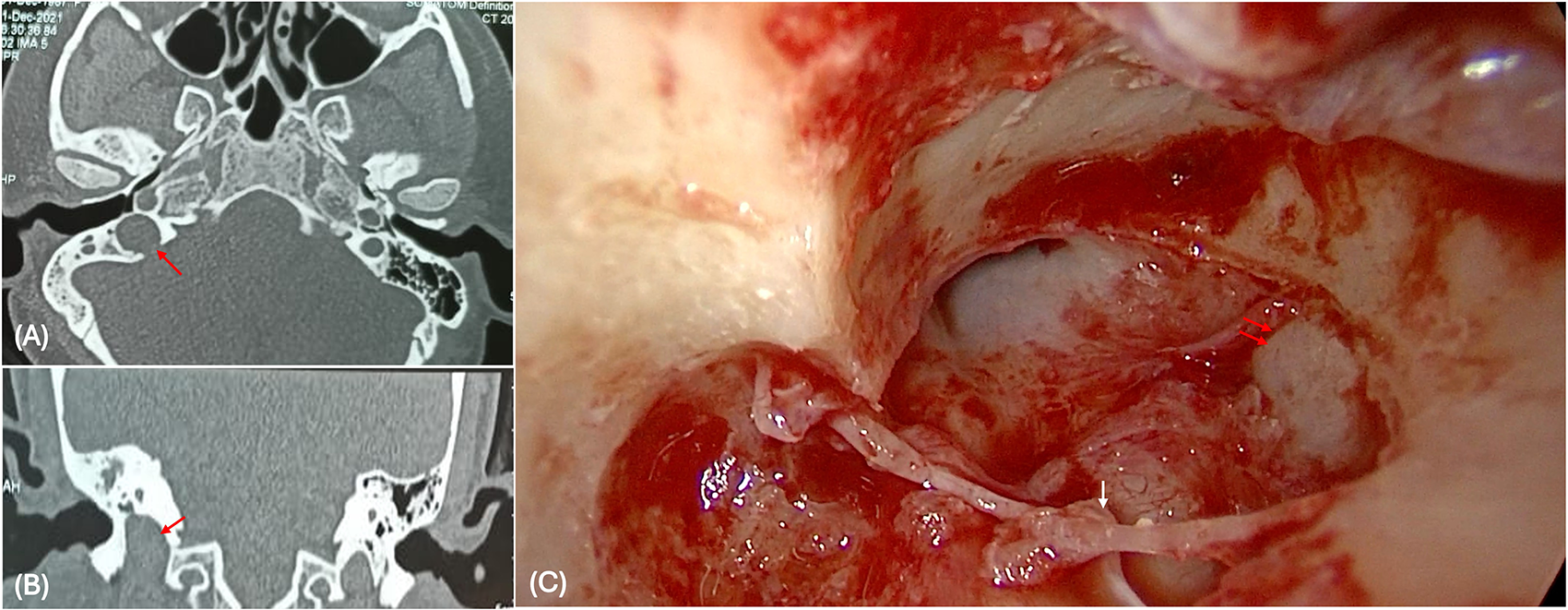
The routine practice of tympanomeatal flap elevation, used in basic otological surgeries and advanced lateral skull base procedures, typically mesotympanum, would be first entered in the inferior quadrant to preserve chorda tympani (e.g. 6-o’clock to the 9-o’clock position on the right side). It is crucial to recognise the importance of this step, particularly in cases classified as Type 2 or higher, especially Type b according to the Manjila et al.Reference Manjila, Bazil, Kay, Udayasankar and Semaan3 classification (Figure 1).
High-resolution CT temporal bone, axial view (A) and coronal view (B) showing the Type 2 jugular bulb right side; (C) intra-operative view of jugular bulb in the hypotympanum (arrow) during tympanomastoid surgery.

The retro facial dissection in the Type B tympanic paraganglioma case is nearly impossible in Type 3 and beyond and may need canal wall down mastoidectomy with or without facial nerve mobilisation for complete exenteration of the tumour. In these cases, a strategic approach may be necessary to avoid potential injury to the jugular bulb, which could occur if the traditional method is followed. Among the many available classifications for the high-riding jugular bulb, the Basava–Prasad grading, based on major endoscopic landmarks such as the inferior tympanic annulus, round window niche, and stapes, is helpful for basic otological cases involving the middle ear.Reference Prasad, Basava, Gopinathan, Induvarsha, Harshita and Ashok6 However, it does not address significant landmarks in lateral skull base surgery and is challenging to apply precisely in radiology. In higher-grade jugular bulb, the surgeon may need to change their position frequently to avoid instrumentation over the high-riding jugular bulb.
Using the current classification, surgeons performing tympanoplasty need not fear a Type 4 high-riding jugular bulb if it is bone-covered. However, any B types and Type 5 should be approached cautiously, although none is a contraindication for surgery.Reference Atmaca, Elmali and Kucuk7 Not all tympanoplasty patients typically undergo pre-operative radiology; therefore, meticulous examination under the microscope, especially of the hypotympanum, is essential after entering the middle ear.Reference Ferri, Fermi, Alicandri-Ciufelli, Villari and Presutti8 These principles also apply to grommet insertions and stapes surgery, but in the latter, the added risk of an overriding bulb hampering the footplate must be ruled out.
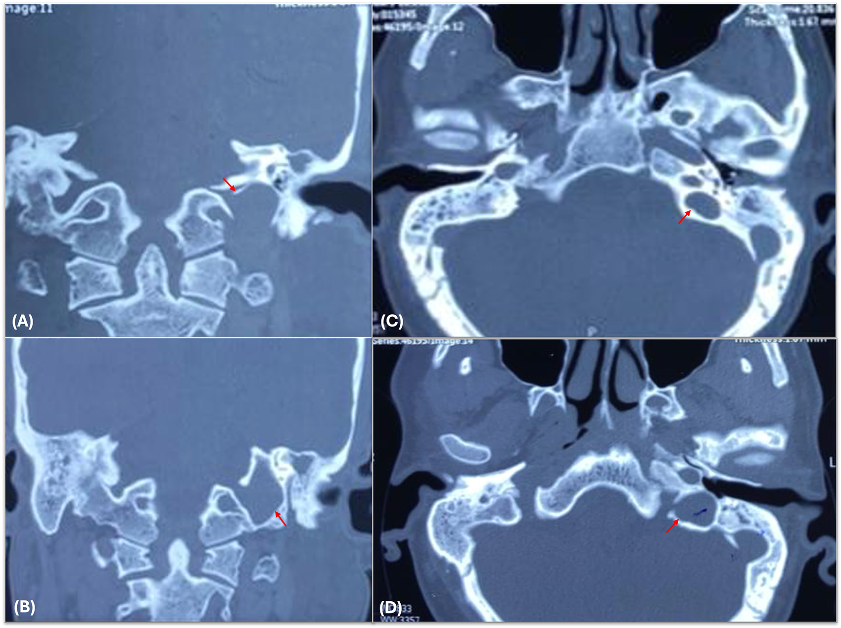
For mastoid exploration, with or without cholesteatoma, a canal wall down procedure requires similar caution, particularly regarding a dehiscent bulb when removing the cholesteatoma sac.Reference Takata, Anzai, Hara, Okada, Matsumoto and Ikeda9, Reference Fox, Nash and Tatler10 Literature suggests that even if dehiscent, the bulb is typically located away from the ossicular chain and sac, with no adhesion between them. In canal wall down procedures, precautions are necessary for a dehiscent bulb, and bulbs classified as greater than Type 3a should be handled with care to avoid injury while lowering the facial ridge near the annulus (Figure 2).Reference Satar, Yazar, Ceyhan, Arslan and Aydin11 In cases of petrous apex cholesteatoma, exenteration of all air cells to the petrous apex poses a risk of injury to bulbs classified as Type 2b and higher when addressing peri-labyrinthine and retro-facial cells.Reference Pandya, Piccirillo, Mancini and Sanna12
High-resolution CT temporal bone, coronal view (A, B) and axial view (C, D) showing the Type 4a jugular bulb in a patient with left chronic otitis media squamous active type.

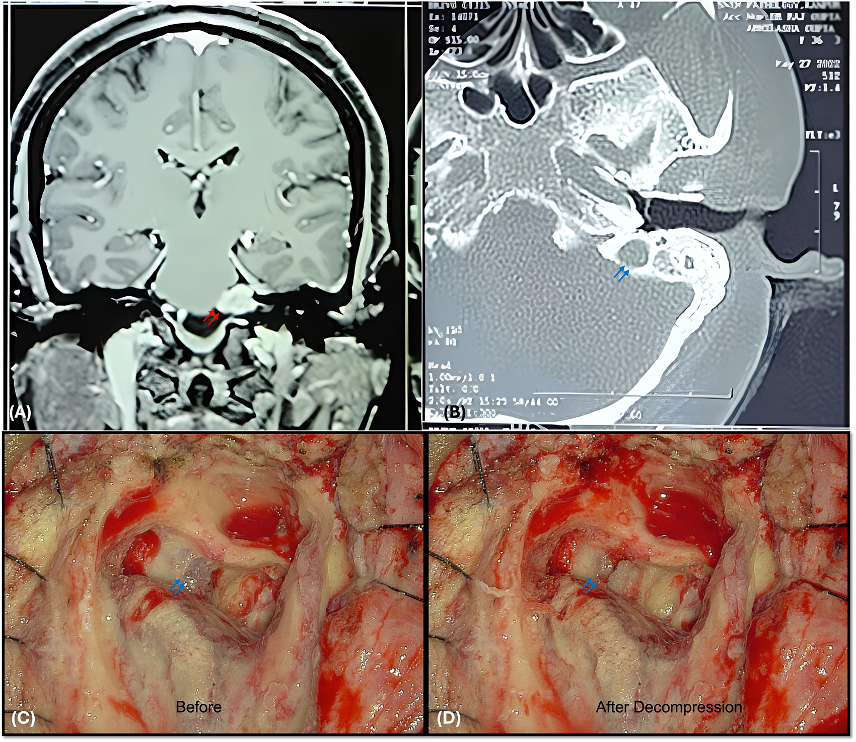
High-riding jugular bulbs pose a challenge in lateral skull base surgeries, as one encounters this structure in the initial steps of the surgery. If not previously diagnosed, a high-riding jugular bulb can lead to significant and life-threatening bleeding if the venous wall is unintentionally opened during the drilling of the posterior wall and the inferior margin of the internal auditory canal. A translabyrinthine approach is commonly preferred by neuro-otologists for the excision of vestibular schwannoma, requiring the creation of superior and inferior troughs around the internal auditory canal (Figure 3).Reference Nickele, Akture, Gubbels and Başkaya13
(A) T1W contrast MRI showing left cerebellopontine angle tumour; (B) high-resolution CT temporal bone, axial view showing a Type 3 jugular bulb on left side; (C, D) intra-operative image showing the translabyrinthine approach to cerebellopontine angle tumour, with jugular bulb being decompressed.

Types 3 and 4 jugular bulb should be managed cautiously by meticulous decompression of the bulb in the subperiosteal plane, with Type 4 considered a relative contraindication, especially if the opposite sinus is hypoplastic. The goal is to achieve adequate internal auditory canal tumour exposure without injuring the jugular bulb. For Types 3 and 4, the bone over the bulb can be made egg-shell thin and compressed to expose the posterior internal auditory canal wall.
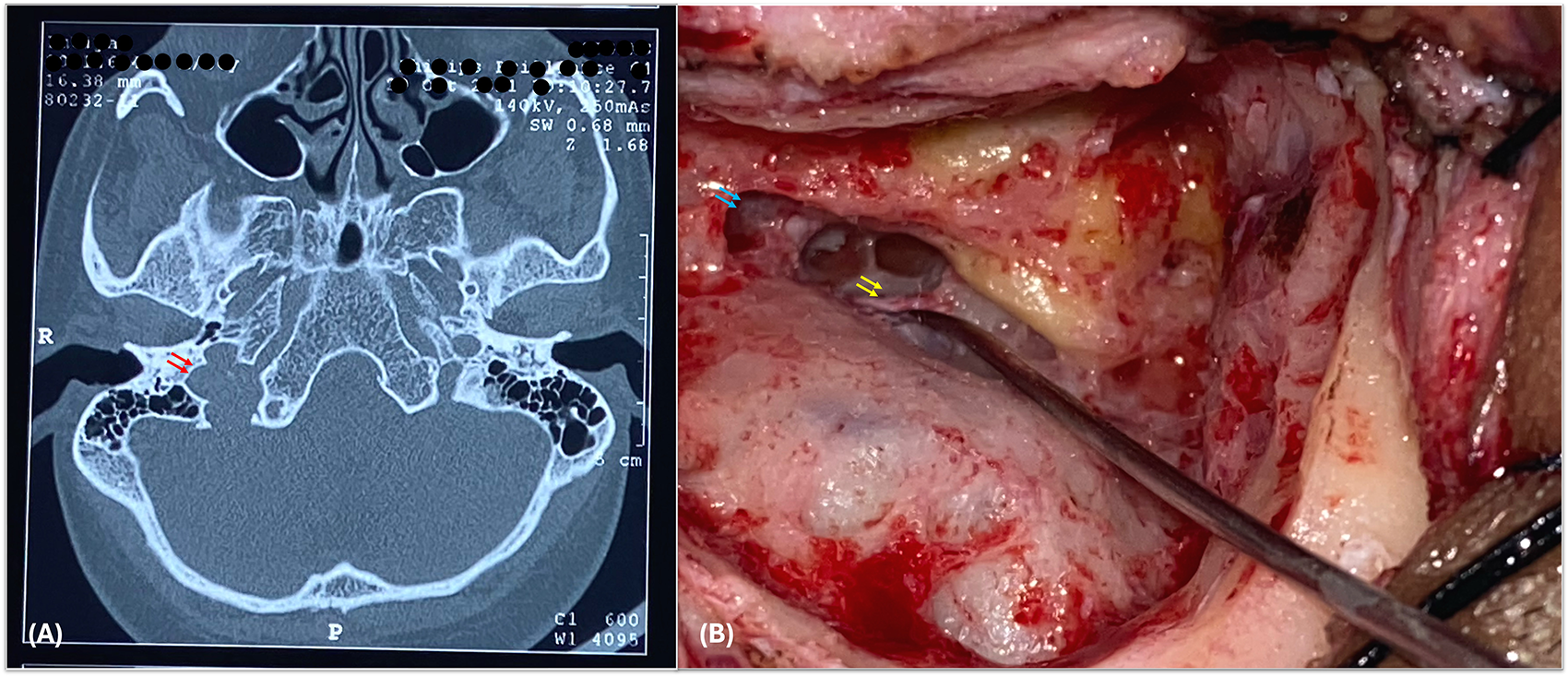
Pre-operative diagnosis of a high-riding jugular bulb often inclines surgeons toward the retrosigmoid approach. Literature indicates that, even the middle fossa approach can be used, offering a superior route to expose the intracanalicular portion of the tumour. However, it presents drawbacks, such as inadequate cerebellopontine angle exposure and more facial nerve manipulation compared to the retro sigmoid approach.Reference Gidley, Passer, Page and DeMonte14, Reference Staecker, JB, Ojeman, Ronner and McKenna15 Even posterior cranial fossa surgery, such as endolymphatic sac decompression, can also be challenging because of a high-riding jugular bulb (Figure 4).
(A) High-resolution CT temporal bone, axial view showing a Type 2 jugular bulb on right side; (B) intra-operative image of endolymphatic decompression surgery showing jugular bulb marked by blue arrows and yellow arrows showing the endolymphatic sac.

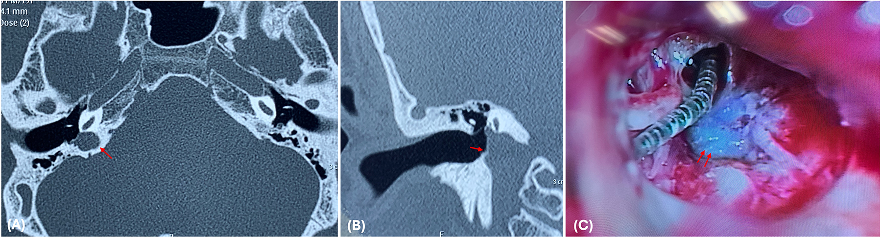
Another common neuro-otological surgery is the cochlear implantation. In posterior tympanotomy for cochlear implantation, a bulb classified as greater than 3a is risky, while the veria technique (transcanal) has no clear contraindications but requires further evaluation of any jugular bulb dehiscence in the middle ear due to potential bleeding during blind manipulation in the facial recess area.Reference Hiremath, Biswas, Mndebele, Schramm, Ertl-Wagner and Blaser16 Irrespective of the surgical technique, tympanomeatal flap elevation should be cautious as the common manipulation sites are retro-tympanum and hypotympanum. An abnormal course of the sigmoid sinus, malformed cochlea, high jugular bulb, low-lying dura, and sclerosed mastoid are highly risky for the posterior tympanotomy approach, making the veria method preferable in such cases (Figure 5).Reference Nagaraj, Singhal, Singh, Sharma, Sharma and Jat17
High-resolution CT temporal bone, axial view (A) and coronal view (B) showing the Type 3b jugular bulb on right side; (C) intra-operative view of Type 3b jugular bulb in the retro-tympanum during cochlear implant surgery.

The clinical grading system for the jugular bulb proposed by Prasad et al. has limited utility, as it primarily focuses on endoscopic landmarks to define the grades of the high-riding jugular bulb.Reference Prasad, Basava, Gopinathan, Induvarsha, Harshita and Ashok6 While the round window is a consistent landmark, the grading system significantly underrepresents the jugular bulb. For a neuro-otologist, the jugular bulb’s extension along the lateral skull base is more relevant than its confinement to the middle ear.
Based on our analysis, we recommend the following: firstly, specify the middle-ear location of the jugular bulb within each subclass, distinguishing between the retro-tympanum and the hypotympanum. Secondly, in cases of retro-tympanic dehiscence, the relationship with the round window should be included in each subclass, providing greater relevance and utility for a skull base surgeon. For example, in the case of a Type 3 hypotympanic bulb, the surgeon can safely execute the posterior tympanotomy for cochlear implantation. Conversely, in a Type 3 retro-tympanic bulb occupying the round window niche, the surgeon should be more cautious about the feasibility of posterior tympanometry and think about alternative surgical strategies for cochlear implantation.
• The jugular bulb, particularly when high riding, complicates lateral skull base surgeries, especially in otological procedures
• The Manjila classification, based on skull base landmarks, aids in pre-operative planning
• A retrospective study validated this classification using high-resolution computed tomography venography, finding substantial concordance with intra-operative observations
• Complications occurred in 15 per cent of cases, with higher jugular bulb grades linked to increased surgical difficulty
• Surgical approaches were modified in 25 per cent of cases, underscoring the need for precise pre-operative planning
• Functional outcomes were positive, with 85 per cent hearing preservation and 98.6 per cent facial nerve function
• The study supports the utility of jugular bulb classification in optimising surgical strategies and reducing complications
Conclusion
In conclusion, the study underscores the importance of a comprehensive jugular bulb classification system, such as the one proposed by Manjila et al.,Reference Manjila, Bazil, Kay, Udayasankar and Semaan3 in otological and lateral skull base surgery. Accurate classification proved essential in guiding surgical planning and intra-operative decision-making, with a significant incidence of high-riding jugular bulb observed among patients. The study demonstrated strong concordance between pre-operative imaging and intra-operative findings, validating the utility of the classification system. Complications, though present in 15 per cent of cases, were effectively managed with appropriate adjustments in surgical approach. Functional outcomes, including hearing preservation and facial nerve function, remained favourable, highlighting the overall success of surgical interventions. Adding further details, such as the position of the bulb in the middle ear and its relation to a round window, will further enhance its utility among neuro-otologists.
Level of evidence
Level 2.
Data availability statement
The datasets used and analysed during the current study are available from the corresponding author on reasonable request.
Funding statement
The authors declare that they have no known competing financial interests or personal relationships that could have appeared to influence the work reported in this paper.
Competing interests
All authors declare that they have no conflicts of interest.
Ethics standards
Approved by Institutional Ethical Committee, Sanjay Gandhi Postgraduate Institute of Medical Sciences.
Consent for publication
Each case was selected after obtaining informed consent from the patient for publication.
Authors’ contributions
All authors were equally involved in conception of the work, data collection, data analysis and interpretation, drafting the article, critical revision of the article, and final approval of the version to be published.
Acknowledgement
Sunil Manjila, McLaren Bay Region Hospital, Bay City, Michigan.