Introduction
Filarial infections are a major health hazard, with important medical, veterinary and economical implications (World Health Organization, 1997). Several species of filariae infect more than 200 million humans, causing serious disabilities, and other zoonotic species can infect a large number of different vertebrates, which can be totally asymptomatic or even die as the consequence of the infection (Urquhart et al., Reference Urquhart, Armour, Duncan, Dunn and Jennings1987). These parasites are transmitted by haematophagous arthropods during a blood meal, when infective larvae actively leave the vector to enter the vertebrate host. The planning of effective control strategies for these potentially zoonotic infections requires the correct identification of the nematode at the species level, both as a developing adult in the vertebrate host and as a larva in the vector. Unfortunately, larval stages cannot be identified by classical morphology (Cancrini & Kramer, Reference Cancrini, Kramer, Simon and Genchi2001), therefore molecular diagnostic approaches, which allow the identification of the parasites in all developmental stages, are very advantageous and particularly suitable for research and surveillance purposes, and significant from a public health perspective (Favia et al., Reference Favia, Lanfrancotti, Della Torre, Cancrini and Coluzzi1996).
The aim of this paper is to report the molecular characterization of filarial nematodes identified morphologically as Micipsella numidica, recorded here for the first time in specimens of Italian Lepus europaeus. The genus Micipsella, a filarial taxon reported as characteristic ‘of the abdominal cavity of rodents’ (Seurat, Reference Seurat1917) and found in lagomorphs only (which until the beginning of the 20th century were considered a superfamily of the order of rodents), belongs to the subfamily Splendidofilariinae (Chabaud & Choquet, Reference Chabaud and Choquet1953), which comprises filariae of reptiles, birds and mammals. Nowadays, three species are recognized in the genus: M. numidica (Seurat, Reference Seurat1917), M. indica (Rao, Reference Rao1938) and M. brevicaudata (Lyons & Hansen, Reference Lyons and Hansen1961), described in Africa, Europe, Asia and the USA. No data are available about intermediate hosts of the genus Micipsella, but experimental results on other Splendidofilariinae, not confirmed by entomological field investigations, suggest the involvement of Culicidae, Simulidae, Psicodidae and Ceratopogonidae (Bain & Chabaud, Reference Bain and Chabaud1986; Anderson, Reference Anderson2000).
Materials and methods
Seven worms, collected by a hunter from the abdominal cavity of four hares killed on the hills of Bologna province (44°23′26″N; 11°27′35″E) during the hunting season 2013, were identified morphologically and then preserved in 70% ethanol for molecular characterization.
DNA was extracted from a section of each worm (250 mg) using a commercial kit (NucleoSpin Tissue, Macherey-Nagel, Duren, Germany), according to the manufacturer's instructions. As previously described for barcoding of filarioids (Ferri et al., Reference Ferri, Barbuto, Bain, Galimberti, Uni, Guerrero, Ferté, Bandi, Martin and Casiraghi2009), 5S rDNA (about 400 bp), cox1 (about 650 bp) and 12S rDNA (about 450 bp) gene fragments were amplified using primers and thermal profiles described in Xie et al. (Reference Xie, Bain and Williams1994), Casiraghi et al. (Reference Casiraghi, Anderson, Bandi, Bazzocchi and Genchi2001) and Casiraghi et al. (Reference Casiraghi, Bain, Guerrero, Martin, Pocacqua, Gardner, Franceschi and Bandi2004), respectively.
Polymerase chain reaction (PCR) amplification was performed in a final volume of 25 μl under the following final conditions: 1 × buffer including 1.5 mm MgCl2, 0.2 mm of each deoxynucleotide triphosphate (dNTP), 1 μm each of forward and reverse primers, and 1 unit of polymerase (BIOTAQ™ DNA Polymerase, Aurogene, Rome, Italy). To test the specificity of the reaction, 2 μl of DNA extracted from Dirofilaria immitis and Enterobius vermicularis were included in each PCR run as positive and negative controls, respectively.
Amplicons, purified using the SureClean kit (Aurogene, Rome, Italy) and following the manufacturer's instructions, were then directly sequenced with PCR primers in both directions by an external sequencing core service (Eurofins MWG Operon, Anzinger, Germany). The resulting chromatograms were analysed and edited.
The sequences obtained were compared to Onchocercidae sequences deposited in GenBankTM and available at the website http://www.ncbi.nlm.nih.gov/genbank/ by using the BLAST application, and aligned using the software CLUSTAL X (www.clustal.org) for comparative analysis.
The maximum likelihood (ML) phylograms based on cox1 and 12S sequence data were reconstructed with MEGA v.5.0 (Tamura et al., Reference Tamura, Peterson, Peterson, Stecher, Nei and Kumar2011) using the Kimura 2-parameter distance model. The robustness of nodes was assessed with 500 bootstrap replicates, and sequences from Ascaris lumbricoides were used as the out-group.
Results and discussion
Parasites were identified as M. numidica, according to previous descriptions by Graber (Reference Graber1972) and Cancrini et al. (Reference Cancrini, Poglayen and Vecchi1988). This species differs from the other two species of Micipsella reported in the hare, especially due to the presence of caudal cuticular protuberances in the male. Furthermore, the M. brevicaudata male has a short and straight tail and has no post-cloacal papillae, whereas the M. indica male has a long tail, three pairs of post-cloacal papillae and the right spicule longer than 110 μm.
The PCR amplification of target genes from each DNA sample yielded amplicons of the expected size, which did not display any significant identity with sequences deposited in GenBankTM. No intraspecific differences were detected among specimens for each gene sequence examined. The BLAST analysis of the cox1, 12S and 5S sequences evidenced the highest nucleotide identity (87%, 86% and 85%, respectively) to those of Chandlerella quiscali available in GenBankTM (accession nos. JQ867051.1, HM773029.1 and HM641830.1).
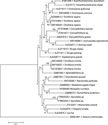
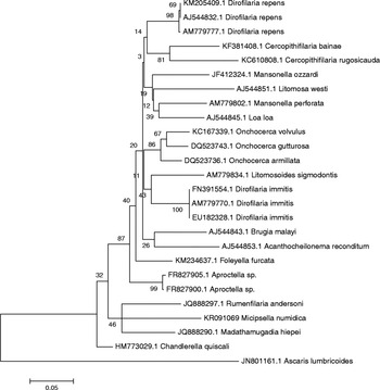
Since 5S rDNA sequences of Spledidofilariinae are available only for C. quiscali, the phylogenetic analysis was conducted using only the mitochondrial gene markers (cox1 and 12S). The analysis based on cox1 sequences yielded a phylogram in which M. numidica is not strongly related to any filarioids and it groups in the same clade as Splendidofilariinae of other mammals (Rumenfilaria andersoni), birds (Chandlerella) and reptiles (Madathamugadia hiepei), in which Mansonella perforata (Onchocercinae) is also present (fig. 1). The tree generated by including sequence data from 12S rDNA of Onchocercidae and M. numidica better evidenced, even if without robust bootstrap values, a close relationship of this filarial species with Splendidofilariinae of mammals and reptiles, and a lower relationship with Splendidofilariinae of birds (Chandlerella and Aproctella) (fig. 2).
A maximum likelihood phylogram derived from cox1 sequences of Micipsella numidica and the Onchocercidae rooted against Ascaris lumbricoides (accession number AB591801.1). Numbers at the nodes indicate percentage bootstrap support from 500 replicates using the Kimura 2-parameter distance model inferred with MEGA v.5.0 (Tamura et al., Reference Tamura, Peterson, Peterson, Stecher, Nei and Kumar2011).

A maximum likelihood phylogram derived from 12S rDNA sequences of Micipsella numidica and the Onchocercidae rooted against Ascaris lumbricoides (accession number JN801161.1). Numbers at the nodes indicate percentage bootstrap support from 500 replicates using the Kimura 2-parameter distance model inferred with MEGA v.5.0 (Tamura et al., Reference Tamura, Peterson, Peterson, Stecher, Nei and Kumar2011).

The nucleotide sequences from the cox1, 12S and 5S partial genes analysed in this paper are available in the GenBankTM database under accession numbers KR232088, KR091069 and KR232089.
We performed the first molecular characterization of M. numidica by amplification, sequencing and analysis of mitochondrial (cox1 and 12S) and ribosomal (5S) target genes. The molecular phylogenetic history inferred using sequences of a barcoding data set and those produced in this study supports the taxonomic position of M. numidica and shows substantial differences between Splendidofilariinae and other Onchocercidae subfamilies. This difference had been also evidenced through the absence (in Aproctella, Rumenfilaria and Chandlerella) of endosymbiotic bacteria of the genus Wolbachia (Lefoulon et al., Reference Lefoulon, Gavotte, Junker, Barbuto, Uni, Landmann, Laaksonen, Saari, Nikander, de Souza Lima, Casiraghi, Bain and Martin2012) present in about all filarial worms and in the Splendidofilariinae M. hiepei. We screened the available specimens of M. numidica for the presence of Wolbachia using primers for the ftsZ gene proposed in the cited study (Lefoulon et al., Reference Lefoulon, Gavotte, Junker, Barbuto, Uni, Landmann, Laaksonen, Saari, Nikander, de Souza Lima, Casiraghi, Bain and Martin2012), with a preliminary result confirming its absence, which needs additional proof through more extensive investigations.
In conclusion, this study, apart from reporting M. numidica for the first time in Italian Lepus europaeus (useful primarily for adding another piece to the map of M. numidica in European hosts), contributes to knowledge of the genetic relationships among filarial nematodes. However, even more significantly, it enables the molecular/genetic diagnosis of filarial parasites of veterinary and medical concern.
Acknowledgements
The authors are grateful to G. Croce and A. Giacomi for their excellent technical assistance.
Financial support
This study has been partially supported by a grant from the Ministry of Health (GR2010-2315984).
Conflict of interest
None.